SOCIEDADE BRASILEIRA DE ORTOPEDIA E TRAUMATOLOGIA
w w w . r b o . o r g . b r
Original
Article
Functional
and
radiological
evaluation
of
acute
acromioclavicular
dislocation
treated
with
anchors
without
eyelet:
comparison
with
other
techniques
夽
Alexandre
Tadeu
do
Nascimento
∗,
Gustavo
Kogake
Claudio
HospitalOrthoservice,GrupodeOmbroeCotovelo,SãoJosédosCampos,SP,Brazil
a
r
t
i
c
l
e
i
n
f
o
Articlehistory:
Received7November2015 Accepted23February2016 Availableonline30August2016
Keywords:
Acromioclavicularjoint Sutureanchors Treatmentoutcome
a
b
s
t
r
a
c
t
Objective:Toassesstherepairresultsofacromioclaviculardislocations(ACJD)gradesIIIand
V,withanchorswithouteyelet,whencomparedwithothertechniques,andtoevaluate factorsthatcanaffectthefinalresult.
Methods:Aretrospectivestudyof36patientswithACJDgradesIIIandVintheRockwood classification,12treatedwithanchorswithouteyelet,11withonetightrope,sixwithtwo tightropes,andsixwithsubcoracoidcerclage,operatedfromSeptember2012toFebruary 2015.PatientswereassessedradiographicallyandthroughDASH,UCLA,thevisualanalog scaleofpain(VAS)andtheShort-Form36(SF-36).Surgicaltimeandthepossibleinfluence ofsomefactorsintheoutcomewerealsoassessed.
Results:ThemeanDASHscorewas6.7;UCLA,32.9;VAS,1.2;andSF-36,79.47. Radiographi-cally,thefinalmeanmeasurementwas9.93mm,withnostatisticaldifferencebetweenthe groups.ThemeansurgicaltimeforGroupIwas31min;GroupII,19min;GroupIII,29min; andGroupIV,59min.TherewasasignificantdifferencebetweenGroupsIIandIVwhen com-paredwiththestudygroup.Theinitialandimmediatepost-operativeACJDmeasurements ACJDwerecorrelatedwiththefinalmeasure.
Conclusion: TherepairofacuteACJDwithanchorswithouteyeletisaseffectiveastheother methods,withsignificantlyshorteroperativetimewhencomparedwiththesubcoracoid cerclagetechnique.Thefinalradiologicalresultisinfluencedbythecoracoclavicularinitial distanceandtheimmediatepostoperativemeasurement.
©2016SociedadeBrasileiradeOrtopediaeTraumatologia.PublishedbyElsevierEditora Ltda.ThisisanopenaccessarticleundertheCCBY-NC-NDlicense(http:// creativecommons.org/licenses/by-nc-nd/4.0/).
夽
StudyconductedattheHospitalOrthoservice,GrupodeOmbroeCotovelo,SãoJosédosCampos,SP,Brazil.
∗ Correspondingauthor.
E-mail:nascimento@icloud.com(A.T.Nascimento).
http://dx.doi.org/10.1016/j.rboe.2016.08.015
visualanalógicadedor(EVA)epeloShort-Form36(SF36).Otempocirúrgicoeapossível interferênciadealgunsfatoresnoresultadofinaltambémforamavaliados.
Resultados: Amédiadosescoresfoide6,7noDASH;32,9noUCLA;1,2naEVAe79,47no SF-36.Radiograficamente,amedidafinalmédiaentreocoracoideeaclavículafoide9,93mm, semdiferenc¸aestatísticaentreosgrupos.Quantoaotempocirúrgico,amédiadogrupoIfoi de31minutos;dogrupoII,19minutos;dogrupoIII,29minutosedogrupoIV,59minutos, houvediferenc¸asignificativaentreosgruposIIeIV,quandocomparadoscomogrupoem estudo.AmedidainicialdaLACeamedidapós-operatóriaimediata(POI)tiveramcorrelac¸ão comamedidafinal.
Conclusão: OreparodaLACagudacomâncorassemeyeletétãoeficazquantooutros méto-dosecomtempocirúrgicosignificativamentemenorquandocomparadocomatécnicade amarrilhosubcoracoide.Oresultadoradiológicofinaléinfluenciadopeladistância coraco-clavicularinicialedoPOI.
©2016SociedadeBrasileiradeOrtopediaeTraumatologia.PublicadoporElsevier EditoraLtda.Este ´eumartigoOpenAccesssobumalicenc¸aCCBY-NC-ND(http:// creativecommons.org/licenses/by-nc-nd/4.0/).
Introduction
The true incidence of acromioclavicular joint dislocations (ACJD)isnotknown,sincemanyaffectedindividualsdonot seektreatment.Approximately12%ofalldislocations involv-ingtheshoulderaffecttheacromioclavicularjoint.
Athleteswho participateincontactsports (e.g.,football, rugby, martial arts) are at higher risk. ACJD is the most commonreasonwhyathletesseekmedicalcarefollowinga traumaticeventintheshoulder;glenohumeraldislocationis thesecondmostfrequentcause.1,2
Menaremorecommonlyaffected,withanapproximate ratioof5:1,3andyoungersubjects(<35years)presentthis
con-ditionmoreoften,mainlyduetotheirgreaterparticipationin high-riskactivities.Malesinthesecondtofourthdecadesof lifehavethehighestfrequencyofACJDandpresent,inmost cases,partialinjuriesoftheligaments.3
Dependingontheseverityofthetrauma,anindividualmay injureoneoralloftheligaments,leadingtodifferentdegrees ofACJD.1 Themostcommonlyusedclassificationisthatof
Rockwood,4whichstratifiesthisconditionintosixtypes.
Themainfunctionoftheacromioclavicularjointandits ligamentsistosustainthescapulaandconnecttheupperlimb totheaxialskeleton.InACJD,thisconnectionislost;dueto gravity,thearmbecomeslowerrelativetotheclavicle,which canleadtogreatercontactoftheacromiononthetendonof thesupraspinatusmuscleandthuscausesymptomsofimpact andtendoninjury,neurologicalsymptomsduetotractionof thebrachialplexus,anddyskinesiaofthescapula.5,6
OneofthefirstmethodsofACJDtreatmentwasfixation withKirschnerwiresafterclosedreduction.Thistechnique givesgoodresults,butithasnotbeenroutinelyuseddueto rarebutpotentiallyfatalcomplicationsthatcanoccurdueto breakageandmigrationofmaterial.7
There are several surgical techniquesfor treating acute ACJD; coracoclavicular fixationwith subcoracoid ligationis oneofthemostcommonlyused.Theliteraturepresents stud-ies that compare the biomechanical differences of several techniques,butfewcompareclinicalandradiological differ-encesintheresultsofthevariousmethods.8
OneoptionforthesurgicaltreatmentofACJDisthe cora-coclavicular stabilization using sutureanchors fixedin the coracoidprocess,tyingtheknotsintheclaviclethroughbone tunnels.9,10
Resultswiththistechniquearedivergentintheliterature duetoapossibleroleoftheanchoreyelet(Fig.1),which pre-cipitates the breakage ofthe wire, thus causing procedure failure.11
Theuseanchorswithouteyelet(Fig.1),inwhichthe high-strengthwireexitsdirectlyfromtheanchoritself,maybea solutiontothisproblem;theanchorismadeofamaterial sim-ilartothatofthewire,avoidingthecontactofthelatterwith amorerigidmaterialthatcouldbreakit.
Material
and
methods
Fig.1–Differencebetweenanchorswitheyelet(arrow)andwithouteyelet.
surgeonatasinglecenterbetweenSeptember2012and Febru-ary2015,wereretrospectivelyreviewed.Age,gender,side,and ACJDclassificationdistributionisshowninTable1.Thestudy includedpatientswithshouldertraumawhohadACJDgrades
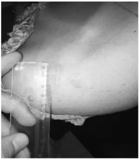
IIIor V,and who were operatedinupto 30daysfrom the timeofinjury.In additiontoconventional radiographs(AP, scapulaprofile,andaxillaryprofile),allradiographsfor diag-nosisweremadeintheorthostaticposition,withaweightof 2.5kgoneachlimb,featuringbothacromioclavicularjointsin sameimage(Fig.2).Theminimumfollow-uptimewassetas sixmonths.Theexclusioncriteriaintheselectionofpatients comprisedcasesofACJDgradeIV,casesassociatedwith frac-turesatothersitesoftheshouldergirdle,andcasesthatwere operated30daysafterinjurydate.
Surgicaltechnique
Surgerywasperformedwithpatientundergeneral anesthe-siaandbrachialplexusblock,inabeachchairposition. An incisionofapproximately 2–3cm(Fig.3)wasmadedirectly onthedistalendoftheclavicle,whichwasosteotomizedin its distal0.5cm and removed together with the meniscus, asdescribedbysomeauthorsinspecificcases.12 Anteriorly
tothe clavicle,thecoracoidwas digitallyidentifiedby pal-pation, i.e., without directvisualization,the authors would positionthe anchorinsertionguide directlyon itssuperior face.Twodouble-loaded2.9-mmanchors(Juggerknot-Biomet) wereusedinallcases.Fourbonetunnelswerecreatedinthe clavicleusinga2-mmdrill,2cmfromtheendoftheclavicle; tunnelsweresquare-shaped, with1cmbetweenthem.Two
Fig.2–Standardstressradiographypresentingthe acromioclavicularjointsinthesameimage,demonstrating anACJDVtotheleft.
wireswerepassedthrougheachofthemtorepairthe dislo-cation.Withthesesamewires,thedeltoidandtrapeziuswere reinserted;theseareoftenaffected,mainlyinACJDgradeV
lesions.
Postoperativeperiod
Patientsremainedincontinuousimmobilizationwithasling forsixweeks,afterwhichrehabilitationwasinitiated. Phys-icaltherapywasinitiallyindicatedonlyforrangeofmotion gain;afterthiswascompleted,muscle-strengtheningphase wasinitiated,lastingaboutthreemonths.
Statisticalanalysis
Theresultsofthescoresofdifferentgroupswereanalyzedin SPSS(IBM)usingtheKruskal–Wallistest,whichissimilarin methodologytotheMann–Whitney,butallowsforthe assess-mentofmorethantwogroupssimultaneously.Surgicaltime and radiographic measurements,which were discrete vari-ables withnormaldistribution,were analyzedbyStudent’s
t-test, comparedinpairs,usingExcel.For alltests,a confi-denceintervalof95%wascalculatedandp-values<0.05were
10 20 Male Right 5
11 23 Male Left 5
12 22 Male Right 5
GroupII
1 34 Male Left 5
2 60 Male Left 5
3 22 Male Left 5
4 32 Male Left 3
5 19 Male Right 3
6 36 Male Left 5
7 28 Male Left 3
8 32 Male Right 5
9 28 Male Left 5
10 22 Male Right 5
11 43 Male Right 3
GroupIII
1 50 Male Left 5
2 29 Male Left 5
3 37 Male Right 3
4 23 Male Right 5
5 35 Male Right 3
6 29 Male Right 3
7 27 Male Right 5
GroupIV
1 27 Male Left 3
2 20 Male Right 3
3 36 Male Left 5
4 69 Male Right 3
5 50 Male Right 5
6 59 Female Left 5
consideredtobesignificant.Thepossiblevariablesthatcould affectthefinalresultwereassessedinExcelusingPearson’s coefficient.Valuesbetween0and0.3wereconsideredtohave aweakcorrelation; between0.3and 0.6,moderate correla-tion;andgreater than0.6,strongcorrelation.Wheninverse relationshipoccurs,valuesarenegativeandwereconsidered usingthesameprinciple.
Results
Themedicalrecordsof36patientsoperatedinthisservicebya singlesurgeonfromSeptember2012toFebruary2015were ret-rospectivelyreviewed.Patientsweredividedintofourgroups accordingtothesurgicaltechniqueused:minimallyinvasive surgeryusinganchorswithouteyelet(GroupI);arthroscopy withuse ofa tightrope(GroupII); arthroscopy withuse of twotightropes(GroupIII);and,openrepairwithsubcoracoid ligationusingfourhigh-resistancewires(GroupIV)(Fig.4).
Themeanageofthepatientswas33.4years,withno signifi-cantdifferencebetweenthegroups(p=0.696).Meanfollow-up was20.2months(6–38.03).RegardingthecausesofACJD,24 (67%)occurredduetosportingaccidents, nine(25%)dueto caraccidents,andthree(8%)duetohouseholdaccidents.As fortheside,15(42%)occurredontherightand21(58%)ofthe left;thedominantsidewasaffectedin16(44%)cases.Mean preoperativedistancebetweenthecoracoidandclaviclewas 19.34mm(10.86–29.38);regardingtheclassification,23cases ofACJDVand13ACJDIII,therewasnosignificantdifference betweengroups(Table2).Meantimebetweentheinjuryand surgerywas7.57days(1–30).
Meantimeofsurgicalprocedure was31mininGroup I, 19mininGroupII,29mininGroupIII,and59mininGroup
Fig.4–Immediatepostoperativeimage.a
aGroupsI,II,III,andIV,fromlefttoright,respectively.
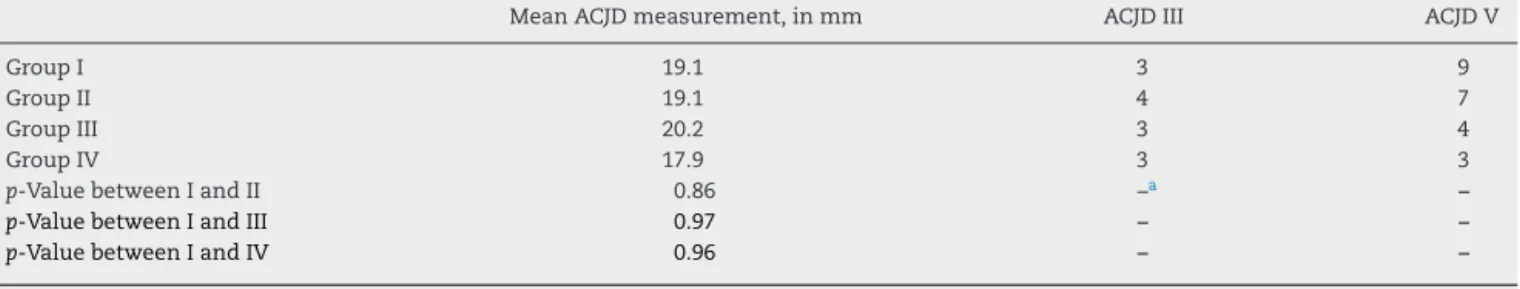
Table2–Measurementsofthedistancebetweenthecoracoidandclavicle,andquantitativeanalysisoftheACJD classification.
MeanACJDmeasurement,inmm ACJDIII ACJDV
GroupI 19.1 3 9
GroupII 19.1 4 7
GroupIII 20.2 3 4
GroupIV 17.9 3 3
p-ValuebetweenIandII 0.86 –a –
p-ValuebetweenIandIII 0.97 – –
p-ValuebetweenIandIV 0.96 – –
a Asthisisascorepresentingnon-normaldistribution,itwasnotpossibletousethet-testtocalculatethep-value.
Table3–Surgicaltimeandpre-andpostoperativemeasurementsofthecoracoclavicularspacewithlong-termlossesof thereductionachievedintheimmediatepostoperativeperiod.a
Surgicaltime inminutes
Pre-op measurement
(mm)
Immediatepost-op measurement
(mm)
Final measurement
(mm)
Immediate post-opreduction
percentageloss
Periodinwhichthe lossofreduction occurred,inweeks
GroupI 31 19.1 4.89 8.23 68% 14.5
GroupII 19 19.1 5.45 11.25 106% 12.7
GroupIII 29 20 4.96 8.17 65% 18.8
GroupIV 59 18 4.27 8.86 107% 20.2
p-Value(betweenIandII) <0.000000001 0.85 0.68 0.6 0.3 0.3
p-Value(betweenIandIII) 0.12 0.97 0.95 0.47 0.4 0.42
p-Value(betweenIandIV) 0.000002 0.96 0.93 0.24 0.2 0.09
a Valuesarepresentedasmeans.Thepercentageoflossofreductionwascalculatedbycomparingtheoutcomeofthemeasurementbetween
thecoracoidandclavicle,withthemeasurementobservedintheimmediatepostoperativeperiod.
lossinmillimetersonthemeasureachievedintheimmediate post-operativeperiod,wassignificantlydifferentforGroupI
inrelationshiptoGroupsIIandIV(Table3).Themomentof lossofreductionwas,onaverage,atthe13thweek,withno differencebetweengroups.
Intheclinicalevaluationatsixmonths,oneyear,andtwo yearsaftersurgery,theDASH,UCLA,VAS,andSF-36scores wereused.MeanDASHscorewas6.7points;meanUCLAwas 32.9,with17(48%)excellentresults,18(50%)good,andone regular(2%);meanVASwas1.2points,with32(91%)casesof
minorpainandthree(9%)casesofmoderatepain;andmean SF-36scorewas79.47,withnosignificantdifferencebetween groups(Tables4and5).
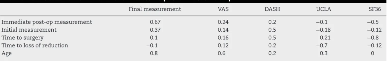
Somefactorsthatcouldbecorrelatedwiththefinalclinical andradiologicaloutcomewereassessed.Astrongcorrelation wasobservedbetweenthereductionachievedinthe imme-diatepostoperativeperiodandatfinalfollow-up,aswellasa moderaterelationshipbetweenthemeasurementatthetime oftheinjuryandfinal measurement(Table6).Fig.5shows the scatter plot for the measurement of the reduction in
Table4–Resultsofclinicalscores(UCLA,DASH,andVAS).a
UCLA DASH VAS
GroupI 32.4±2.5(26–35) 7.7±7.1(0.83–25) 1.2±1.3(0–4)
GroupII 33.4±2.3(27–35) 5.9±9.8(0–34) 1.2±2.0(0–7)
GroupIII 32.0±2.0(29–35) 5.8±9.0(0.83–25.83) 1.8±1.2(0–4) GroupIV 29.4±1.9(30–35) 6.5±19.1(0–47.5) 0.9±1.2(0–3)
Kruskal–Wallistest(p-value) 0.33 0.31 0.16
r
e
v
b
r
a
s
o
r
t
o
p
.
2
0
1
6;
5
1(5)
:561–568
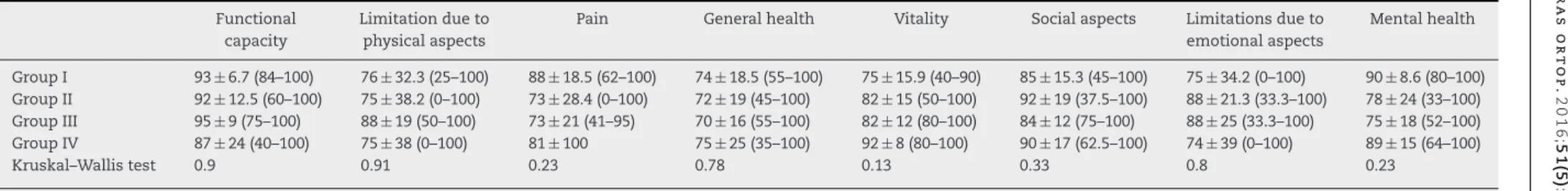
Table5–SF-36score,stratifiedbyitsareas.a
Functional capacity
Limitationdueto physicalaspects
Pain Generalhealth Vitality Socialaspects Limitationsdueto emotionalaspects
Mentalhealth
GroupI 93±6.7(84–100) 76±32.3(25–100) 88±18.5(62–100) 74±18.5(55–100) 75±15.9(40–90) 85±15.3(45–100) 75±34.2(0–100) 90±8.6(80–100) GroupII 92±12.5(60–100) 75±38.2(0–100) 73±28.4(0–100) 72±19(45–100) 82±15(50–100) 92±19(37.5–100) 88±21.3(33.3–100) 78±24(33–100) GroupIII 95±9(75–100) 88±19(50–100) 73±21(41–95) 70±16(55–100) 82±12(80–100) 84±12(75–100) 88±25(33.3–100) 75±18(52–100) GroupIV 87±24(40–100) 75±38(0–100) 81±100 75±25(35–100) 92±8(80–100) 90±17(62.5–100) 74±39(0–100) 89±15(64–100)
Kruskal–Wallistest 0.9 0.91 0.23 0.78 0.13 0.33 0.8 0.23
Table6–Correlationofvariableswiththeoutcome(Pearson’scoefficient).
Finalmeasurement VAS DASH UCLA SF36
Immediatepost-opmeasurement 0.67 0.24 0.2 −0.1 −0.5
Initialmeasurement 0.37 0.14 0.5 −0.18 −0.12
Timetosurgery 0.1 0.16 0.5 0.21 −0.8
Timetolossofreduction −0.1 0.12 0.2 −0.7 −0.12
Age 0.8 0.6 0.2 0.3 0
Association between immediate postoperative period measurement and final measurement
25 20 15 10 5 0
0 2 4 6
Immediate postoperative period measurement
Final measurement
8 10 12 14
Fig.5– Scatterplot.
theimmediatepostoperativeperiodandfinalmeasurement, showingastrongcorrelationbetweenthesemeasures.
Therewas a symptomaticlossofreduction inone(2%) casefrom GroupII,whichoccurred at14 weeks postopera-tively,requiring surgicalapproachandtreatedasachronic ACJDusingasemitendinosusgraft.Allpatientswhopracticed sportswithuseoftheupperlimb(16patients)wereableto returntothesamelevelofactivitypriortoinjury,exceptfor onepatientfromGroupII,whowasaswimmer.Therewere nosignificantcomplicationsinanyofthegroups.
Discussion
The high rate of complications associated with the vari-etyofmethodsdescribed inliteratureforthe treatmentof ACJDreflectstheinefficiencyinrestoringtheanatomyofthe acromioclavicularregion.Provisionalfixationwithpinsor cer-clageis notrecommended, dueto the increasedincidence ofdegenerativechangesoftheacromioclavicularjoint,bone erosion,andpinbreakageormigration.13Theconceptof
trans-ferofthecoracoacromialligament(Weaver-Dunnprocedure), withitsvariousmodifications,isthatsuchatransferwould withstandtensileforces asthenative ligamentdoes. How-ever,ithasbeenproventhatthecoracoacromialligamentis biomechanicallyinferiorincomparisonwiththe reconstruc-tion with semitendinosus tendon graft, leading tochronic subluxationordislocationofthe acromioclavicularjoint in 30%ofcases.14
Treatment principle for ACJD cases is reduction of the injured joint and maintenance of this reduction until the softtissuehealsandthedistalclaviclestabilizes.Suetal.10
used an anchor in place of a screw, as a modification of the Bosworth technique, and obtained satisfactory results in11 patients operated due toACJD. They concludedthat this procedure is simple,and anatomically reproduces the coracoclavicularligamentstoprovideverticalandhorizontal stabilityincasesofACJD.Theadvantagesofusinganchors
insteadofsubcoracoidligationincludeshortersurgicaltime, whichwasalsodemonstratedinthepresentstudy,andless riskofnerveandvascularinjuries,asitisnotnecessaryto addressthemedialaspectofthecoracoid.15,16Furthermore,
thenewgenerationofanchorswithouteyelethasthe poten-tial advantage of not having implant material, which can causebreakageofthehigh-strengthwireupontheircontact.11
Breslow et al.,17 in a cadaveric study, compared the
mechanicalstabilityachievedaftercoracoclavicular stabiliza-tionwiththetechniqueofsubcoracoidligaturewiththesuture anchors technique. Although the group with anchors has shownslightlybetterresults,bothmethodswere provento bestatisticallysimilar.Thesuggestedhypothesiswasthatthe ligaturehassomeaccommodationofmovementinthe sub-coracoidregion, and that it would generatelower stability. Anotherstudy,whichcomparedthebiomechanicalstrength ofEndobuttons,anchors,andhookplates,demonstratedthat thefirsttwohavebetterstabilityandresistance.18
Thelossofinitialreductionhasbeendescribedinthe liter-ature;theinaccurateinsertionsiteoftheanchorshasbeenthe reasonpointedoutbysomeauthors.9,10Thepresentauthors
believethattheobservedlossismorecloselyrelatedtothe qualityofscartissue,occurringwhenthereisaruptureofthe wiresduetofatigueandthistissuehastoassumetheroleof jointstabilizer;itisimportanttonotethatthisisahypothesis, andtodatetherearenostudiesthatcorroborateit.However, this wasobservedinthe newsurgicalapproachtothe sin-glecasethatrequiredanother surgeryduetosymptomatic lossofreduction.Inthepresentstudy,weobservedthatin allcasesfromthefourgroups,therewasalossofreduction comparedtowhatwasachievedintheimmediate postopera-tiveperiod;thislossofreductionoccurred aroundthe13th week.Theauthorsalsohypothesizedthatthequalityofscar tissueisdirectlydeterminedbythestabilityachievedbythe fixationmethod,whichwasverifiedinthepresentstudy,as thesmallestlosses,inastatisticallysignificantmanner,were observedpreciselyinthemethodsthatpresentedgreater sta-bilityinbiomechanicalstudies.17,18Assomelossofreduction
isexpectedtooccur,toagreaterorlesserdegree,theauthors soughttoperformahyper-reductioninallcasesinthepresent study. A strong correlation between the immediate post-operativemeasurementandthefinalmeasurewasobserved. Thegreaterthehyper-reduction,thesmallerthefinal radio-graphic measurementofthe coracoclavicular region. Thus, the final resultwasestheticallyand radiologically satisfac-tory,withoutimpactingthefunctionalresult.Studiesinthe literatureshowthatthelossofreductiondoesnotaffectthe clinicaloutcomeofthetreatment.19–21Thiswasalsoobserved
alwaysbeattempted,aimingformorefavorableradiographic andestheticresults.
Conflicts
of
interest
Theauthorsdeclarenoconflictsofinterest.
r
e
f
e
r
e
n
c
e
s
1. LapradeRF,SurowiecRK,SochanskaAN,HentkowskiBS, MartinBM,EngebretsenL,etal.Epidemiology,identification, treatment,andreturntoplayofmusculoskeletal-basedice hockeyinjuries.BrJSportsMed.2014;48(1):4–10.
2. LynchTS,SaltzmanMD,GhodasraJH,BilimoriaKY,Bowen MK,NuberGW.Acromioclavicularjointinjuriesinthe NationalFootballLeague:epidemiologyandmanagement. AmJSportsMed.2013;41(12):2904–8.
3. RockwoodCAJr,GreenDP,BucholzRW,HeckmanJD.Fractures inadults.4thed.Philadelphia:Lippincott-Raven;1996.
4. RockwoodCJ,WilliamsGDY.Disordersofthe
acromioclavicularjoint.In:RockwoodC,MatsenFI,editors. Theshoulder.2nded.Philadelphia:WBSaunders;1998. p.483–553.
5. GuminaS,CarboneS,PostacchiniF.Scapulardyskinesisand SICKscapulasyndromeinpatientswithchronictypeIII acromioclaviculardislocation.Arthroscopy.2009;25(1):40–5.
6. KiblerWB,McMullenJ.Scapulardyskinesisanditsrelationto shoulderpain.JAmAcadOrthopSurg.2003;11(2):142–51.
7. SethiGK,ScottSM.Subclavianarterylacerationdueto migrationofaHagiepin.Surgery.1976;80(5):644–6.
8. LädermannA,GueorguievB,StimecB,FaselJ,RothstockS, HoffmeyerP.Acromioclavicularjointreconstruction:a
Bras.2011;1999(3):141–4.
12.RockwoodCAJr,MatsenFA3rd,WirthMA,LippittSB, FehringerEV,SperlingJW.Rockwoodtheshoulder.4thed. Philadelphia:SaundersElsevier;2009.
13.MazetRJ.MigrationofaKirschner-wirefromtheshoulder regionintothelung:reportoftwocases.JBoneJointSurg. 1943;25:477–83.
14.WeaverJK,DunnHK.Treatmentofacromioclavicularinjuries, especiallycompleteacromioclavicularseparation.JBoneJoint SurgAm.1972;54(6):1187–94.
15.BaumgartenKM,AltchekDW,CordascoFA.Arthroscopically assistedacromioclavicularjointreconstruction.Arthroscopy. 2006;22(2):228.e1–6.
16.WellmannM,ZantopT,PetersenW.Minimallyinvasive coracoclavicularligamentaugmentationwithaflip button/polydioxanonerepairfortreatmentoftotal
acromioclavicularjointdislocation.Arthroscopy.2007;23(10), 1132.e1-5.
17.BreslowMJ,JazrawiLM,BernsteinAD,KummerFJ,RokitoAS. Treatmentofacromioclavicularjointseparation:sutureor sutureanchors?JShoulderElbowSurg.2002;11(3): 225–9.
18.NüchternJV,SellenschlohK,BishopN,JauchS,BriemD, HoffmannM,etal.Biomechanicalevaluationof3
stabilizationmethodsonacromioclavicularjointdislocations. AmJSportsMed.2013;41(6):1387–94.
19.Fraser-MoodieJA,ShorttNL,RobinsonCM.Injuriestothe acromioclavicularjoint.JBoneJointSurgBr.
2008;90(6):697–707.
20.SimovitchR,SandersB,OzbaydarM,LaveryK,WarnerJJ. Acromioclavicularjointinjuries:diagnosisandmanagement. JAmAcadOrthopSurg.2009;17(4):207–19.
21.KwonYW,IannottiJP.Operativetreatmentof