Labial salivary glands transplantation
in the treatment of dry eye in dogs by autograft
Transplante de glândulas salivares labiais no tratamento
de olho seco em cães pela autoenxertia
Leticia Séra Castanho
1, Hamilton Moreira
2, Carmen Austrália Paredes Marcondes Ribas
3, Antônio Felipe Paulino
de Figueiredo Wouk
4, Manuella Sampaio
1, Tatiana Giordano
51 Postgraduate (M.Sc.) Student, Evangelical University of Paraná/Evangelical University Hospital of Curitiba, Curitiba/PR, Brazil. 2 Ph.D. in Surgery, Professor, Evangelical University of Paraná, Curitiba/PR, Brazil.
3 Professor, Federal University of Paraná, Curitiba/PR, Brazil.
4 Ph.D. in Veterinary Surgery and Ophthalmology, Professor, Federal University of Paraná, Curitiba/PR, Brazil 5 M.Sc. in Veterinary Anaesthesiology, Evangelical University of Paraná, Curitiba/PR, Brazil.
Work conducted at the Postgraduate Programme on Principles of Surgery, Evangelical University of Paraná/Evangelical University Hospital of Curitiba, Curitiba/PR, Brazil.
A
BSTRACTObjective: To evaluate the clinical effects of lips salivary gland secretion as ocular lubricant for dry eye relief in mild cases, severe and refractory to medical treatment, through the transposition technique of salivary glands autograft to the conjunctival fornix. Methods:
Seventeen dogs exhibiting autoimmune dry eye with no satisfactory response to clinical treatment were selected. Lacrimal Schirmer Test and Tear Film break-up time (BUT) preoperative tests were performed to estimate the quantity and the quality of produced tear. Animals were submitted to complete ophthalmic exams routine preoperative, each 15 days for two months and then each 30 days for more two months after surgery, totalizing six returns. Photos were taken before and after surgical procedure for photo archive. Photoshop software was utilized for corneal neovascular evaluation. Results: Mucopurulent secretion, conjunctival hyperemia and blepharospasm diminished in all cases, as well as occurred stabilization of pre existent damages with important reduction of corneal neovascularization. The transposition resulted on break-up time tests improvement but no significant changes on Schirmer tests. Conclusion: This technique is simple, quick and effective, accessible to any veterinary ophthalmologist surgeon and is of great value for moderate and severe cases of dry keratoconjunctivitis not responsive to medications.
Keywords: Keratoconjunctivitis sicca/therapy; Dogs; Salivary glands; Transplantation, autologous
R
ESUMOObjetivo: Avaliar os efeitos clínicos da secreção das glândulas salivares labiais como alternativa de lubrificação ocular para alívio
do olho seco, em casos moderados, severos e refratários ao tratamento clínico, através da técnica de transposição de glândulas salivares labiais para o fórnice conjuntival pela autoenxertia. Métodos: Foram selecionados 17 cães os quais apresentavam olho seco autoimune sem reposta satisfatória ao tratamento clínico. O teste lacrimal de Schirmer e o tempo de ruptura do filme lacrimal foram realizados no pré-operatório para avaliar a quantidade e a qualidade da lágrima produzida. Os pacientes foram submetidos aos exames oftálmicos completos no pré-operatório, a cada 15 dias por dois meses e a cada 30 dias por mais dois meses, totalizando seis retornos pós-operatórios. No pré-operatório e em todos os pós-operatórios fotografias digitais foram tiradas para o arquivo fotográ-fico. Utilizou-se o programa photoshop para avaliação e marcação dos neovasos corneanos em todos os retornos. Resultados: Houve redução em todos os casos da secreção mucopurulenta, hiperemia conjuntival e blefarospasmo, bem como estabilização de lesões pré-existentes e redução importante do número de neovasos corneanos. A transposição resultou na melhora do tempo de ruptura do filme lacrimal, porém sem alterações significativas no teste de Schirmer. Conclusão: O transplante das glândulas salivares labiais para o fórnice conjuntival é um procedimento de fácil execução, rápido, eficaz, acessível a qualquer cirurgião veterinário oftalmolo-gista e de grande valia para casos moderados e severos de ceratoconjuntivite seca não responsivos às medicações existentes.
Descritores: Ceratoconjuntivite seca/terapia; Cães; Glândulas salivares; Transplante autólogo
The authors declare no conflicts of interest
I
NTRODUCTIONL
acrimal gland dysfunction syndrome, known as dry eye syndrome, has a high prevalence in humans, affecting 14-33% of the world population.It is a multifactorial disorder of the tears and ocular surface associated with discomfort or visual disturbances. Symptoms include burning, itching, conjunctival hyperaemia, tearing, foreign body sensation, and photophobia(1).
The condition is a major source of frustration for both patients and ophthalmologists, who often fail to alleviate symptoms despite their diagnostic and therapeutic efforts. The condition is particularly important as dry eyes are one of the most common complaints in ophthalmic practice. This is also true in veterinary medicine, and the clinical signs in animals tend to be much more severe(2).
In humans, the most common disorders that can cause dry eye syndrome include rheumatoid arthritis and Sjögren’s syndrome. Rheumatoid arthritis (RA) is an inflammatory autoimmune disease of unknown origin characterised by symmetrical erosive arthritis, and it often includes extra-articu-lar symptoms. The eye is one of the extra-articuextra-articu-lar structures affected by the condition, with keratoconjunctivitis sicca being the most common manifestation. Patients with RA and keratoconjunctivitis sicca may have a dry eye alone or as part of secondary Sjögren’s syndrome, with progressive infiltration of exocrine glands by lymphocytes and plasma cells(3).
Another ophthalmic concern is related to blepharoplasty. This is one of the most common cosmetic surgeries worldwide, and dry eye syndrome is a feared and well known complication, with an incidence of 8-21%(1).
Currently, due to the great importance of pets in human life, there is growing concern with animal welfare and life expectancy. Eye disorders are among the many conditions affecting pets, and dogs in particular(4).
Veterinary ophthalmology has been growing in recent years, with increasingly accurate diagnosis and increasingly effective and early treatment.
Diseases of the lacrimal and nasolacrimal system are not uncommon, and tear deficiencies — keratoconjunctivitis sicca ( KCS) or dry eye syndrome — are often found in clinical practice. KCS is a common eye disease in dogs, resulting from deficiencies in the aqueous component (quantitative disorder) or other tear components (qualitative disorder), usually the mucin layer, resulting in dryness and inflammation of the conjunctiva and cornea, eye pain, progressive corneal disease and visual loss. This is common in dogs and less frequent in cats(4).
Drug therapy is not always effective, especially for immune-mediated causes; for this reason, new treatment alternatives are needed.
Transplantation of labial salivary glands into the conjunctival fornix to treat severe dry eye syndrome has shown satisfactory results in experiments in humans, with statistically-significant clinical improvement(5).
Thus, we used this new treatment alternative in dogs with KCS, assessing its effects and efficacy based on the clinical signs of KCS: ocular discharge, conjunctival hyperaemia, blepharospasm, corneal luster, corneal neovascularisation, quantification of tear production (Schirmer test), and tear film break-up time (BUT).
The aim of this study was to assess the clinical effects of autograft transplantation of labial salivary glands into the conjunctival fornix, using their secretion as an alternative ocular
lubricant to treat dry eye syndrome.
M
ETHODSThe study was developed at the Batel Veterinary Hospital and the Evangelical University of Paraná, Faculty of Veterinary Medicine, Curitiba/PR, Brazil, after approval by the institution’s Ethics Committee on Animal Research.
A total of 17 dogs of different breeds, ages and weights were included in the study after screening at the research site. The animals underwent a complete ophthalmic examination and specific tests such as the Schirmer test and tear film BUT to confirm the diagnosis of keratoconjunctivitis sicca. We included dogs whose KCS was immune-mediated and unresponsive to medical treatment. All dogs had owners or, if belonging to NGOs, an official of the organisation was responsible for postoperative care and the necessary follow-up visits.
The owners gave their informed consent to treatment and committed to return for postoperative follow-up visits every 15 days for two months, and then every 30 days for another two months, totalling six follow-up visits. The owners were aware of their responsibilities and of the possibility that transplantation might not lead to the expected effects.
The animals underwent the following tests to confirm the diagnosis of KCS: Schirmer test, tear film BUT, and ophthalmoscopy. These tests were performed during screening, before the experiment, and postoperatively during the four-month follow-up period.
The Schirmer test is a semi-quantitative method that measures the production of precorneal tear film. It uses sterile, individually-packaged strips of absorbent paper (Whatman paper 41) with a notch 5 mm from one end. Each strip is folded at the notch and inserted in the medial portion of the lower eyelid for 60 seconds. The distance from the notch to the wet end of the paper is measured immediately after the strip is removed from the eye. Results were interpreted based on the following criteria: 1) e”15 mm/min: normal tear production, 2) 11-14 mm/min: incipient KCS; 3) 6-10 mm/min: moderate KCS, 4) 0-5 mm/min: severe KCS.
We included animals whose Schirmer test values were below normal, i.e., under 14 mm/min.
Fluorescein dye on sterile strips was used to assess tear film BUT. The dye was placed on the ocular surface, the dog blinked once and then, using the cobalt light of a direct ophthalmoscope, we counted the time until the film began to break up, with eyelids held open. Normal values for dogs should be between 15 and 20 seconds. All animals included in the study had a BUT under 10 seconds preoperatively.
Subjective parameters were also assessed by the same observer on all follow-up visits, including: 1) corneal luster: absent or present; 2) mucus discharge: severe, moderate, mild, or absent; 3) blepharospasm: absent or present; 4) conjunctival hyperaemia: severe, moderate, mild, or absent; 5) clinical neovascularisation: severe, moderate, mild, or absent.
Figure 1. Marking corneal vessels using Photoshop software: A, unmarked image; B, neovessels marked with red brush tool.
Figure 2. A, Irritation of the inner part of the lip near the labial commissure with iodine for better visualisation of labial salivary glands; B, elliptical 5 mm incision in the labial commissure, in the inner portion of the lip, to obtain a labial salivary gland graft.
Figure 3. A, Subconjunctival injection with saline to locate the site where a fusiform incision will be made to prepare the recipient site; B, graft suture using simple isolated nylon 6-0 sutures
A
A
A
B
B
B
Transplantation technique
All animals were hospitalised, assessed by an anaesthesiologist, and then submitted to the procedure. The anaesthetic technique consisted of pre-anaesthetic medication (acepromazine 0.02 mg/kg and meperidine 5mg/kg), induction with propofol 5 mg/kg, and maintenance with isoflurane.
The operation had three stages:
1) Obtaining the graft: Based on anatomical location and irritation with iodine for better visualisation, we decided to re-move labial glands from the inner portion of the upper or lower lip, 5 mm from the labial commissure, in a elliptical cut with a scalpel blade B number 15. The incision was deepened until the muscle layer was reached and then, using blunt-pointed scissors, the graft was obtained by dissection, following the areolar layer separating the gland mantle and the muscle layer. Graft size was approximately 6 x 4 mm in a single piece, consisting of the mucosa and underlying salivary glands (Figure 2). The graft was then immersed in sterile saline until the upper conjunctival fornix ipsilateral to the incised labial mucosa was prepared to receive it. The donor area was sutured with 4-0 vicryl in a simple continuous pattern.
2) Preparing the recipient site in the conjunctival fornix: A fusiform wound was created on the posterior face of the upper eyelid. To prepare the recipient site we first applied sterile saline in the subconjunctival space using a 1 ml syringe to separate the mucosa from the underlying muscle layer and expose the conjunctiva to be incised (Figure 3A). We made a horizontal incision of approximately 5 mm in the temporal region of conjunctiva, in the bottom of the fornix. The goal was to create a fusiform wound in the posterior face of the upper eyelid.
3) Suture of the gland-mucosal graft: The graft was applied to the recipient site with the gland face in contact with the wound surface. The graft mucosa was then sutured to the conjunctiva in a simple interrupted pattern; we used approximately six 6-0 nylon sutures (Figure 3B).
Postoperatively, the animals received an oral nonsteroidal anti-inflammatory agent (meloxicam 0.1 mg/kg [Meloxivet™, Duprat Laboratório, RJ, Brazil]) in a single dose; antibiotic eyedrops (moxifloxacin 0.5% [Vigamox™, Alcon Laboratório, SP, Brazil) four times a day for seven days; artificial tears (aminomethylpropanol, boric acid, hydroxypropyl guar, polyquaternium-1, preservative, 0.001% potassium chloride, purified water, sodium chloride, and sorbitol); and hydrochloric acid and/or sodium hydroxide for pH adjustment (Systane™, Alcon Laboratório, SP, Brazil), three times a day. The animals returned for follow-up visits every 15 days for two months and then every 30 days for another two months, totalling six visits.
Statistical analysis
The Shapiro-Wilk test was used to assess the normality of sample distribution in relation to the quantitative variables. In the Schirmer test, tear film BUT, and counting of corneal vessels, we assumed that in hypothesis 1 the sample had a parametric distribution and in hypothesis 2 it had a nonparametric distribution. After checking the results of the Shapiro-Wilk test, the Wilcoxon test was used to analyse results in the pairing scheme. To compare the time points with respect to the dichotomous nominal variables (discharge, hyperaemia, blepharospasm, clinical neovascularisation, and corneal luster) the McNemar test was used, where in hypothesis 1 there was no difference before and after treatment while in hypothesis 2 there was a difference. p-values less or equal than 0.05 indicated statistical significance.
R
ESULTSThe surgical procedures were uneventful, without intra- or postoperative complications; surgical time and inhalation anaesthesia averaged 40 minutes for each dog.
Significant clinical improvement was observed postoperatively in all dogs, with reduction in the mucopurulent discharge that was present in all cases of moderate and severe KCS; reduced conjunctival hyperaemia, blepharospasm, and number of corneal vessels; recovery of corneal luster; and increased BUT.
Table 1
p-values for the comparison of hyperaemia on day 0 (preoperatively) with postoperative days 15, 45, and 120
Comparison McNemar chi-square p-value
0 and 15 days 4,1667 0,0412
0 and 45 days 3,1250 0,0771
0 and 120 days 9,6000 0,0019
45 and 120 days* 4,0000 0,0455
Figure 4. Break-up time of the tear film, with means and standard deviations for all follow-up visits
Figure 5. Number of eyes with ocular discharge preoperatively (day 0) and on postoperative follow-up visits; note the reduction in the number of eyes with moderate and severe ocular discharge The Shapiro-Wilk test was used to assess the normality of
sample distribution for the Schirmer test.This showed that the sample had a non-parametric distribution. Thus, statistical significance was assessed using the Wilcoxon test for paired data, comparing day 0 (preoperatively) with PO days 15, 45, and 120. The results of the Schirmer test had p>0.05, therefore they were not significant.
Saliva efficiently lubricated the ocular surface without producing major changes in the Schirmer test. Despite the clinical improvement observed in dogs after transplantation, there were no significant changes in Schirmer test values, i.e. clinical improvement was not associated with an increase in tear production. The initial values were 4.4 mm/min on average, with a slight improvement to 7.2 mm/min on PO day 60 followed by a reduction to 6.5 mm/min on PO day 120, but with a positive clinical response. We also found a high dispersion of results, with high standard deviations.
A significant reduction of mucopurulent discharge and increased BUT was seen in all animals.
The Shapiro-Wilk test (used to evaluate the normality of the sample distribution for BUT) showed that the sample had a non-parametric distribution. Therefore, statistical significance was assessed using the Wilcoxon test for paired data. The results for BUT had p<0.05, therefore they were statistically significant.
Figure 4 shows BUT values on all follow-up visits. Mean preoperative BUT was 6.8 seconds, increasing to 7.9 seconds on PO day 45 and to 8.9 seconds on PO day 120. This indicates that the secretion produced by labial salivary glands maintained tear film stability for a longer time.
The McNemar chi-square test was used to assess ocular discharge, comparing preoperative values to those of PO days 15, 45, and 120; a significant reduction in ocular discharge was found at all time points, with p<0.05.
Figure 5 shows the number of dogs with conjunctival discharge from preoperative values to PO day 120. Significant improvement was found, with the number of eyes with severe manifestations falling from 13 preoperatively to zero by PO day 120. The number of cases with moderate manifestations also decreased from 13 preoperatively to only 4 on PO day 120. Six animals had no discharge on PO day 120, and 18 dogs had mild discharge.
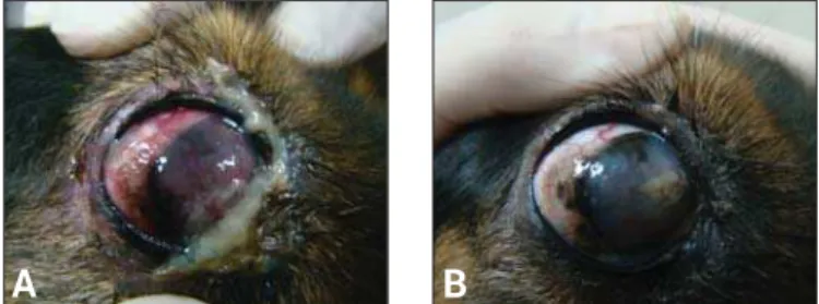
Figure 6 shows the postoperative improvement of a dog with severe KCS that did not respond to medical therapy; the dog had severe mucopurulent discharge, conjunctival hyperaemia, eye discomfort, absence of corneal luster, and intense neovascularisation. By PO 30 there was a very significant reduction in discharge, hyperaemia, and corneal neovascularisation, and a recovery of corneal luster.
The symptoms of KCS improved as early as the second
postoperative week, with recovery of corneal luster and decreased irritation, photophobia, and blepharospasm. The improvement was statistically significant for blepharospasm and corneal luster. To assess blepharospasm and corneal luster the McNemar chi-square test was used, comparing preoperative values to those of PO days 15, 45, and 120; a significant improvement was found at all time points, with p<0.05.
A significant reduction was also found in corneal neovascularisation and in the inflammatory process. As the sample was not normally distributed, statistical significance was assessed using the Wilcoxon test for paired data. The results had p-values <0.05, therefore the reduction in the number of corneal vessels was statistically significant. Figure 7 shows the progression in the number of corneal neovessels from the preoperative period to PO day 120. There was a significant reduction in the number of neovessels, from an average of 17.7 per eye preoperatively to 12.2 on PO day 45 and 8.4 on PO day 120. The reduction of neovascularisation also indicates a significant reduction in the inflammatory and irritative process.
Figure 6. A, An animal with mucopurulent discharge, conjunctival hyperaemia, and severe neovascularisation before surgery; B, 30 days after the transposition of labial salivary glands, absence of mucopurulent discharge, reduced conjunctival hyperaemia, and recovery of corneal luster
Figure 8. A labial salivary gland graft completely embedded into the conjunctival fornix; the graft regained its original colour with no signs of rejection. Full recovery of luster and stabilisation of the clinical signs of keratoconjunctivitis sicca Figure 7. Values , means and standard deviations for the number of corneal neovessels in the right and left eyes, showing an important reduction in all follow-up visits
A
B
D
ISCUSSIONTransplantation of labial salivary glands into the conjunctival fornix to treat severe dry eye syndrome has shown satisfactory results in experiments with humans, with statistically significant clinical improvement(5). Although there are several
alternatives for the treatment of dry eye syndrome, some cases represent a therapeutic challenge, particularly those with immune-mediated causes, prompting the need for new treatment alternatives.
Lubrication of the ocular surface by salivary secretion was shown to be effective, well tolerated, and constant in humans(5).
This prompted the present study in dogs, a species where dry eye syndrome can have autoimmune causes making it a good model for comparative ophthalmology.
Labial salivary glands are the main source of immunoglobulin A in the saliva, producing about 1/3 of the IgA in
the entire oral cavity. The high concentration of IgA is important for regulating the levels of microorganisms in the oral cavity. In dogs, labial salivary glands are numerous small glands measuring 1-3 mm in diameter and located in the submucosa of the upper and lower lips. They form a nearly continuous layer between the mucosa and the orbicularis oris muscle and can be palpated by moving the tongue over the inner surface of the lips(6).
Saliva produced by the labial glands has four times the concentration of IgA than that produced by the parotid gland(6).
This is an advantage for lubrication of the ocular surface, favouring labial gland transposition instead of parotid duct gland transposition, the older technique.
Saliva has a defence function due to its antimicrobial proteins such as lysozymes, lactoferrins, apolactoferrin, IgA, fibronectin, histatins, â2-microglobulin, salivary agglutinins, and mucins(7). The significant improvement in the infectious and
inflammatory processes from PO day 45 found in this study — as evidenced clinically by reduced conjunctival hyperaemia and mucopurulent discharge and absence of blepharospasm — can be partly attributed to the composition of the saliva produced by the transplanted labial glands.
Saliva has a lubricating function do to its higher amount of mucin(7). The Schirmer test measures the aqueous component of
tears. Saliva has no fat content and its aqueous content is less significant than its mucous content. Thus, saliva effectively lubricates the ocular surface without causing significant changes in the Schirmer test. Clinical improvement was observed in all dogs included in the study but without significant changes in the Schirmer test, confirming the results of a study by Lawrence(7).
The clinical improvement observed after grafting associated with lubricating eye drops is due to the fact that lacrimal gland secretion contains elements which maintain and protect the ocu-lar surface, especially a higher concentration of mucin, which increases the viscosity of the “salivary tears”. The more viscous secretion decreases evaporation, forming a more permanent and stable wet layer(5). In our study, we found a significant reduction
of mucopurulent discharge and increased BUT, promoting stability of the tear film and reducing evaporation.
The symptoms of KCS were relieved as early as the second postoperative week, with recovery of ocular surface luster and a significant decrease in irritation, photophobia, and blepharospasm(8). We found a statistically significant improvement
in blepharospasm and corneal luster, indicating postoperative ocular comfort.
Dry eye syndrome is often associated with ocular discomfort, mucoid or mucopurulent discharge, ocular surface dryness, conjunctival hyperaemia, corneal neovascularisation, and pigmentation(9). The normal cornea is avascular and transparent,
and its nutrition depends mainly on the blood vessels of the limbus, the aqueous humuor, and tears; however, in the terminal stages of several conditions including KCS, chronic hypoxia and prolonged inflammation stimulate the proliferation of limbus vessels, leading to a loss of transparency and consequent loss of visual acuity(10,11). In our study, all animals with moderate to
severe KCS had important inflammation and a large number of corneal vessels, which were significantly reduced after transplantation.
The labial salivary gland grafts were pale only in the first hours after the procedure, but with no signs of distress; it is essential that the grafts be incorporated into the recipient site and regain their original pink colour by the second PO day(5).
of symptoms and improved vision. Graft incorporation occurred in all dogs, with slight pallor in the first hours after the procedure and return to normal pink colour after 12 hours. The donor sites also healed fully, resulting in a barely noticeable linear scar.
C
ONCLUSIONWe found a significant clinical improvement in cases of moderate to severe KCS, as well as those which were non-responsive to medical treatment, as evidenced by clinical examination and statistical tests. Transplantation of labial salivary glands showed that lubrication of the ocular surface by salivary secretion is stable and effective.
R
EFERENCES1. Silvério J, Lucci LM, Fonseca Júnior NL, Rehder JR. Efeitos da blefaroplastia na síndrome da disfunção lacrimal. Rev Bras Oftalmol. 2011;70(3):151-6.
2. Fridman D, Freitag MM, Kleinert F, Lavinsky J. Olho seco: conceitos, história natural e classificações. Arq Bras Oftalmol. 2004;67(1):181-5. 3. Gehlen ML, Skare TL, Silva MB, Antero DC, Miyazaki F, Parra AG. Olho seco e Sjögren secundário na artrite reumatoide. Rev Bras Oftalmol. 2012;71(1):36-9.
4. Gellat NK. Doença e cirurgia dos sistemas lacrimal e nasolacrimal do cão. In: Gellat NK.Manual de oftalmologia veterinária. 3a ed. São Paulo: Manole; 2003. p. 73-83.
5. Soares EJ, França VP. Transplante de glândulas salivares labiais no tratamento do olho seco grave. Arq Bras Oftalmol. 2005;68(4):481-9. 6. Localização e comparação morfo-funcional dos diferentes tipos de glândulas salivares menores [Internet]. [citado 2012 Fev 9]. Disponível em: http://www.ebah.com.br/content/ABAAABTZQAB/ histologia
7. Lawrence HP. Salivary markers of systemic disease: noninvasive di-agnosis of disease and monitoring of general health. J Can Dent Assoc. 2002;68(3):170-4.
8. Angélico GT, Ranzani JJ, Brandão CV, Schellini SA, Padovani CR, Sereno MG, et al. Transplante de glândulas salivares menores no tratamento da ceratoconjuntivite seca em cães. Arq Bras Med Vet Zootec. 2011;63(5):1087-92.
9. Pigatto JA, Pereira FQ, Almeida AC, Redaeli R, Faganello CS, Franzen AA. Ceratoconjuntivite seca em cães e gatos. Acta Sci Vet. 2007;35(Supl 2):S250-1.
10. Trincão F, Feijão J, Maduro V, Alves N, Batalha C, Candelária P. Tratamento da neovascularização da córnea com injeção de bevacizumab intraestromal. Oftalmologia. 2011;35(2):129-34. 11. Gehlen ML, Moreira H, Moreira L, Sabag FP, Repka JC. Avaliação
espectrofotométrica do azul de Evans na reação inflamatória da córnea: estudo experimental em coelhos. Arq Bras Oftalmol. 2004;67(2):219-25.
Corresponding author:
Leticia Séra Castanho