Abstract
Submitted: May 14, 2016 0RGL¿FDWLRQ6HSWHPEHU Accepted: October 28, 2016
supported monolithic crowns in
zirconia-reinforced lithium silicate
Zirconia-reinforced lithium silicate (ZLS) is a ceramic that promises to have better mechanical properties than other materials with the same indications as well as improved adaptation and fracture strength. Objective:
thermal-mechanical aging (TMA) of monolithic ZLS and lithium disilicate (LDS) crowns were evaluated. Material and methods: Crowns were milled using a computer-aided design/computer-aided manufacturing system. Marginal gaps (MGs), absolute marginal discrepancy (AMD), axial gaps, and occlusal gaps were measured by X-ray microtomography (n=8). For fracture load testing, crowns were cemented in a universal abutment, and divided into four groups: ZLS without TMA, ZLS with TMA, LDS without TMA, and LDS with TMA (n=10). TMA groups were subjected to 10,000 thermal cycles
(5-subjected to compressive strength testing in a universal testing machine at a crosshead speed of 1 mm/min until failure. Student’s t-test was used to
calculated (D=0.05). The materials were analyzed according to Weibull
and AMD (p=0.003) values were greater in ZLS than in LDS crowns. TMA did not affect the fracture load of either material. However, fracture loads of ZLS
moderately correlated with MG (r=-0.553) and AMD (r=-0.497). ZLS with TMA was least reliable, according to Weibull probability. Conclusion: Within the limitations of this study, ZLS crowns had lower fracture load values and
within acceptable limits.
Ke yw or ds: Dental materials. Dental prosthesis. X-Ray microtomography.
Rafael Soares GOMES1
Caroline Mathias Carvalho de SOUZA2
Edmara Tatiely Pedroso BERGAMO1
Dimorvan BORDIN1
Altair Antoninha DEL BEL CURY1
http://dx.doi.org/10.1590/1678-7757-2016-0233
1 Universidade Estadual de Campinas, Faculdade de Odontologia de Piracicaba, Departamento de
Prótese e Periodontia, Piracicaba, SP, Brasil.
2 Universidade Estadual de Campinas, Faculdade de Odontologia de Piracicaba, Departamento de
Odontologia Restauradora, Piracicaba, SP, Brasil.
Introduction
The evolution of ceramic systems has been guided
by efforts to enhance their strength and aesthetics32.
The use of zirconia seemed to solve the problem of
resistance in these systems, but the aesthetic quality
of this material is less than desirable2. In the effort
to obtain an aesthetic and strong material, a ceramic with 10% zirconia added to lithium silicate was recently
developed and released19. Named zirconia-reinforced
lithium silicate (ZLS), this material was designed for
exclusive use with computer-aided design/computer-aided manufacturing (CAD/CAM) systems. Its
manufacturer claims that it is an outstanding aesthetic
material, with more strength and easy milling ability compared with lithium disilicate (LDS), generating
optimized edge stability. Nevertheless, few studies
supporting these features have been published6,19,30.
LDS monolithic crowns have been used with success26. Monolithic crowns withstand greater
resistance than bi-layered crowns, and can be used
in regions with greater masticatory forces24. With
the application of extrinsic staining techniques, LDS has been established as an aesthetic and strong
material7. The use of ZLS in combination with CAD/
CAM technology appears to be another option for
restorative treatments with similar indications and requirements as for LDS30.
CAD/CAM technology has facilitated restorative
prosthetic treatment for clinicians and patients,
decreasing restoration placement and overall chair times12. Although the use of this technique has widely
made with the same system and impression technique
due to material composition1,12,14,16. Material hardness
susceptible to small fractures in very thin regions, such
as cervical areas14,17,25,27; thick and irregular cement 11,28. In addition, some
studies have shown that the worse the adaptation of
the crown, the lower its resistance28,33.
remain a matter of debate. The most commonly
3,9,15. Some
authors have claimed that thick cement layers may
lead to increased cement dissolution, microleakage,
localized stress accumulation, and reduction of fracture
strength20,21,28.
The clinical success of a restoration can depend
on many factors, including fracture load, which can
withstand cyclic loading1,31,33. These properties need to
be investigated in ZLS; studies testing the advantages
attributed to this material remain scarce. This study
fracture load with and without thermal-mechanical
aging (TMA), and reliability of ZLS compared with LDS.
ZLS would be superior to those of LDS.
Material and methods
Specimens fabrication
Using a three-dimensional (3D) optical scanning
device (Ceramill Map400, Amann Girrbach, Koblach, Vorarlberg, Austria), a 3D digital model of a morse
taper universal abutment (Munhão Universal, Intraoss,
Itaquaquecetuba, São Paulo, Brazil) with a 4.5 mm
diameter, 6 mm height, and 2.5 mm collar height was obtained. From this model, and regarding the anatomy
drawn using CAD software (Ceramill Mind, Amann
Girrbach). From this CAD model, 20 ZLS crowns (Suprinity, Vita Zahnfabrik, Bad Säckingen,
Baden-Württemberg, Germany) and 20 LDS crowns (IPS
e.max CAD, Ivoclar Vivadent, Schaan, Liechtenstein)
were milled (Ceramill Motion 2, Amann Girrbach). Integrity of crown margins was examined by scanning
electron microscopy (SEM) (JSM-5600LV, Jeol, Boston,
Massachusetts, USA).
material was evaluated using X-ray microtomography
USA). Each crown–universal abutment set was twice
wrapped for an adhesive tape that ran through on
the occlusal surface to the abutment’s base, avoiding
any displacement, and positioned perpendicular to the X-ray source. The parameters used for image
acquisition were 80 kV, 1400 ms exposure time,
each scan and reconstructed using NRecon software
coronal and sagittal slices were isolated from the
reconstructed images. Using CTAn software (Bruker),
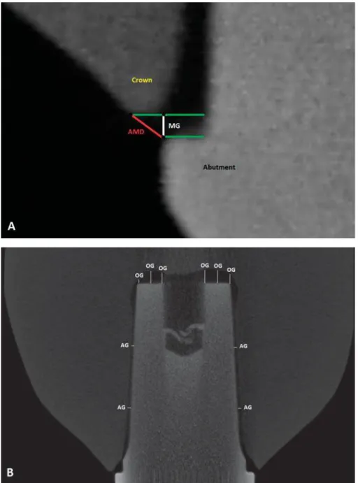
the averages of the values obtained on each axis were calculated. Following Holmes, et al.8 (1989),
four locations (centers of the buccal, lingual, mesial,
and distal faces) were selected for the evaluation
of marginal gaps (MGs) and absolute marginal discrepancy (AMD; Figure 1A). Ten locations on
each of the two slice types [four on the axial walls
for the evaluation of axial gaps (AGs) and six on the occlusal walls for the evaluation of occlusal gaps (OGs)
(Figure 1B)] were used for the evaluation of internal
3,12,13.
Fracture load
Prior to cementation, the crowns (n=20 of each material) were rinsed in 98% alcohol for 1 minute
in an ultrasonic bath. Their external surfaces were
then protected with wax (New Wax, Technew, Rio
de Janeiro, Rio de Janeiro, Brazil), and the intaglio surfaces were conditioned with 5% hydrofluoric
acid (Condac porcelana, FGM, Joinville, SC, Brazil)
for 20 seconds. The excess gel was removed with a water jet, and the crowns were washed again in the
ultrasonic bath with 98% alcohol for 3 minutes. A thin
layer of silane coupling agent (Prosil, FGM) was then
applied to the intaglio surfaces and allowed to act for 60 seconds; excess silane was volatilized with an air
jet. The crowns were cemented into abutments that
had been tightened into implant analogs28 (Titaoss Max
CM Analog, Intraoss) with 32 N.cm torque (TQ8800,
Lutron, Taipei, Taiwan) using a dual-cure resin composite cement (Panavia F, Kuraray Noritake Dental
Inc., Okayama, Tokyo, Japan), and photopolymerized
for 20 seconds/face using an LED source with 1000
mW/cm² light intensity (VALO, Ultradent Products Inc, South Jordan, Utah, USA).
The analogs were embedded in polyurethane resin
(F160, Axson Technologies, Saint Ouen I’Aumône,
10
in a metal matrix (20 mm diameter, 20 mm height).
Twenty crowns were used as the experimental groups
and the other 20 crowns served as controls. Ten crowns of each material were subjected to 1 million
mechanical cycles (200 N load, 3.8 Hz frequency;
ER-1300, ERIOS, São Paulo, SP, Brazil), with 10,000
thermal cycles (MSCT-3e, Elquip, São Carlos, SP,
Brazil) in alternating water baths with temperatures
interval), amounting to about 65 seconds per cycle.
For fracture load testing, a mechanical load
was applied (Instron 4411, Instron, Norwood,
M a s s a c h u s e t t s , U SA ) w i t h a s t a i n l e s s - s t e e l hemispherical indenter (5 mm diameter) to the
occlusal surface of each crown at a crosshead speed of
or catastrophic fracture of the crown. After fracture, all samples were submitted to fractographic analysis
by SEM to identify the origin of failure4,22,23.
Statistical analysis
the IBM SPSS Statistics 20 software (IBM Corp.,
.
were separately compared between materials using
Student’s t-tests. The means of fracture loads in the
two groups were examined using two-way ANOVA. The power obtained with the current sample size in
both analyses exceeded 90%. Pearson’s correlation
load. The Weibull distribution was examined using SAS
software (SAS Institute Inc., Cary, North Carolina,
the reliability of sample survival based on the applied
load.
Results
lower for LDS crowns than for ZLS crowns. No
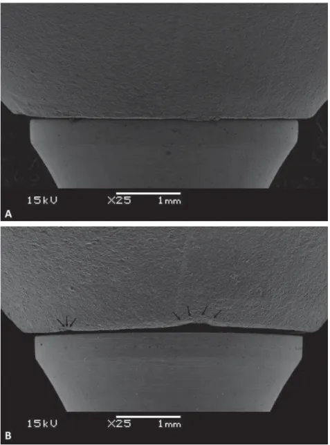
(Table 1). Chipping in the cervical region occurred during the milling process in some crowns (Figure
2A and B).
Group MG AMD AG OG
LDS 41.45±18.11a 180.67±23.23a 96.07±30.19a 255.80±65.05a
ZLS 101.86±32.12b 235.54±35.75b 100.09±23.83a 252.68±35.18a
/'6 OLWKLXP GLVLOLFDWH =/6 ]LUFRQLDUHLQIRUFHG OLWKLXP VLOLFDWH 'LIIHUHQW VXSHUVFULSWHG OHWWHUV LQGLFDWH VLJQL¿FDQW GLIIHUHQFHV EHWZHHQ PDWHULDOV6WXGHQW¶VWWHVWS
Table 1-0DUJLQDOJDS0*DEVROXWHPDUJLQDOGLVFUHSDQF\$0'D[LDOJDS$*DQGRFFOXVDOJDS2*YDOXHVPQ PHDQVWDQGDUG
deviation)
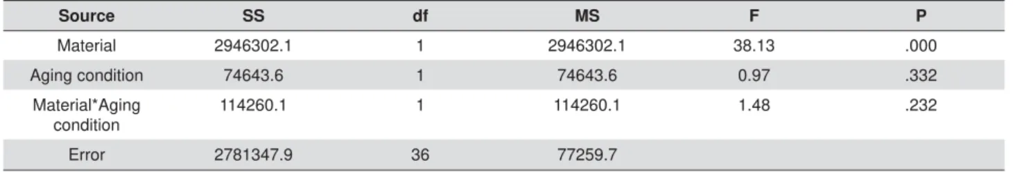
Source SS df MS F P
Material 2946302.1 1 2946302.1 38.13 .000 Aging condition 74643.6 1 74643.6 0.97 .332
Material*Aging condition
114260.1 1 114260.1 1.48 .232
Error 2781347.9 36 77259.7
666XPRI6TXDUHVGIGHJUHHIUHHGRP060HDQ6TXDUH
Table 2- Two-way ANOVA (2x2) of material, aging condition, and interactions between these variables
Figure 3- Mean fracture loads of LDS and ZLS crowns before and after thermal-mechanical aging. Bars indicate standard deviations and
fracture load (Table 2). Mean fracture load values
respectively, for ZLS crowns (Figure 3). Fracture load
p>0.05) or AG (r=-0.237, p>0.05), but it showed
moderate negative correlations with MG (r=-0.553, p=0.026) and AMD (r=-0.49, p=0.05).
Crowns presented catastrophic failure, exposing
the abutment. Hackle lines, commonly formed when
cracks grow rapidly, and arrest lines, which indicate
the direction of crack propagation, showed that crack
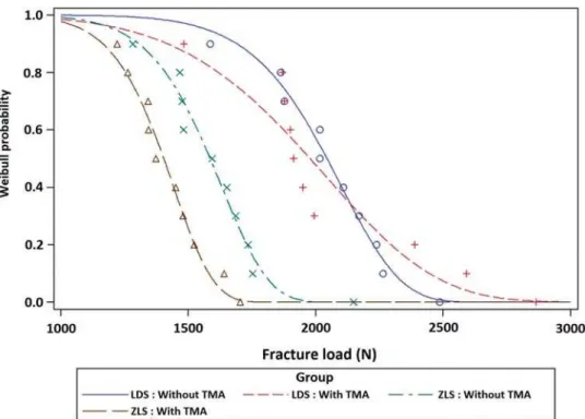
propagation originated from the load point (Figure 4). The Weibull distribution showed overlapping of the
two LDS aging conditions, and higher reliability than
observed for ZLS crowns in both conditions (Figure 5).
Discussion
Studies on internal and marginal misfit of
monolithic ZLS crowns are scarce. The use of a
non-Figure 4- Fractographic image showing fracture origin (O), direction of crack propagation (dcp), arrest lines (A), and hackle lines (H)
the great advantage of enabling evaluation of not only
the marginal area, but also the intaglio surface, while preserving samples for further analyses (e.g., fracture
load) with no effect on the results. It may, however,
be considered a costly and slow technique13,16.
in the cervical region than did LDS crowns, which
presented misfit values compatible with those
published previously12-14
cervical region could be that a small amount of internal
space obstructs the settling of the crown1, but this was
not observed in this study, since the two materials
produced similar internal adaptation values. A second hypothesis is that this difference could be due to the
occurrence of very small fractures during milling of
the cervical region in ZLS crowns14. The results of this
study support this hypothesis, as SEM images showed more irregularity in the cervical regions of ZLS crowns
compared with LDS crowns, which may have increased
study was considered that there are no differences among abutment sizes, which could interfere also in
the measurements outcomes.
Margin inaccuracy may be related to the increased
brittleness index and chipping factor of ZLS, resulting in greater marginal misfit, probably due to the
presence of zirconia in the microstructure19,27. The
chipping factor (%) is the ratio of the total amount
of chipping around the marginal circumference of the restoration multiplied by 10027. It is positively related
to the brittleness index, which is a ratio of hardness
and fracture toughness. A higher hardness value and
lower fracture toughness value increase the brittleness index of a material, indicating that it is more prone
to chipping27. According to the manufacturers of the
materials used in this study, ZLS has a higher hardness value than does LDS in crystallized mode (7000 vs.
5600 MPa), and a lower fracture toughness value (2.00
vs. 2.25 MPa m-0.5). However, no study has examined
these properties in the pre-crystalized phase, in which these restorations are milled.
The occlusal and axial walls of ZLS and LDS crowns
areas may be less vulnerable to microfracture, thus supporting the hypothesis that thin regions are more
susceptible to damage during the milling process.
Although ZLS presented a greater difference in
μm according to most studies1,3,11-16,21,28. In addition,
the greatest discrepancy did not seem to compromise the mechanical behavior of the material a lot, since
moderate negative correlations were observed
between fracture load and MG and AMD.
μ
reduce the fracture strength of a crown16,17,25,27. In
the present study, AG values for both materials were
μ
load. Another area of internal analysis is the occlusal
μ
cementation due to polymerization shrinkage16. In this
μ
not differ between materials12.
Fracture loads were lower for ZLS crowns than for LDS crowns, with and without TMA. The presence
of zirconia in the microstructure seems to increase
material hardness, making it more prone to chipping
during milling19. Chipping can worsen the adaptation
of the material and indirectly compromise fracture
loads20
and fracture loads was detected in this study. TMA
resistance of dental ceramics, although the Weibull
probability curves showed that ZLS is affected more
than LDS5,31,33.However, comparison of the present
results with those of other studies should be done
with caution, given the poor standardization of
loads, frequency, number of cycles, and substrates.
In addition, Weibull probability can be unreliable in
18, although
Both materials tested in this study resisted loads larger than maximum relative values of bite force
found in the literature (~880 N)29. However, the
fracture load test does not mimic failures occurring
when crowns are in clinical use4,5,33. The fractographic
analysis conducted in the present study showed crack
propagation from the load point to the cervical area in
all samples, opposite the direction of propagation in
clinical situations4
results of the present study represent the real clinical
behavior of these materials.
reliability. However, the effectiveness of this material
Conclusion
Within the limitations of this study, ZLS crowns had lower fracture load values and greater marginal
all values were within the limits considered to be acceptable. Thus, both materials comply with the
indication criteria.
References
1- Anadioti E, Aquilino SA, Gratton DG, Holloway JA, Denry IL,
computer-aided manufacturing ceramic crowns made from digital and conventional impressions. J Prosthet Dent. 2015;113(4):304-9.
2- Baldissara P, Llukacej A, Ciocca L, Valandro FL, Scotti R. Translucency of zirconia copings made with different CAD / CAM systems. J Prosthet
Dent. 2010;104(1):6-12.
3- Borba M, Miranda WG Jr, Cesar PF, Griggs JA, Della Bona A. Evaluation
CT technology. Braz Oral Res. 2013;27(5):396-402.
4- Campos RE, Soares P V, Versluis A, Júnior OB, Ambrosano GM, Nunes IF. Crown fracture: failure load, stress distribution, and fractographic
analysis. J Prosthet Dent. 2015;114(13):447-55.
5- Coelho PG, Bonfante EA, Silva NRF, Thompson VP. Laboratory
simulation of Y-TZP all-ceramic crown clinical failures. J Dent Res. 2009;88(4):382-6.
6- Elsaka SE, Elnaghy AM. Mechanical properties of zirconia reinforced lithium silicate glass-ceramic. Dent Mater. 2016;32(7):908-14.
7- Herrguth M, Wichmann M, Reich S. The aesthetics of all-ceramic veneered and monolithic CAD/CAM crowns. J Oral Rehabil.
2005;32(10):747-52.
8- Holmes JR, Bayne SC, Holland GA, Sulik WD. Considerations in
fused-to-metal and two types of ceramic crown. J Prosthet Dent. 1990;63(1):26-31.
10- International Organization for Standardization. ISO 14801: dynamic fatigue test for endosseous dental implants. Geneva: The
Organization; 2007.
11- Liu B, Lu C, Wu Y, Zhang X, Arola D, Zhang D. The effects of
adhesive type and thickness on stress distribution in molars restored with all-ceramic crowns. J Prosthodont. 2011;20(1):35-44.
12- Mously HA, Finkelman M, Zandparsa R, Hirayama H. Marginal and internal adaptation of ceramic crown restorations fabricated with
CAD/CAM technology and the heat-press technique. J Prosthet Dent. 2014;112(2):249-56.
13- Neves FD, Prado CJ, Prudente MS, Carneiro TA, Zancopé K, Davi LR,
disilicate crowns fabricated by using chairside CAD / CAM systems or the heat-pressing technique. J Prosthet Dent. 2014;112(5):1134-40.
fabricated with digital and conventional methods. J Prosthet Dent. 2014;112(3):555-60.
15- Park JK, Lee WS, Kim HY, Kim WC, Kim JH. Accuracy evaluation of
metal copings fabricated by computer-aided milling and direct metal laser sintering systems. J Adv Prosthodont. 2015;7(2):122-8.
16- Pimenta MA, Frasca LC, Lopes R, Rivaldo E. Evaluation of
x-ray microtomography (micro-CT) technology. J Prosthet Dent. 2015;114(2):223-8.
17- Quinn GD, Giuseppetti AA, Hoffman KH. Chipping fracture resistance of dental CAD/CAM restorative materials: Part I, procedures
and results. Dent Mater. 2014;30(5):e112-23.
18- Quinn JB, Quinn GB. A practical and systematic review of Weibull
statistics for reporting strengths of dental materials. Dent Mater. 2010;26(2):135-47
19- Ramos NC, Campos TM, La Paz IS, Machado JPB, Bottino MA, Cesar
PF, et al. Microstructure characterization and SCG of newly engineered dental ceramics. Dent Mater. 2016;32(7):870-8
20- Rojpaibool T, Leevailoj C. Fracture resistance of lithium disilicate ceramics bonded to enamel or dentin using different resin cement types
21- Rungruanganunt P, Kelly JR, Adams DJ. Two imaging techniques
J Dent. 2010;38(12):995–1000.
22- Scherrer SS, Quinn JB, Quinn GD, Kelly JR. Failure analysis of ceramic clinical cases using qualitative fractography. Int J Prosthodont.
2006;19(2):185-92.
23- Scherrer SS, Quinn JB, Quinn GD, Wiskott HW. Fractographic
ceramic failure analysis using the replica technique. Dent Mater. 2007;23(11):1397-404.
24- Schultheis S, Strub JR, Gerds TA, Guess PC. Monolithic and
bi-prostheses: Comparison of fracture loads and failure modes after fatigue. Clin Oral Investig. 2013;17(5):1407-13.
25- Song X, Ren H, Yin L. Machinability of lithium disilicate glass ceramic in in v it r o dental diamond bur adjusting process. J Mech Behav Biomed
Mater. 2016;53:78–92.
26- Sulaiman TA, Delgado AJ, Donovan TE. Survival rate of lithium
disilicate restorations at 4 years: a retrospective study. J Prosthet Dent. 2015;114(3):364-6.
27- Tsitrou EA, Northeast SE, van Noort R. Brittleness index of machinable dental materials and its relation to the marginal chipping
factor. J Dent. 2007;35(12):897-902.
28- Tuntiprawon M, Wilson PR. The effect of cement thickness on the
fracture strength of all-ceramic crowns. Aust Dent J. 1995;40(1):17-21. 29- Van der Bilt A. Assessment of mastication with implications for oral
rehabilitation: a review. J Oral Rehabil. 2011;38(10):754-80. 30- Weyhrauch M, Igiel C, Scheller H, Weibrich G, Lehmann KM.
Fracture strength of monolithic all-ceramic crowns on titanium implant abutments. Int J Oral Maxillofac Implants. 2016;(31)2:304-9
31- Yang R, Arola D, Han Z, Zhang X. A comparison of the fracture resistance of three machinable ceramics after thermal and mechanical
fatigue. J Prosthet Dent. 2014;112(4):878-85.
resistance of monolithic ceramics. Dent Mater. 2013;29(12):1201-8. 33- Zhang Y, Sailer I, Lawn BR. Fatigue of dental ceramics. J Dent.