Abstract
Submitted: September 2, 2016
Accepted: December 18, 2016
proliferation and differentiation of
odontoblast
collagen (PCOL) on odontoblast-like cells in v it r o. Material and Methods:
Rat odontoblast-like cells (MDPC-23 cells) were inoculated and cultured on Fn-coated or type I collagen-coated substrates. Proliferation assay, alkaline phosphatase activity (ALP activity), mRNA expression of hard tissue-forming markers, and Alizarin red staining were investigated over a period of 10 days. Results: Cells maintained a high proliferation activity on Fn and PCOL even at a low seeding concentration (0.5×10⁴/mL) as demonstrated by CCK-8 assay. The proliferation activity of cells on Fn increases in a concentration-dependent manner while it reached a plateau after 10 μg/mL. Cells adopted
were observed in MDPC-23 cells cultured on Fn and PCOL. ALP activity was markedly up-regulated on Fn and PCOL-coated surfaces. Importantly, gene
substrates as compared with control; moreover, expression of integrin beta 1 (ITGB1), an ubiquitous cell surface receptor was augmented in Fn(10-50) and PCOL groups simultaneously. In accordance with the ALP activity and
PCOL was observed as well. Conclusion: Despite the limitation of this study,
differentiation and mineralization of odontoblast-like cells by activation of integrin beta 1 (ITG B1). The promoting effects of Fn on MDPC-23 cells were achieved at a comparatively lower coating concentration than type I
μ
concentration of Fn to be 10 μg/mL.
Keyw ords: Fibronectin. Odontoblasts. Cell differentiation. Cell proliferation
Jia TANG1
Takashi SAITO1
http://dx.doi.org/10.1590/1678-7757-2016-0442
1Health Sciences University of Hokkaido, School of Dentistry, Department of Oral Rehabilitation, Division of Clinical Cariology and Endodontology, Hokkaido, Japan.
Corresponding address: Jia Tang Division of Clinical Cariology and Endodontology, Department of Oral Rehabilitation, School of Dentistry, Health Sciences University of Hokkaido Kanazawa, 1757 - Ishikari-Tobetsu, 061-0293
Introduction
Fibronectin (Fn) is a dimeric multi-domain
glycoprotein (about 450kDa per dimer) that is found in circulation or tissue extracellular matrix (ECM).
Two major types of Fn are present in vertebrates
arising from alternative splicing of its pre-mRNA:
soluble plasma Fn (corresponds to the aforementioned CIg) and insoluble cellular Fn. Both of them contain
various adhesive domains for cells and other proteins
9,21, the synergic site of RGD:
PHSRN4,18 9,
Arg-Glu-Asp-Val (REDV)10 15, and
17. The plasma form of
Fn (pFn) is predominantly synthesized by hepatocytes,
circulates in blood and deposits rapidly upon tissue
injury to initiate hemostasis, this deposition process is independent of other hemostasis factors, such as
platelet30. Cellular Fn (cFn) is mainly produced by
epithelial cell25, macrophage2, and endothelial cells20.
cFn contributes to support the extracellular structure
framework by actively binding with cells and other
matrix proteins as mentioned above.
Previous histological localization study in tooth
germ revealed that Fn, which is present in the
mesenchymal tissue, basement membrane, and
pre-dentine, was not detected in late pre-dentine and mineralized dentine. Further, epithelial tissues of
tooth germ were negative for Fn except in the stellate
reticulum12,28. Tooth development or odontogenesis is
a complex process, which needs reciprocal interaction between epithelium and mesenchyme. Various growth
factors, paracrine signal molecules27 and extracellular
matrix (ECM) proteins6 are believed to be essential
for this process. Among those, the importance of ECM proteins is becoming increasingly apparent,
since serious changes were noted in oral cancer as
compared with normal tissue because of the alteration
of ECM26. As a ubiquitous ECM, it is reported that
Fn is required for calvarial osteoblast differentiation
and mineralization16. Since Fn is expressed in early
pre-dentine and disappeared in mineralized mature dentine, it is thus reasonable to conceive that Fn
might play some roles in the differentiation of dental
mesenchyme into dentine-forming odontoblast.
Previously, Mizuno, et al.14 (2008) reported that Fn
enhanced the osteocalcin (OCN) and osteopontin
(OPN) gene expression in human dental pulp cells. However, the precise effect of Fn on cells of dental
mesenchymal origin is still unclear to date. Hence,
the current experiment seeks to uncover the potential
interaction between Fn and odontoblast-like cells. To better imitate the real scenario that cells are
surrounded by extracellular matrix proteins in sit u, we examined the cell proliferation, differentiation and mineralization behavior in Fn-coated substrate using
porcine type I collagen as a comparison ECM.
Material and methods
Cell culture
Rat odontoblast-like cell (MDPC-23 cell), a
spontaneously immortalized cell line originally isolated from molar papillae, were cultured in
Gaithersburg, MD, USA) supplemented with 5% fetal
CO2
with PBS, trypsinized (TrypLETM Express 1×, Phenol
Red) (12605-010, Invitrogen, Carlsbad, CA, USA),
and seeded at pre-determined concentrations. The medium was changed every other day. The cells used
in this study were obtained from 25 to 33 passages.
Protein coating on non-treated tissue culture
plates
Human plasma Fn was purchased from Gibco (33016-015), reconstituted in phosphate buffered
saline (PBS) (Gibco) at a stock concentration of 1 mg/
skin type I collagen was obtained from Nitta gelatin
(3 mg/mL, Cell matrix type I-C, 140820). Non-tissue
culture grade plates (96-well plate, 351177, Falcon;
24-well plate, 1820-024, Iwaki; 12-well plate, 351143, Falcon) coated with Fn were prepared by soaking the
plates in a series of concentrations (0.1, 1, 10, 20, 30, 40, 50 μg/mL, herein referred to as Fn0.1, Fn1, Fn10, Fn20, Fn30, Fn40, Fn50, respectively), collagen was coated at the concentration of 300 μg/mL (PCOL300) as recommended by manufacturer. After overnight
coating, the solution was aspirated and the wells were
washed twice with PBS. The cells were rinsed with PBS,
or 1×10⁴/mL; 100 μL culture media/well), 24-well
plate (5×10³ or 1×10⁴/mL; 1 mL culture media/well)
and the 12-well plate (1×10⁴/mL; 2 mL culture media/
well). The cells were cultured in DMEM supplemented
with 5% FBS for the experiment. Inducer medium μ
acid) (Wako, Osaka, Japan)19 was added to the culture
medium from day 5. Cells seeded in wells coated with
PBS served as the control.
Cell morphology observation
Light microscopy observation: cells were inoculated
at the concentration of 1×10⁴/mL. Photos of the cells
were taken under light microscopy (Olympus) after 20
hours culture in 12 well plate (non-treated, 351143, Falcon).
Immunofluorescence staining: MDPC-23 cells
was seeded into 24 well plate (non-treated,
1820-024, IWAKI) at the concentration of 5×10³/mL.
3. F-actin and nucleus were visualized by staining with
Alexa Fluor 568® phalloidin (A12380) (Invitrogen) and
DAPI (D9542) (Sigma, St Louis, MO, USA), respectively.
Waltham, MA, USA) for 15 minutes, permeabilized
with Triton-X-100 (0.1%, v/v, in PBS) (T8787-100mL)
(Sigma) for 5 minutes. Alexa Fluor 568® phalloidin
was reconstituted in 1.5 mL methanol to generate
the stock solution (200 U/mL). The stock solution was subsequently diluted (0.01 U/μL) and added into 24 well plate (200 μL/well). Tween 20 (0.05%, v/v) (Kanto Chemical) in PBS (PBST) was used to wash the cells
after 1 hour incubation in room temperature. Finally,
the cells were counterstained with DAPI (300 nM in PBS) for 5 minutes at room temperature and washed
were taken using EVOS® FLoid® Cell Imaging Station
(Advanced Microscopy Group, Mill Creek, WA, USA).
Cell proliferation assay
Cells were seeded into a 96-well plate (non-treated,
351172, Falcon) at the concentration of 5×10³ or
1×10⁴/mL. After incubation for 1, 2, 4, 6 days, cell
counting kit-8 (CCK-8) reagent (Dojindo, Kumamoto,
Japan) was added to each well to a volume of 10%
(10 μL/well), followed by incubation for another 1
2
atmosphere. The optical density was measured at 450
nm using a microplate reader (Bio-Rad, Hercules, CA,
USA).
activity
Cells (1×10⁴/mL) were seeded into Fibronectin
and collagen-coated 12 well plates (non-treated)
with 5% FBS. On day 5, cells were removed from the culture plate using Triton-X-100 (0.1%, w/w, in
distilled water) and sonicated (Bioruptor®, Diagenode,
Seraing, Belgium) for ten minutes on ice. The lysates
(Hitachi Koki, Tokyo, Japan). The resulting supernatant
was diluted and assayed for ALP activity (100-times
dilution) (Wako, Osaka, Japan) and BCA protein
Rockford, IL, USA) according to the manufacturers’
instructions. Absorbance was read at 405 nm and
570 nm for the ALP assay and the protein assay, respectively.
Quantitative reverse transcription-polymerase
chain reaction (qRT-PCR)
Cells (1×10⁴/mL) were seeded into 12 well plates
(non-treated). RNA was isolated from aliquots of cells harvested on day 7. The mRNA levels of bone
sialoprotein (BSP), OCN, integrin beta 1 (ITGB1),
ALP, OPN, and dentine matrix protein-1 (DMP-1)
were measured using a quantitative RT-PCR machine
(LightCyclerTM Nano, Roche, Basel, Switzerland).
The mRNAs were converted to cDNA using M-MLV
reverse transcriptase (Invitrogen). Real time RT-PCR was carried out in a 20 μL reaction system [cDNA: 1 μL; forward primer: 1 μL; backward primer: 1 μL;
FastStart Essential DNA Green Master PCR grade H2O
(Roche): 7 μL; FastStart Essential DNA Green Master
2× conc. (Roche): 10 μL]. The 2 method was
used to calculate relative gene expression. The gene
level. Real time RT-PCR primer sequences and reaction conditions are listed in Figure 1 and 2, respectively.
Primers were generated from Invitrogen.
Alizarin red staining
In the same manner, cells were cultured in 12-well plates to day 10 at the concentration of 5×10³/mL (Low
concentration) and 1×10⁴/mL (High concentration).
stained with Alizarin red S solution (1%, w/v, in
water, pH 4.0; Wako). Photographs were taken using
a digital imaging system (Funakoshi, Tokyo, Japan)
incorporating an inverted digital camera (Canon, Tokyo, Japan).
Data analysis
Cell proliferation, ALP activity, and real time
RT-PCR data were independently subjected to one-way analysis of variance. Once the null hypothesis of
absence of differences among groups was rejected,
Tukey’s multiple comparison tests were applied for
set for all analysis.
Results
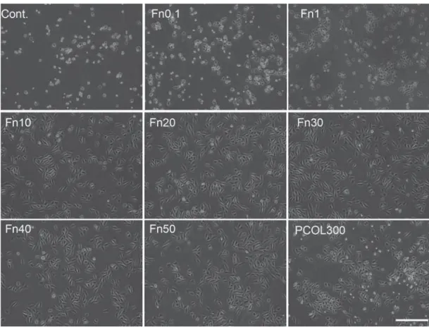
Fn promoted MDPC-23 cells spreading and
proliferation
Photographs of light microscopy cells indicated that
cells adopted well spread shape in Fn (10-50) and
PCOL300 after 20 hours (Figure 3), but those cultured
on control, Fn (0.1-1) were poorly spread, moreover,
the attached number of cells in the three groups were
much lower than Fn (10-50) and PCOL300. Given
that cells growth and spreading remain comparatively unchanged in Fn with concentration over 10 μg/mL, only the immunostaining of cells grown on Fn10 was
shown in Figure 4. In Figure 4, regular and parallel
on Fn (10-50) and PCOL300; however, cells assumed a polygonal or round shape while growing on Fn1.
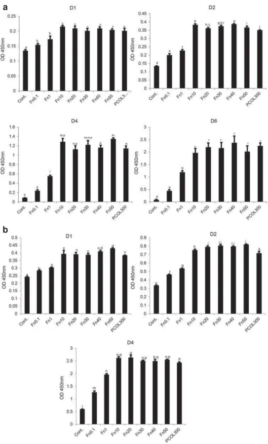
To quantify the cell proliferation activity, MDPC-23
cells were cultured in 96 well plate coated with Fn
or PCOL. To further evaluate the biocompatibility of Fn, cells were inoculated at two concentrations (Low
concentration: 5×10³/mL and high concentration:
1×10⁴/mL). Cells fail to grow in low concentration
group in control well (Figure 5a); however, those
cultured in Fn exhibited concentration-dependent
fashion (0.1-10) of growth for the points tested four
times, when the coating concentration of Fn was over 10 μg/mL, proliferation activity was maintained at a constant level; moreover, the proliferation activity
in PCOL300 was quite similar to that in Fn (10-50),
Fn groups and PCOL300. In the higher concentration
Gene name Forward Backward Fragment size(bp)
Rat DMP-1 (NM_203493.3)
CGTTCCTCTGGGGGCTGTCC CCGGGATCATCGCTCTGCATC 577
Rat ALP (NM_013059.1)
GGAAGGAGGCAGGATTGACCAC GGGCCTGGTAGTTGTTGTGAGC 338
Rat BSP (NM_012587.2)
CTGCTTTAATCTTGCTCTG CCATCTCCATTTTCTTCC 211
Rat OCN (NM_013414.1)
AGCTCAACCCCAATTGTGAC AGCTGTGCCGTCCATACTTT 190
Rat OPN (NM_012881.2)
TTTCCCTGTTTCTGATGAACAGTAT CTCTGCTTATACTCCTTGGACTGCT 228
Rat ITGB1 (NM_017022.2)
ACAAGAGTGCCGTGACAACT AGCTTGATTCCAAGGGTCCG 325
(NM_031144.3)
AACCCTAAGGCCAACAGTGAAAAG TCATGAGGTAGTCTGTGAGGT 241
Figure 1- Real time RT-PCR primer
Gene name Initialization Denaturation Annealing Elongation Cycle
DMP-1 95°C 10 min 95°C 15 s 60°C 30 s 72°C 30 s 50
ALP 95°C 10 min 95°C 15 s 55°C 30 s 72°C 30 s 45
BSP 95°C 10 min 95°C 15 s 55°C 15 s 72°C 30 s 50
OCN 95°C 10 min 95°C 15 s 55°C 30 s 72°C 30 s 50
OPN 95°C 10 min 95°C 15 s 55°C 30 s 72°C 30 s 45
ITGB1 95°C 10 min 95°C 15 s 59.9°C 30 s 72°C 40s 45
95°C 10 min 95°C 15 s 53°C 30 s 72°C 40s 50
Figure 3- Cell morphology observation by light microscopy
MDPC-23 cells were seeded to 12 well plate (non-treated tissue culture polystyrene) at the concentration of 1×10⁴/mL in DMEM supplemented with 5% FBS. The photograph was taken 20 hours after inoculation. Cells adopted well spread and spindle shape in Fn(10-50) and PCOL-300, while those in control and Fn(0.1-1) were in round shapes. (Scale bar: 400 μm)
Figure 4-
group (Figure 5b); cells did increase in control well,
whereas the growth speed was rather low when
compared with the Fn and PCOL.
Fn increased ALP activity and the expression
of odontogenic differentiation markers
Alkaline phosphatase was established to be one of the phenotypic markers of odontoblastic differentiation
a
b
Figure 5- Cell proliferation activity
U/μg protein) at day 5 (Figure 6), while that of μ
μ μ
μ μ
μ
the mRNA expression of phenotypic markers (Figure
7). Among the markers tested, we observed that
especially BSP (three fold) and OCN (2.5-fold) were
was also higher than PCOL300 (2.2-fold in BSP;
1.6-fold in OCN). ITGB1, ALP, and DMP1 were slightly
up-regulated in Fn (10-50) and PCOL300. OPN was
of Fn but not PCOL300.
was incorporated into culture media from 5 days. Similarly, two concentrations equal to the proliferation
experiment were used for cell inoculation. After 10
days, calcifying nodules appeared in Fn (10-50)
and PCOL300 in both low (Figure 8 upper) and high concentration groups (Figure 8 lower); however, higher
inoculation number leads to more calcifying nodules.
Discussion
Fn is an adhesive extracellular matrix glycoprotein
that is involved in a variety of physiological and pathological processes. Small interfering RNA
against Fn abrogated cleft formation and branching
morphogenesis, while exogenous Fn facilitated and
accelerated the branching and cleft formation. More importantly, Fn was found to mediate the conversion
process of cell-cell adhesion to cell-matrix adhesion
in human salivary epithelial cells24. Although Fn has
been extensively studied in the development of tissues and organs, its role in the regulation of odontoblast
activity remains elusive. To determine the precise
role of Fn in the regulation of odontoblast activity, we evaluated proliferation and differentiation of
MDPC-23 cells in Fn-coated substrates using type I
collagen as a comparison with matrix protein. This
study showed that culturing MDPC-23 cells in the presence of Fn can direct MDPC-23 cells down lineage
markers of odontogenic differentiation (intracellular
ALP activity, phenotypic gene expression) were greater with MDPC-23 cells cultured in Fn. Furthermore, Fn
concentration when exposed to mineralization factors.
relationship exists between Fn and odontoblast-like
cell. The study highlighted natural cues for odontoblast
differentiation within the local microenvironment in v it r o.
Cells started to spread merely one hour after
inoculation in Fn (10-50) (coated in either non-treated
Figure 6- ALP activity
polystyrene or tissue culture polystyrene), which is much earlier than those grown on control (2 days) and
type I collagen (18 hours) (data not shown). Actin is
It is comprised of free monomer G-actin (globular) and linear polymer F-actin (filamentous). Actin
is a mechanosensing tool7 and transduces the
extracellular signals to change the shape of the
cell, direct cell migration and differentiation. F-actin
were successfully initiated and activated on Fn10 and PCOL300, indicating that mechanosensing and
signaling transduction was promoted in cells. Next,
using CCK-8 assay. We found that Fn (0.1-50) and
compared with the control. The activity exhibited
concentration-dependent trend when concentration of Fn was lower than 10 μg/mL. It reached a state of plateau when the coating concentration of Fn was over
Figure 7- Real time RT-PCR
MDPC-23 cells were seeded to 12 well plate (non-treated tissue culture polystyrene) at the concentration of 1×10⁴/mL in DMEM
10 μg/mL. Further, we showed that the non-treated polystyrene was not able to support the growth of MDPC-23 cells when the cell concentration was only
5×10³/mL. When inoculation number increased to
1×10⁴/mL, the control did maintain a certain level of
cell growth, the cell viability was markedly lower than that in Fn and PCOL300 though. The proliferation data
revealed that Fn is highly biocompatible, coating of
Fn to hydrophobic polystyrene facilitated cell adhesion
and growth. Indeed, although Fn was not expressed in mineralized dentin and epithelial tissue in the
mouse tooth germ, a strong staining of Fn using its
antibody was detected between dental epithelium and
mesenchyme, Thesleff, et al.28 (1979) hence inferred
that Fn may be a potential anchorage site for cells
of mesenchymal origin to differentiate into mature
with the proliferation data of this study provide a direct evidence of Fn as a potent odontoblast-like
cell-adhesive, which allows MDPC-23 cells to attach,
spread and proliferate rapidly.
Both calcium deposition and elevation of ALP activity were observed by Fn exposure. The results
indicate that Fn promoted mineralization and is
associated with increased ALP activity, since ALP
1, an early
enhancement of ALP activity denotes accelerating
differentiation process toward mineralizing
tissue-forming cells. Besides the enhanced ALP activity, the markedly increased expression of mineralized
tissue-forming genes was detected as well. BSP, OPN
and DMP1 are Small Integrin Binding Ligand N-linked
Glycoprotein (SIBLING) members. They possess
functions. BSP is a component of mineralized tissues
and is suggested to constitute approximately 8% of all non-collagenous proteins found in bone and
initial stages of connective tissue mineralization5 and
is strongly expressed in the odontoblast-like cells of
reparative dentine11. OPN, a highly phosphorylated
glycoprotein, is important for type I collagen secretion
in reparative dentine formation by newly differentiated
odontoblast-like cells23. DMP1 is an RGD containing
incisor cDNA library; it is an inductive factor for dental
pulp stem cells to differentiate into odontoblast3. OCN,
a non-collagenous protein found in both dentin and bone, is found to be strongly expressed at 2 and 3
days post-tooth preparation, its expression correlates
with reactionary dentine formation8. The strongest
expression of OCN in Fn provides evidence of an earlier differentiation of MDPC-23 cells on the substrates.
ITGB1, also known as CD29, is able to associate with
a number of alpha integrin to form various types of heterogeneous integrin dimers, a recent paper
ITGB1 is essential for ameloblast differentiation and
enamel formation22. In particular, among the genes
tested, BSP and OCNwere found to be enhanced by a
much higher fold change on Fn(10-50) than PCOL300,
which indicated that Fn possesses a stronger capacity of
initiating odontogenic differentiation as compared with PCOL300. Indeed, a previous study using dog dental
to exhibit odontoblastic phenotype in response to
absence of exogenous inductive molecules29. The
Figure 8- Alizarin red staining
Cells were inoculated in 24 well plate (non-treated tissue culture polystyrene) at the concentration of 5×10³/mL (upper) or 1×10⁴/mL
up-regulation of those mineralizing tissue markers
suggested that Fn is a suitable cell expansion matrix
to retain high odontogenic differentiation potential and provide a synergistic effect in the presence of
in v it r o. Furthermore, concomitant increased expression of
ITGB1 suggests that the up-regulation of odontogenic markers might be mediated, at least partially, by
activation of ITGB1.
Cells secrete a wide variety of extracellular matrix that is crucial for the maintenance of normal
function. ECM plays the role not only as a mechanical
and structural framework, it also modulates many
aspects of cell behavior, such as adhesion, migration, differentiation, and mineralization. To gain further
insight into the in v it r o
nodule. Alizarin red s was used as a vital stain for mineralization.
Fn and PCOL were pre-coated in a sterile
environment in 24 well plate (non-treated polystyrene)
days, calcifying nodules appeared in Fn (10-50) and
PCOL300-treated groups in both cell concentration
groups, however, the number of nodules in groups with high number of cells was observed to be comparatively
higher than that of low cell number groups. This
phenomenon correlates well with previous studies
using umbilical cord stem cells31 and human bone
marrow stromal cells13, indicating that not only the
ECM but also initial seeding density is a critical factor
in determining the late stage mineralization intensity of
cells. In addition, cells started to detach from control and Fn (0.1-1) at around 8 days in culture, which is the
reason we do not show the alizarin staining of control
and Fn (0.1-1). This has prompted us to think that cell attachment strength in Fn (10-50) is higher than
that on control and Fn (0.1-1). Further detachment
strength test between cells and Fn or collagen needs
to be carried out.
Conclusion
The proliferation of MDPC-23 cells cultured on
polystyrene preadsorbed with Fn was promoted.
Further, an early differentiation marker of odontoblast,
grown on Fn-treated polystyrene. The expression
of odontogenesis’ markers during the differentiation
and mineralization phase was increased by coated Fn. Finally, the mineralization of cells was facilitated by
Fn as well. The results suggest that, a surface coating of polystyrene with Fn at the concentration of 10 μg/ mL or more effectively supported the proliferation, differentiation and mineralization of
odontoblast-like cells; moreover, this inductive effects of Fn was
achieved at a coating concentration markedly lower
than type I collagen (300 μg/mL).
Acknowledgements
The study was supported by a grant-in-aid for
the Promotion of Science (Grant No.15K15702 and
No.15H05024) and a research grant (May 2016-March
2017) for young Chinese researchers in Japan from The
Japan China Medical Association (JCMA). The authors
References
1- Ahlers J. The mechanism of hydrolysis of beta-glycerophosphate
by kidney alkaline phosphatase. Biochem J. 1979;149(3):535-46. 2- Alitalo K, Hovi T, Vaheri A. Fibronectin is produced by human
macrophages. J Exp Med. 1980;151(3):602-13.
3- Almushayt A, Narayanan K, Zaki AE, George A. Dentin matrix protein
1 induces cytodifferentiation of dental pulp stem cells into odontoblasts. Gene Ther. 2006;13(7):611-20.
4- Aota S, Nomizu M, Yamada KM. The short amino acid sequence
Pro-J Biol Chem. 1994;269(40):24756-61.
5- Chen J, Shapiro HS, Sodek J. Development expression of bone sialoprotein mRNA in rat mineralized connective tissues. J Bone Miner
Res. 1992;7(8):987-97.
6- Fukumoto S, Yamada Y. Review: extracellular matrix regulates tooth
morphogenesis. Connect Tissue Res. 2005;46(4-5):220-6.
7- Hayakawa K, Tatsumi H, Sokabe M. Mechano-sensing by actin
filaments and focal adhesion proteins. Commun Integr Biol. 2012;5(6):572-7.
8- Hirata M, Yamaza T, Mei YF, Akamine A. Expression of osteocalcin and Jun D in the early period during reactionary dentin formation after
tooth preparation in rat molars. Cell Tissue Res. 2005;319(3):455-65. 9- Humphries JD, Byron A, Humphries MJ. Integrin ligands at a glance.
J Cell Sci. 2006;119(Pt 19):3901-3.
10- Humphries MJ, Akiyama SK, Komoriya A, Olden K, Yamada
1986;103(6 Pt 2):2637-47.
of TGF-beta1 on the expression of BSP, DSP, TGF-beta1 receptor I and Smad proteins during reparative dentinogenesis. J Mol Histol.
2008;39(2):153-60.
12- Kubler MD, Lesot H, Ruch JV. Temporo-spatial distribution of matrix
13- Lode A, Bernhardt A, Gelinsky M. Cultivation of human bone
marrow stromal cells on three-dimensional scaffolds of mineralized
osteogenic differentiation. J Tissue Eng Regen Med. 2008;2(7):400-7. 14- Mizuno M, Banzai Y. Calcium ion release from calcium hydroxide
differentiation of dental pulp cells to mineralized tissue forming cells
sequence for the integrin alpha 4 beta 1 in the COOH-terminal
heparin-16- Moursi AM, Globus RK, Damsky CH. Interactions between
differentiation in v it r o. J Cell Sci. 1997;110(Pt 18):2187-96.
17- Moyano JV, Carnemolla B, Domínguez-Jiménez C, García-Gila
M, Albar JP, Sánchez-Aparicio P, et al. Fibronectin type III5 repeat contains a novel cell adhesion sequence, KLDAPT, which binds
activated alpha4beta1 and alpha4beta7 integrins. J Biol Chem. 1997;272(40):24832-6.
18- Ochsenhirt SE, Kokkoli E, McCarthy JB, Tirrell M. Effect of RGD secondary structure and the synergy site PHSRN on cell
2006;27(20):3863-74.
19- Park JB. The effects of dexamethasone, ascorbic acid, and
estrogen receptor and osteopontin expression. J Surg Res.
2012;173(1):99-104.
20- Peters JH, Sporn LA, Ginsberg MH, Wagner DD. Human endothelial
alternatively spliced type III homology (ED1). Blood.
1990;75(9):1801-8.
21- Pierschbacher MD, Ruoslahti E. Cell attachment activity of
molecule. Nature. 1984;309(5963):30-3.
22- Saito K, Fukumoto E, Yamada A, Yuasa K, Yoshizaki K, Iwamoto
for tooth development. PLoS One. 2015;10(4):e0121667.
23- Saito K, Nakatomi M, Ida-Yonemochi H, Ohshima H. Osteopontin is essential for type I collagen secretion in reparative dentin. J Dent
Res. 2016;95(9):1034-41.
24- Sakai T, Larsen M, Yamada KM. Fibronectin requirement in
branching morphogenesis. Nature. 2003;423(6942):876-81. 25- Seebacher T, Marianne M, Kornblihtt AR, Bade EG. Fibronectin is
induced by epidermal growth factor, but not by dexamethasone or cyclic AMP in rat liver epithelial cells. FEBS Lett. 1988;239(1):113-6.
26- Sharma M, Sah P, Sharma SS, Radhakrishnan R. Molecular changes in invasive front of oral cancer. J Oral Maxillofac Pathol.
2013;17(2):240-7.
27- Thesleff I, Mikkola M. The role of growth factors in tooth development. Int Rev Cytol. 2002;217:93-135.
28- Thesleff I, Stenman S, Vaheri A, Timpl R. Changes in the matrix
germ. Dev Biol. 1979;70(1):116-26.
29- Tziafas D, Alvanou A, Kaidoglou K. Dentinogenic activity of allogenic
95.
30- Wang YM, Reheman A, Spring CM, Kalantari J, Marshall AH, Wolberg
thrombosis. J Clin Invest. 2014;124(10):4281-93.
31- Zhou H, Weir MD, Xu HH. Effect of cell seeding density on
proliferation and osteodifferentiation of umbilical cord stem cells