43 Case Report
International Braz J Urol
Official Journal of the Brazilian Society of Urology
Vol. 29 (1): 43-44, January - February, 2003
LEIOMYOSARCOMA OF THE RENAL VEIN
GUSTAVO C. LEMOS, OMAR R. EL HAYEK, MARCELO APEZZATO
Albert Einstein Jewish Hospital, São Paulo, SP, Brazil
ABSTRACT
Leiomyosarcoma of the renal vein is a rare tumor of complex diagnosis. We presented a case of renal vein leiomyosarcoma detected in a routine study. The primary treatment was complete surgi-cal removal of the mass. In cases where surgisurgi-cal removal is not possible the prognosis is poor, with high rates of local recurrence and distant spread.
Key words: kidney; renal veins; muscle neoplasms; leiomyosarcoma Int Braz J Urol. 2003; 29: 43-4
INTRODUCTION
There are approximately 30 cases of leiomyo-sarcoma of the renal vein reported in literature. They affect primarily the inferior vena cava in more than 50% of the cases (1). Pre-operative diagnosis is diffi-cult due to its low incidence, and since it presents a slow growing rhythm and nonspecific symptoms. We present a case of primary leiomyosarcoma of the left renal vein.
CASE REPORT
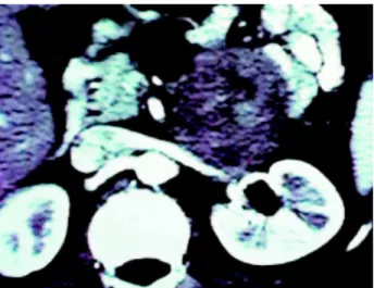
Patient with 47 years, male, white came to the urologist to routine exam. In physical examination identified a mass in left flank, painless and non mo-bile, by palpation. An abdominal ultrasound showed a tumor without not related to renal parenchyma and excretory route but in close contact with the renal vein (Figure-1).
During the surgery we found that the tumor had its origins in the renal vein and the patient was submit-ted to radical left nephrectomy (Figure-2). The histo-logical study disclosed renal vein leiomyosarcoma.
DISCUSSION
Leiomyosarcoma is an uncommon soft tissue tumor, generally occurring in myometrium, respira-tory tract and retroperitoneal organs. It seldom origi-nates in vascular structures, and the inferior vena cava responds for more than 50% of the cases (2). Lei-omyosarcoma of the inferior vena cava was first de-scribed in 1871 (1), and its diagnosis and treatment is still challenging. These tumors attain women over 30 years in 85% of the cases (2). More frequently they are left-sided (64%). Symptoms are nonspecific, such as mild lumbar and abdominal pain, and wasting. He-maturia and palpable mass are rare (2).
Until the 80’s, approximately 50% of the cases were autopsy findings. Presently they are incidentally found in routine studies (2). Clinical context and ul-trasound and computed tomography studies are non-specific and do not allow an adequate differential di-agnosis with other retroperitoneal solid tumors (3).
approxi-44
LEIOMYOSARCOMA OF THE RENAL VEIN
mately half of the cases present metastatic disease or are nonresectable due to local invasion, presenting therefore a poor prognosis (3).
The best treatment for leiomyosarcoma is sur-gery with total tumor removal. This is the option that offers the best chances of local control and 5 years survival. Studies performed at Memorial Sloan Kettering, New York, showed that the major prog-nostic factor is total surgical resection. When it is complete, 5 years survival free of disease is of ap-proximately 60%, vs. just 30 to 35% when it is par-tial. Once total removal is performed, major prog-nostic factor becomes histological grade, with 5 years free of disease survival of 90 to 95% for low grade tumors, and of 30 to 35% for high grade tumors.
Radiotherapy and adjuvant chemotherapy have limited effects due to toxicity on contiguous structures. Adjuvant therapy is generally used to high grade tumors, with partial resection (3).
REFERENCES
1. Bhathena D, Vasquez M: Primary renal vein leiomyo-sarcoma. Cancer 1972; 30: 541-4.
2. Brandes SB, Chelsky MJ, Petersen RO, Greenberg RE: Leiomyosarcoma of the renal vein. J Surg Oncol. 1996; 63: 195-200.
3. Bevilacqua RG, Rogatko A, Hadju SI, Brennan MF: Prognostic factors in primary retroperitoneal soft-tis-sue sarcomas. Arch Surg. 1991; 126: 328-34.
Received: August 12, 2002 Accepted after revision: January 9, 2003
Correspondence address: Dr. Gustavo Caserta Lemos Rua Jayme de Almeida Paiva, 81 São Paulo, SP, 05657-170, Brazil Fax: + 55 11 3747-3333
E-mail: [email protected]
Figure 1 – Abdominal computed tomography showing a bulky mass near the left renal hilum, invading the renal vein.
Figure 2 – Surgical specimen. A) - Place of tumor origin near the left renal vein wall. B) - Macroscopic view of the solid mass.