PENILE FRACTURE - EXPERIENCE IN 56 CASES
LEANDRO KOIFMAN, ANDRÉ G. CAVALCANTI, CARLOS HENRIQUE MANES, DAIBES R.
FILHO, LUCIANO A. FAVORITO
Division of Urology, Souza Aguiar Municipal Hospital, Rio de Janeiro, RJ, Brazil
ABSTRACT
Objective: The aim of this work is to report the diagnostic and therapeutic options for 55 patients with clinical diagnosis of penile fracture.
Material and Methods: The patients were retrospectively assessed between 1982 and 2002. The primary diagnostic evaluation method for 55 patients (56 fractures) was clinical history and physical exam. Ten (17.8%) cases required complementary exams. Ultrasound (US) was performed in 2 cases, and magnetic resonance imaging (MRI) in 1 case. Retrograde urethrocystogram was per-formed in suspicious urethral injury, which happened for 7 patients.
Results: Of 56 assessed cases, 49 (89.5%) were submitted to surgical exploration, and only 7 were conservatively conducted. Surgical treatment was performed in 48 patients (49 fractures), in these cases, 47 (95.9%) presented tunica albuginea disruption and solely 2 (4.1%) evidenced lesion of dorsal vein. Ultrasonography confirmed disruption of tunica albuginea in 1 (50%) case, and in the other it was not possible to determinate the origin of the lesion, and the patient was submitted to surgical exploration, which confirmed the condition. MRI was used only in 1 case, confirming the lesion. Among 7 patients submitted to conservative management, until now, 3 (42.8%) required surgi-cal intervention to correct penile chordee.
Conclusions: Penile fracture is an entity of eminently clinical diagnosis, which management should be surgical and immediate, avoiding thus complications related to erectile dysfunction. When suspecting an associated urethral injury, Urethrocystogram is recommended. In cases where there is diagnostic uncertainty, ultrasound and/or MRI may be used to reveal the condition.
Key words: penis; urethra; fractures; therapeutics; surgery Int Braz J Urol. 2003; 29: 35-9
INTRODUCTION
Penile fracture is one of the less frequent uro-logical traumas. There are 183 reports about this sub-ject published, with 1,331 cases described from 1935 to 2001 (1). It is defined as a rupture of the corpus cavernosum due to a blunt trauma in an erect penis. Lesions on a flaccid penis or lesions in the suspensor ligament of the penis are not included in this defini-tion (2).
Vaginal intercourse is the most common cause of penile fractures (1,3-5), masturbation is also re-ported as a cause of penile fracture (6). In lower
inci-dence, the lesion could occur during a nocturnal erec-tion due to the patient rolling over his own body.
Penile fracture has a quite typical clinical pre-sentation. Patients report hearing a snap sound fol-lowed by pain, penile detumescence, and late appear-ing swellappear-ing, hematoma and penile deformity (7-9). In the presence of associated urethral injury, happen-ing in 10% to 20% of the cases, findhappen-ings as urethral bleeding, hematuria and difficulty voiding may be observed (8,10).
ultrasound and MRI use as diagnostic tools are un-common.
The aim of this paper is to report the experi-ence of 56 cases assessed in 55 patients, admitted to our facility with a clinical diagnosis of penile frac-ture in the last 20 years, and discuss the therapeutic and diagnostic options to this type of lesion.
MATERIALS AND METHODS
In the period between January 1982 and May 2002, 55 patients (56 fractures—the same patient having had 2 fractures in a 90 days interval) with clini-cal diagnosis of penile fracture were admitted in our facility and retrospectively assessed. Patients’ age ranged from 18 and 63 years (mean 33 years). Sexual trauma was the most common cause corresponding to 53 (94.7%) cases, followed by lesion due to penis manipulation in 3 (5.3%) cases. Time elapsed form the trauma to the arrival at the hospital ranged from 2 hours to 3 weeks (mean 14 hours).
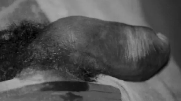
Primary diagnostic assessment was clinical history and physical exam (Figure-1). In 10 (17.8%) cases, complementary exams were required. Ultra-sound scan was used in 2 cases and MRI in 1 case. Retrograde urethrocystogram was performed solely when urethral injury was suspected, what happened to 7 patients.
SURGICAL TECHNIQUE
The surgical technique used consisted of a subcoronal incision, with penile degloving and ex-posure of the corpora cavernosum and urethra. Blad-der catheterization was routinely performed, except for the cases where a urethral injury was suspected. All corpora cavernosa lesions identified during sur-gical exploration were treated by interrupted polyglactine 3-0 sutures. Urethral lesions were pri-marily corrected with interrupted absorbable polyglactine 5-0 sutures. Bladder catheter was main-tained during 12 hours after the surgical procedure conclusion for patients without urethral lesions; 7-10 days in patients with partial urethral injury; and 14-21 days in patients with total urethral section.
Only 1 patient required Penrose #1 drain owing to the severity of the lesion (bilateral albug-inea disruption and total urethral section), and the presence of a large hematoma. The drain was with-drawn at hospital discharge.
RESULTS
From 56 cases assessed, 49 (87.5%) were submitted to surgical exploration, and only 7 (12.5%) were conservatively managed. Table-1 shows lesions observed during surgical exploration.
From 48 patients (49 fractures) submitted to surgical procedure, 47 (95.9%) presented disruption of tunica albuginea and only 2 (4.1%) showed lesion of the dorsal vein (Figure-2). In 6 (12.2%) patients urethral injury occurred, and in all these we found associated corpus cavernosum lesions.
Figure 1 - Patient with penile fracture, with an hematoma ex-tending through the whole penis.
From 47 penile fracture cases, with albug-inea disruption, 45 (95.7%) presented unilateral le-sion, and 2 (4.3%) bilateral lesion. Both cases of bi-lateral corpora cavernosa lesion were associated to urethral injury. Lesion size ranged from 0.3 cm to 4.0 cm (mean 1.5 cm).
In the group of patients submitted to surgical exploration, 32 had follow-up longer than 1 year. In this group, there was no complaint about erectile dys-function after the trauma, and only 2 (6.2%) patients developed slight penile curvature, without sexual function impairment.
We have observed urethral bleeding and dif-ficulty voiding in 7 (12.5%) cases, for which
Urethrocystogram was performed, evidencing con-trast medium leakage in 6 cases. After their surgical exploration, the urethral injury was confirmed in all 6 cases, demonstrating the exam accuracy. Of 6 pa-tients with confirmed diagnosis of urethral injury, only 1 had total lesion. Among these patients, 4 presented unilateral corpus cavernosum lesion, and 2 presented bilateral lesion.
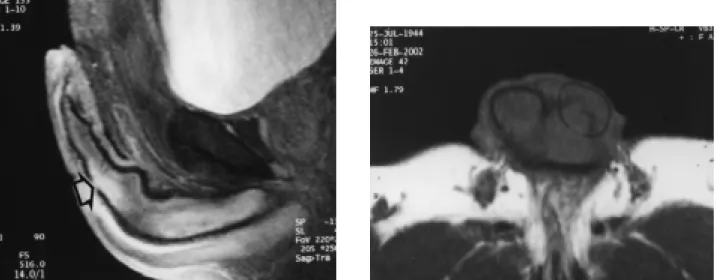
Ultrasound study confirmed an albuginea dis-ruption in 1 (50%) case, in the other, it was not pos-sible to determine the lesion, and the patient was sub-mitted to surgical exploration that confirmed the con-dition. MRI was used in only 1 case, confirming the lesion (Figure-3).
Among 7 patients conservatively managed (treatment decided by the urologist during the admis-sion), until nowadays 3 (42.8%) needed surgical pro-cedures to correct the penile chordee.
DISCUSSION
There are few reports with a significant se-ries about penile fracture in the literature (11). Penile fracture is an entity that generally has its diagnosis confirmed by its clinical presentation. The typical history, associated with physical exam findings, ex-empts performing complementary exams. For the rare exceptions where there is diagnostic uncertainty, some imaging methods may be used.
Figure 3 - A) Sagital view of MRI showing rupture in the tunica albuginea (arrow). B) Frontal view of the same patient showing disruption in the tunica albuginea of the corpus cavernosum (circle).
Table 1 – Lesions found in patients presenting penile frac-tures submitted to surgery. Patients with urethral lesion presented associated corpus cavernosum lesion, and in 2 cases, lesions were bilateral.
Type of Lesion Cases %
Ultrasound has a limited role in the diagno-sis of penile fracture (12-14). As it is an examiner-depending method, for which interpreting depends on the examiner’s experience, rareness of these le-sions often precludes an accurate diagnosis (12). Small albuginea disruptions and the presence of clots at the place where fracture site occurred may easily be unperceived (12-15).
In both cases assessed by ultrasound, albug-inea disruption was observed only in 1 patient. How-ever, as it is a non-invasive method, with low cost and accessible in the great majority of institutions, it may help to evaluate uncertain cases.
Magnetic resonance imaging has been also used for demonstrating corpus cavernosum lesions (11,12,16,17). Its high accuracy can discriminate the intensity of corpora cavernosa vascular sinusoids (high intensity) relative to tunica albuginea (low in-tensity), allowing for accurate diagnosis (12,16,17). Nevertheless, it is a high-cost exam, and it is not avail-able at most institutions. In our series, only 1 patient (with diagnostic uncertainty) was submitted to this exam, with an accurate confirmation of the lesion.
In suspicion of urethral injury, an urethrocystogram shall be performed. As it is a low-cost exam, easy to perform and highly accurate, all patients with an urethral injury suspected were sub-mitted to the exam. Of 7 cases assessed, 6 presented contrast medium leakage. After surgical exploration, urethral injury was confirmed for all 6 cases. How-ever, it is worth remembering that, in other published series, Urethrocystogram did not demonstrate this ef-ficiency, being thus criticized by some authors (8,18). Previous studies report 10% to 41% compli-cations rates from conservative management of pe-nile fracture, and surgical treatment is, thus, the main option for this type of trauma (4,6,7,11,12,18-20). We have observed medical complications for 37.5% of conservatively managed patients, supporting the data presented in previous publications. In our series, none of surgically treated patients presented penile curva-ture during pos-operative period, a relatively infre-quent complication that generally does not affect sexual intercourse (21).
During sexual intercourse the rupture of dor-sal vein of the penis may occur, leading to a clinical
presentation similar to penile fracture (22). Differen-tiating these 2 types of trauma sometimes is possible only through surgical exploration, which is the treat-ment of choice for both conditions. Among 56 cases in our series, only 2 presented dorsal vein of penis lesion.
Disagreement about the type of incision to be used in treating penile fracture remains. Longitu-dinal incisions over the area where the fracture is sus-pected, parapenile incisions exposing shaft or even inguino-scrotal incisions were proposed (23-25). Pe-nile deglovement, however, offers a better exposure, in addition to allowing evaluation of both corpora cavernosa and corpus spongiosum.
CONCLUSIONS
Penile fracture is an entity which diagnosis is eminently clinical, and its management should be surgical and immediate, avoiding thus complications related to erectile dysfunction.
In suspicious of urethral injury, Urethrocystogram is recommended. In cases where there is diagnostic uncertainty, an ultrasound and/or magnetic resonance imaging may be use to reveal the condition.
REFERENCES
1. Eke N: Fracture o the penis. Br J Surg. 2002; 89:555-65.
2. El-Sherif AE, Dauleh M, Allowneh N, Vijayan P: Man-agement of fracture of the penis in Quatar. Br J Urol. 1991; 68:622-5.
3. Schonberger B: Verletzungen der mannlichen Genitalorgane. Z Urol Nephrol. 1982; 5:879-83. 4. Nicoliasen S, Melamud A, McAninch JW: Rupture of
the corpus cavernosum: Surgical management. J Urol. 1983; 130:917-20.
5. Klein FA, Smith V, Miller N: Penile fracture: Diagno-sis and management. J Trauma 1985; 25:1090-2. 6. Taha SA, Sharayah A, Kamal BA, Salem AA, Khwaja
S: Fracture of the penis: Surgical manegement. Int Surg. 1988; 73:63-4.
7. Meares EM: Traumatic rupture of the corpus cavernosum. J Urol. 1971; 105:407-9.
cavernosography imaging in a small series of penile fractures: a comparison with surgical findings. Urol-ogy 1998; 51:616-9.
9. Ruckle CH, Hadley HR, Lui PD: Fracture of the pe-nis: Diagnosis and manegement. Urology 1992; 40:33-5.
10. Tsang T, Demby AM: Penile fracture with urethral in-jury. J Urol. 1992; 147:466-8.
11. Zargooshi J: Penile fracture in Kermanshah, Iran: re-port of 172 cases. J Urol. 2000; 164:364-6.
12. Miller S, McAninch JW: Penile Fracture and Soft Tis-sue Injury. In: Traumatic and Reconstructive Urology. Edited by J.W. McAninch. Philadelphia, W.B. Saunders, 1996; chapt. 59, pp.693–8.
13. Dierks PR, Hawkins H: Sonography and penile trauma. J Ultrasound Med. 1983; 2: 417-9.
14. Martinez Perez E, Arnaiz Esteban F: Fracture of the penis: two new cases. Review of the literature: useful-ness of ecography. Arch Esp Urol. 1997; 50:1099-102. 15. Hoekx L: Fracture of the penis: role of ultrassonography in localizing tear. Acta Urol Belg. 1998; 66:23-5.
16. Fedel M, Venz S, Andressen R, Sudhoff F, Loening S: The value of magnetic ressonance imaging in the di-agnosis of suspected penile fracture with atypical clini-cal findings. J Urol. 1996; 155:1924-7.
17. Susuki K, Shimizu N, Kurokawa K, Susuki T, Yamanaka H: Fracture of the penis: magnetic ressonance imaging of the rupture of the corpus cavernosum. Br J Urol. 1995; 76:803-4.
18. Gianini PTR, Piovesan ACP, Mesquita JLB, Romao RLP, Arap S: Long-term outcome of penile fracture treatment. Int Braz J Urol. 2001; 27:46-9.
19. Pryor JP, Hill JT, Packham DA, Yates-Bell AJ: Penile injuries with particular reference to injury to erectile tissue. Br J Urol. 1981; 53:42-6.
20. Orvis BR, McAninch JW: Penile rupture. Urol Clin North Am. 1989; 16:369-75.
21. Uygur MC, Gulerkaya B, Altug U, Germiyanoglu C, Erol D: 13 years experience of penile fracture. Scand J Urol Nephrol. 1997; 31:265-6.
22. Babu N: Rupture of the dorsal vein mimicking frac-ture of the penis. BJU Int. 1999; 84:179-80.
23. Ozen HA, Erkan I, Alkibay T, Kendi S, Remzi D: Frac-ture of the penis and long-term results of surgical treat-ment. Br J Urol. 1986; 58:551-2.
24. Mellinger BC, Douenias R: New surgical approach for operative management of penile fracture and penetra-tions trauma. Urology 1992; 39:429-32.
25. Seftel AD, Haas CA, Vafa A, Brown SL: Inguinal scro-tal incision for penile fracture. J Urol. 1998; 159:182-4.
Received: August 8, 2002 Accepted after revision: February 4, 2003
Correspondence address:
Dr. Luciano Alves Favorito Rua Professor Gabizo, 104 / 201 Rio de Janeiro, RJ, 20271-320, Brazil Fax: + 55 21 3872-8802