45
LAPAROSCOPIC APPROACH IN THE OVARIAN VEIN SYNDROME Case Report
International Braz J Urol
Official Journal of the Brazilian Society of Urology
Vol. 29 (1): 45-47, January - February, 2003
LAPAROSCOPIC APPROACH IN THE OVARIAN VEIN SYNDROME
ARAKÉN ALMEIDA, FRANCISCO CAVALCANTI, SÁVIO BARBOSA, ROBERTO COHEN,
AMAURY MEDEIROS
Oswaldo Cruz University Hospital, Pernambuco University, Recife, Pernambuco, Brazil
ABSTRACT
Introduction: The main objective of this article is to describe ureterolysis and ovarian vein resection laparoscopic technique.
Surgical technique: With the patient in a 45o flank position, 3 trocars are used, 1 of 12 mm in the umbilicus for the optic passage, and 2 of 5 and 10 mm inserted in right hypochondrium and iliac fossa, respectively, for the forceps and stapler passage. The ureter and ovarian vein are identified after the mobilization of the colon. Both structures are dissected, with one ovarian vein segment is resected between metallic clips.
Comments: the ureteral approach by transperitoneal laparoscopy and colon mobilization facilitates its dissection, identify its relation to other structures, as well as making possible the con-comitant treatment of gynecological diseases. For the ovarian syndrome treatment, ureterolysis and ovarian vein resection are performed, using only 3 trocars. Owing to its simplicity, low morbidity, and good results obtained, this procedure represents a good option for the surgical management of this syndrome.
Key words: laparoscopy; ovary; veins; syndrome; therapeutics Int Braz J Urol. 2003; 29: 45-7
INTRODUCTION
The therapeutic procedure for the ovarian vein syndrome varies according to the intensity and urgency of the clinic presentation. In nulliparous women, non-pregnant multiparous women and in children, the treatment is conservative. Yet, pregnancy may promote an intensification of this condition, lead-ing to the necessity of a surgical intervention (1). We present case report of 1 ovarian vein syndrome treated by laparoscopic approach, with surgical technique description.
CASE REPORT AND SURGICAL
TECHNIQUE
A 26 years-old patient, 62 kg, 1.68 m (BMI = 22), with right lumbar pain and 8 months recurrent
urinary tract infections complaints, also with obstet-ric history of 3 gestations, was submitted to ureteroly-sis and right ovarian vein laparoscopic resection in our Unit, using the technique described as follows. Diagnosis was established by intravenous pyelogram and computed tomography (Figures-1 and 2).
decu-46
LAPAROSCOPIC APPROACH IN THE OVARIAN VEIN SYNDROME
bitus. The incision in the Toldt line is performed, mobilizing the colon medially. The ovarian vein plexus is identified, considering its course and rela-tion to the ureter. Both structures are dissected, re-leasing the ureter and isolating the ovarian vein plexus, in a sufficient extension for its transection between metallic clips, approximately 2 cm above and below where it cross the ureter. The segment of the ovarian vein is pulled out, and then both trocars are removed under direct vision.
The pneumoperitoneum is reverted and the 10 mm aponeurosis incisions are sutured with absorp-tive suture. The adhesive plaster is placed over the incisions after the skin is sutured with intradermic stitches.
The procedure was performed in 45 minutes. Per- and postoperative period were uneventful, and the patient progressed well, without analgesic require-ments in the immediate postoperative period, being
discharged 36 hours after surgery. In a follow-up of 18 months, the patient remained asymptomatic, with negative uroculture results every 3 months and with no radiologic signs of ureteral obstruction (Figure– 3).
COMMENTS
In the ovarian vein syndrome, the symptoms appear usually in multiparous women, being unusual in nuliparous women and children. It may also occur during or immediately after labor, and in non-preg-nant women before menstrual period (2).
In 1996, Elashry et al. (3) published the re-sults of 6 patients with benign extrinsic ureteral ob-struction, submitted to successful ureterolysis by
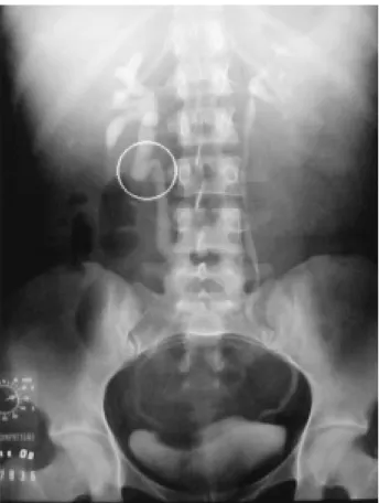
Figure 1 – Intravenous pyelogram showing dilation and tortu-osity of the upper ureter (circle) and dilation of the right pyelocalyceal system.
47
LAPAROSCOPIC APPROACH IN THE OVARIAN VEIN SYNDROME
laparoscopic approach. In 4 cases, patients presented retroperitoneal fibrosis, one case was due to ovarian disease, and one case was the ovarian vein syndrome, the latter was the first case reported in literature on a
patient submitted to a laparoscopic surgery manage-ment.
In 1998, Marcovich & Wolf Jr. (4) published the second case of ovarian vein syndrome managed by ureterolysis and ovarian vein laparoscopic resec-tion, highlighting the benefits of this technique, such as the diagnostic value of direct inspection of patho-logic anatomic conditions, the low postoperative mor-bidity related to pain reduction, and the excellent cos-metic results.
Owing to its simplicity, low morbidity, and good results attained, the laparoscopic approach rep-resents a good option for the surgical management of this syndrome.
REFERENCES
1. Hubmer G: The ovarian vein syndrome. Eur Urol. 1978; 4:263-8.
2. Dykhuizen RF, Roberts JA: The ovarian vein syn-drome. Surgery Gynec Obstet. 1970; 130:443-52. 3. Elashry OM, Nakada SY, Wolf Jr JS, Figenshau RS,
McDougall EM, Clayman RV: Ureterolysis for ex-trinsic ureteral obstruction: a comparison of laparoscopic and open surgical techniques. J Urol. 1996; 156:1403-10.
4. Marcovich R, Wolf Jr JS: Laparoscopy for the treat-ment of positional renal pain. Urology 1998; 52:38-43.
Figure 3 – Intravenous pyelogram within 18 months of follow-up, showing a reduction in the right ureter dilation and in the pyelocalyceal system, and the presence of metallic clips.
Received: October 10, 2002 Accepted after revision: January 24, 2003
Correspondence address:
Dr. Arakén Almeida Rua do Paissandu, 667 / 13 Recife, PE, 52010-000, Brazil Fax: + 55 81 3222-2024