einstein. 2013;11(4):545-6
LEARNING BY IMAGES
Disseminated fusariosis with endophthalmitis in a
patient with hematologic malignancy
Fusariose disseminada com endoftalmite em paciente com neoplasia hematológica
Guilherme Fleury Perini1, Luis Fernando Aranha Camargo1, Claudio Luiz Lottenberg1, Nelson Hamerschlak1
1 Hospital Israelita Albert Einstein, São Paulo, SP, Brazil.
Corresponding author: Guilherme Fleury Perini – Hospital Israelita Albert Einstein, Instituto Israelita de Ensino e Pesquisa, Avenida Albert Einstein, 627/701, building A – Morumbi – Zip code: 05652-900 – São Paulo, SP, Brazil – Phone: (55 11) 2151-8709 – E-mail: guiperini@einstein.br
Received on: Aug 2, 2012 – Accepted on: Feb 24, 2013
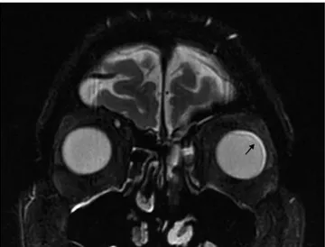
Figure 1. Orbit computerized tomography scan showing anterior, medial and
lateral delamination of left eye, which was compatible with endophthalmitis Figure 2. Anatomopathological exam of left eye showing intraocular abscess
einstein. 2013;11(4):545-6
546 Perini GF, Camargo LF, Lottenberg CL, Hamerschlak N
A 68-year-old patient previously diagnosed with acute myeloid leukemia had fever and myalgia during chemotherapy. Despite broad spectrum antibiotics, fever persisted and, after 3 days, skin lesions compatible with Fusarium infection were seen on patients’ lower limbs. Dyspnea and hypoxia were observed, and computerized tomography showed extensive pulmonary infiltrates; blood cultures were positive for Fusarium sp. A diagnosis of disseminated fusariosis was done, and lipossomal amphotericin, voriconazole and granulocyte infusion were initiated.
The patient had complete regression of skin lesions and pulmonary infiltrates, but a week later he complained of visual blurring in the left eye. An orbital magnetic resonance imaging showed enhancement of left ocular globe with a lateral, medial and anterior delamination that was compatible with endophthalmitis (Figure 1). An intraocular treatment with voriconazole was applied and a little improvement was seen. Fusarium sp
endophthalmitis affecting his right eye was diagnosed, which justified his visual loss. Despite treatment, a progressive worsening of bilateral endophthalmitis occurred and, to control the disease, the eye was enucleated. The pathological examination of the enucleated eye showed an intraocular abscess adjacent to the retina (Figure 2). In a higher magnification,
Fusarium hyphae could be identified (Figure 3).
Fusarium species cause a broad spectrum of infections in humans, including superficial, locally invasive, and disseminated infections. Immunocompromised patients are at higher risk, especially those with prolonged and severe neutropenia and/or severe T-cell immunodeficiency(1).
Patients with acute leukemia and patients undergoing hematopoetic stem cell transplantation are particularly at risk, especially to the invasive and disseminated(2)
forms. The typical pattern of disseminated disease is a
combination of cutaneous lesions (often with external necrosis in the center of the lesion), positive blood cultures, and with or without involvement at other sites (sinuses, lungs, and others)(3).
Fusarium endophthalmitis in the immunocompromised host usually results from hematogenous seeding(4,5).
Intraocular and systemic therapies often have poor responses. In order to avoid central nervous system involvement, the evisceration of the eye may be necessary(6). Few case reports describe successful
treatment of Fusarium sp endophthalmitis with voriconazole alone or in combination with caspofungin and posaconazole(7,8).
REFERENCES
1. Boutati EI, Anaissie EJ. Fusarium, a significant emerging pathogen in patients with hematologic malignancy: ten years’ experience at a cancer center andimplications for management. Blood. 1997;90(3):999-1008.
2. Nucci M, Anaissie E. Cutaneous infection by Fusarium species in healthy and immunocompromised hosts: implications for diagnosis and management. Clin Infect Dis. 2002;35(8):909-20.
3. Nucci M, Anaissie E. Fusarium infections in immunocompromised patients. Clin Microbiol Rev. 2007;20(4):695-704.
4. Dursun D, Fernandez V, Miller D, Alfonso EC. Advanced fusarium keratitis progressing to endophthalmitis. Cornea. 2003;22(4):300-3.
5. Rezai KA, Eliott D, Plous O, Vazquez JA, Abrams GW. Disseminated Fusarium infection presenting as bilateral endogenous endophthalmitis in a patient with acute myeloid leukemia. Arch Ophthalmol. 2005;123(5):702-3. 6. Tiribelli M, Zaja F, Filì C, Michelutti T, Prosdocimo S, Candoni A, et al.
Endogenous endophthalmitis following disseminated fungemia due to Fusariumsolaniin a patient with acute myeloid leukemia. Eur J Haematol. 2002; 68(5):314-7.
7. Tu EY, McCartney DL, Beatty RF, Springer KL, Levy J, Edward D. Successful treatment of resistant ocular fusariosis with posaconazole (SCH-56592). Am J Ophthalmol. 2007;143(2):222-7.