Hindawi Publishing Corporation Dermatology Research and Practice Volume 2010, Article ID 569345,4pages doi:10.1155/2010/569345
Case Report
Indeterminate Cell Histiocytosis in Association with
Acute Myeloid Leukemia
Filipa Ventura,
1Teresa Pereira,
1Maria da Luz Duarte,
1Herlander Marques,
2Fernando Pardal,
3and Celeste Brito
11Dermatology and Venereology Department, Braga Hospital, Apartado 2242, 4701-965 Braga, Portugal 2Oncology Department, Braga Hospital, 4701-965 Braga, Portugal
3Pathology Department, Braga Hospital, 4701-965 Braga, Portugal
Correspondence should be addressed to Filipa Ventura,filipamanuelventura@hotmail.com
Received 21 March 2010; Accepted 25 May 2010
Academic Editor: Bernhard W. H. Zelger
Copyright © 2010 Filipa Ventura et al. This is an open access article distributed under the Creative Commons Attribution License, which permits unrestricted use, distribution, and reproduction in any medium, provided the original work is properly cited.
Indeterminate cell histiocytosis (ICH) is a rare proliferative disorder, in which the predominant cells share morphologic and immunophenotypic features from both Langerhans and non-Langerhans cell histiocytosis. We describe a 62-year-old man presenting a 2-month history of firm nodular lesions on the upper lip. Histopathology, immunohistochemical, and ultrastructural analysis showed typical findings of ICH. The patient was treated with thalidomide and almost complete regression of the lesions was reached within 7 months. Nevertheless, one month after remission, he developed an acute myeloid leukemia of the subtype monocytic leukemia (M5). The patient’s condition rapidly worsened and he died due to a respiratory failure four weeks later. We present this case because apart of being rare it joins the effectiveness of thalidomide and the association with an acute monocytic leukemia. A review of the literature is made.
1. Introduction
Indeterminate cell histiocytosis (ICH) is a rare proliferative disorder, in which the predominant cells share morphologic and immunophenotypic features from both Langerhans and non-Langerhans cell histiocytosis. Clinically, most patients present multiple asymptomatic papulonodules with a colour variation from pink to brownish, covered by intact skin. Solitary lesions may also occur like soft nodules mainly on the trunk and arms. ICH lesions are usually restricted to the skin [1]. The major part of these cases have an indolent and benign course, not requiring aggressive treatment.
2. Case Report
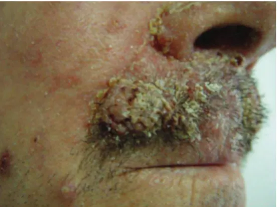
A 62-year-old Caucasian man presented a 2-month history of firm nodular lesions on the upper lip (Figure 1). These lesions were isolated and confluent; some of them were cov-ered by hemorrhagic crusts. The mucous membranes were not involved and the lesions were painless and nonpruritic. He was not taking any medication. Skin biopsy was taken and
sent for histopathology and cultures. Histopathology showed
a diffuse, monomorphous, nonepidermotropic infiltrate of
large “histiocytic” cells with eosinophilic cytoplasm and irregularly shaped nuclei throughout the entire dermis. These cells were mixed with lymphocytes, plasma cells, and
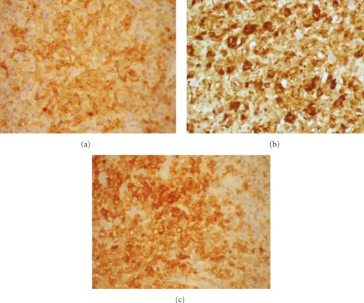
eosinophils (Figures2(a)and2(b)). Immunohistochemical
examination revealed that the predominant cells coexpressed
CD1a (Figure 3(a)), CD68 markers (Figure 3(b)), and
S100 protein (Figure 3(c)). They were negative for CD3,
2 Dermatology Research and Practice
Figure1: Nodular lesions located on the upper lip.
(a)
(b)
Figure2: (a) Histopathology revealed a diffuse, monomorphous,
nonepidermotropic infiltrate of large epithelioid cells throughout the entire dermis. (stain type—Hematoxylin and eosin; origi-nal magnification: ×100). (b) Histopathology revealed a diffuse, monomorphous, nonepidermotropic infiltrate of large epithelioid cells throughout the entire dermis. (stain type—Hematoxylin and eosin; original magnification:×400).
(Figure4). No adverse effects from treatment were observed on the clinical and electrophysiological examinations. How-ever, at this time, the laboratory findings showed
hyper-leucocytosis (white blood count 72,9 × 10⁹/L) with 67%
blasts, anemia (hemoglobin 6,1 g/dL), thrombocytopenia (platelet count 34×109/L), hyperuricemia (serum acid uric 12,4 mg/dL, normal), high levels of lactate dehydrogenase
(1087 U/L, normal), and β2-microglobulin (2610 ng/mL,
normal). The bone marrow aspirate was hypercellular with
extensive infiltrate by the blasts. The blasts were positive for
α-naphthylacetate esterase, myeloperoxidase, and Periodic
Acid-Schiff(PAS). Flow cytometry analysis revealed a blast
population with monocytic differentiation expressing
CD-11B, CD14, CD15, CD33, CD36, CD45, CD56, CD64, and HLA-DR, but lacking CD34 expression. These findings support a diagnosis of an acute monocytic leukemia by the World Health Organization (WHO). Cytogenetic analysis showed two abnormal clones on the chromosomes 7 and 18. Treatment with hydroxyurea had been initiated; however patient’s condition deteriorated and he died 1 month later due to a respiratory failure.
3. Discussion
Histiocytoses are a heterogeneous group of disorders charac-terized by the proliferation and accumulation of cells of the mononuclear-macrophage system and the dendritic cells [2]. All these cells derive from neutrophil/macrophage lineage progenitor cells in bone marrow [3,4]. There are reported in the literature different opinions trying to classify these challenging but poorly understood disorders.
In what concerns to the indeterminate cells’ origin and the ICH there is still a lot of controversy. Indeterminate cells are defined to be closely related to Langerhans cells, histologi-cally and immunocytochemihistologi-cally, but do not contain Birbeck granules. Some authors have reported that indeterminate cells may represent precursors of Langerhans cells which acquire Birbeck granules as they transit from dermal to epidermal sites, possibly the result of the interaction between their cell receptors and epidermis-specific ligands [1,2,5]. Others suggest that indeterminate cells represent members of the dermal/epidermal dendritic cell system that migrate from the skin to the regional lymph nodes [1,2,5]. Hence ICH seems to be a disorder of proliferating indeterminate cells which have been locally arrested before they can leave the skin travelling as veiled cells via the lymphatics to the T-cell-dependent paracortical areas of the regional lymph nodes [1,2,5].
More recently, it has been suggested that what today is described as ICH could be a variant of non-Langerhans’ cell histiocytosis (NLCH) rather than an overlap between
Langerhans cell histiocytosis (LCH) and NLCH [2]. In
fact ICH shows mainly features of NLCH except for the consistent expression of S-100 protein. This is not totally surprising because aberrant S-100 protein has been reported before [2]. Other authors believe that there are disorders of Langerhans cells and macrophages; both differ according to the time cycle of the lesions. Late stages of LCH may completely lose Langerhans cells markers and then consist mainly of xanthomatized macrophages. On the other hand, macrophage disorders have a polymorphous presentation which also includes their immunoprofile.
Nowadays it is not clear that ICH is a separate entity or if represents various macrophage disorders identified at different periods in the inflammatory response.
Dermatology Research and Practice 3
(a) (b)
(c)
Figure3: Immunohistochemical examination showed positivity for CD1a (a), CD68 (b), and S100 protein (c).
Figure4: Regression of the lesions after 7 months of treatment with
thalidomide.
The majority of cases have a benign clinical behaviour and self-healing skin lesions that completely or partially regress spontaneously, requiring no aggressive treatments [5,6]. Chemotherapy is necessary for aggressive cutaneous ICH, defined as ICH involving over 50% of the body surface and lasting at least 6 months with no tendency to spontaneous resolution and with new crops of lesions
continuing to appear [6]. In our case in spite of localized lesions without visceral involvement, it was a disfigurating cutaneous lesion and we have decided to treat the patient with thalidomide 200 mg/day, with almost complete regres-sion of the leregres-sions after 7 months. Our deciregres-sion was based
on the predominantly cutaneous pharmacological effect,
immunomodulatory and anti-inflammatory properties of this drug. It has also been shown that thalidomide modulated the function properties of monocyte-derived dendritic cells [7].
An association between ICH and malignancies has been described in four patients: twice with low grade B-cell
lymphomas [2], once with mast cell leukemia [8], and
another with myelomonocitic leukemia [1]. Our patient
presented a monocytic leukemia 7 months after the onset of the skin lesions.
While antineoplastic drugs are used, such as cyclophos-phamide, vinblastine, etoposide, and 2-chlorodeoxyadeno-sine, to treat aggressive diffuse forms of ICH, the possibility of this treatment being related on the etiology of oncohema-tological disorders cannot be excluded [6]. It does not occur in our case because it was used thalidomide, with proven
effectiveness on multiple myeloma and myelodysplastic
syndrome [9].
4 Dermatology Research and Practice
concerns to our case, the association between indeterminate cell histiocytosis and monocytic leukemia was probably greater than that by chance alone. We present this case because apart of being rare it joins the effectiveness of thalidomide and the association with an acute monocytic leukemia.
References
[1] C. Vener, D. Soligo, E. Berti et al., “Indeterminate cell histiocy-tosis in association with later occurrence of acute myeloblastic leukaemia,”British Journal of Dermatology, vol. 156, no. 6, pp. 1357–1361, 2007.
[2] G. Ratzinger, W. H. C. Burgdorf, D. Metze, B. G. Zeiger, and B. Zelger, “Indeterminate cell histiocytosis: fact or fiction?” Journal of Cutaneous Pathology, vol. 32, no. 8, pp. 552–560, 2005.
[3] B. Newman, W. Hu, K. Nigro, and A. C. Gilliam, “Aggressive histiocytic disorders that can involve the skin,”Journal of the American Academy of Dermatology, vol. 56, no. 2, pp. 302–316, 2007.
[4] E. Dzierzak, “Embryonic beginnings of definitive hematopoi-etic stem cells,”Annals of the New York Academy of Sciences, vol. 872, pp. 256–264, 1999.
[5] A. Sidoroff, B. Zelger, H. Steiner, and N. Smith, “Indeterminate cell histiocytosis—a clinicopathological entity with features of both X- and non-X histiocytosis,”British Journal of Dermatol-ogy, vol. 134, no. 3, pp. 525–532, 1996.
[6] R. Caputo, A. V. Marzano, E. Passoni, and M. Bellinvia, “Chemotherapeutic experience in indeterminate cell histiocy-tosis,”British Journal of Dermatology, vol. 153, no. 1, pp. 206– 207, 2005.
[7] E. Mauro, C. Fraulini, G. M. Rigolin, R. Galeotti, R. Spanedda, and G. Castoldi, “A case of disseminated Langerhans’ cell histiocytosis treated with thalidomide,” European Journal of Haematology, vol. 74, no. 2, pp. 172–174, 2005.
[8] G. Kolde and E.-B. Brocker, “Multiple skin tumors of indeter-minate cells in an adult,”Journal of the American Academy of Dermatology, vol. 15, no. 4, pp. 591–597, 1986.