54
TESTICULAR METASTASIS OF PROSTATE CANCER Case Report
International Braz J Urol
Official Journal of the Brazilian Society of Urology
Vol. 31(1): 54-56, January - February, 2005
SINGLE TESTICULAR METASTASIS MIMICKING PRIMARY
TESTICULAR NEOPLASM: A RARE MANIFESTATION OF PROSTATE
CANCER
CARLOS M. N. JESUS, JOSE GOLDBERG, JOAO L. V. CAMARGO
Departments of Urology and Pathology, Botucatu Medicine School, Paulista State University, UNESP, Botucatu, Sao Paulo, Brazil
ABSTRACT
The incidence of secondary testicular tumors ranges from 0.02 to 2.5% among autopsies in general. With the exception of leukemias and lymphomas, prostate cancer is the most common pri-mary site. It is diagnosed in autopsies or incidentally, following therapeutic orchiectomies in more advanced stages of the disease.
In the present report, we show a case of testicular metastasis derived from prostate neoplasm whose clinical presentation as a single metastasis was similar to a primary testicular neoplasm. The diagnosis was evidenced after orchiectomy by histological examination and immunohistochemical tests.
Key words: testis; neoplasm metastasis; prostatic neoplasms
Int Braz J Urol. 2005; 31: 54-56
INTRODUCTION
Prostate adenocarcinoma is the most fre-quently diagnosed neoplasm in men and its natural history is largely known. The most common meta-static sites are iliac lymph nodes, bones and lungs, then followed more rarely by bladder, liver, adrenal and brain (1,2). The testis is a rare site for prostate cancer metastases and is usually found in autopsies or incidentally after therapeutic orchiectomies for advanced disease. In this paper, we report one case of testicular metastasis from prostate cancer mimick-ing a primary testicular tumor in a young patient.
CASE REPORT
A 45-year old Caucasian patient presented increased volume and sensation of heaviness in his left testis for 2 months. Upon physical examination, he presented increased volume in the left testis with
hardened consistency, smooth surface and absence of inflammatory signs. Two years earlier, the patient had undergone radical prostatectomy due to Gleason 9 prostate adenocarcinoma, and one year earlier he showed an increase in prostate specific antigen (PSA) levels, which was interpreted as a progression of dis-ease, and hormone therapy with cyproterone acetate was used to control the disease. Bone scintigraphy and chest X-radiography were normal.
A scrotal ultrasound was performed, which showed an enlarged testis (5 x 3 x 3 cm) with hetero-geneous parenchyma and multiple calcifications. The spermatic cord and epididymis had normal shape and outline.
55
TESTICULAR METASTASIS OF PROSTATE CANCER
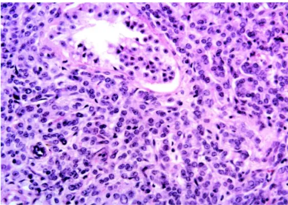
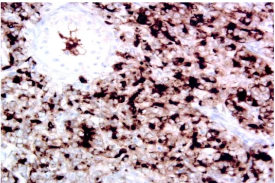
revealed a solid, non-differentiated tumor, which did not suggest a primary testicular tumor. There was tu-bular infiltration as well in the rete testes and epid-idymis (Figure-1). The immunohistochemical panel confirmed the prostate as a primary site due to the positivity of cytokeratins AE1/AE3 and PSA and the negativity of classical markers of germinative tumors such as the antigen CD30, human chorionic gonadot-ropin (β-HCG), alpha-fetoprotein (AFP) and placen-tal alkaline phosphatase (Figure-2).
Serum levels of β-HCG, AFP and lactic de-hydrogenase were normal. The pre-operative PSA, which was 4.13 ng/mL decreased to 1.22 ng/mL within 2 months of the procedure. The patient has been fol-lowed with no progression of the disease found to the present moment.
COMMENTS
Testicular metastases are uncommon phe-nomena that are present in 0.02 to 2.5% of autopsies, with approximately 200 cases being reported in the
Figure 1 – Neoplasm infiltrating the testis. The seminiferous tubule is involved with non-differentiated neoplastic cells (HE, X250). literature (1). Tumors that most commonly send me-tastases to the testis are prostate, lung, melanoma and kidney. Testicular metastases are rarely derived from stomach, pancreas, bladder, rectum or penis (2).
This case shows a rare finding of testicular metastasis deriving from a prostate neoplasm, which was clinically observed through an increase in left scrotal volume associated with young age, a fact that is uncommon in this neoplasm. Most cases of me-tastases from prostate cancer are evidenced by au-topsies or palliative bilateral orchiectomies aimed to control advanced disease in elderly patients. Addi-tionally, the metastasis was single, with no metastases detected in other organs, stressing its rarity.
56
TESTICULAR METASTASIS OF PROSTATE CANCER
Figure 2 – Positive expression of prostate specific antigen (PSA) by metastatic cells involving a seminiferous tubule (Avidin-biotin peroxidase, anti-PSA antibody 1:20, X250).
REFERENCES
1. Dutt N, Bates AW, Baithun SI: Secondary neoplasms of the male genital tract with different patterns of in-volvement in adults and children. Histopathology. 2000; 37: 323-31.
2. Patel SR, Richardson RL, Kvols L: Metastatic cancer
to the testes: a report of 20 cases and review of the literature. J Urol. 1989; 142: 1003-5.
3. Askari A, Faddoul A, Herrera H: Metastatic carcinoma to testicle. Urology. 1981; 17: 601-3.
Received: June 28, 2004 Accepted after revision: December 10, 2004
Correspondence address:
Dr. Carlos M. Nóbrega de Jesus
Dept. Urologia, Fac. de Medicina de Botucatu Distrito de Rubião Júnior, S/N