USE OF THE MONTI PRINCIPLE FOR CONSTRUCTING A CONTINENT
GASTROSTOMY
LUIZ A. P. ARAUJO, CARLOS T. BRANDT, SALVADOR V. C. LIMA, FABIO O. VILAR,
ANDRE A. P. ARAUJO
Divisions of Pediatric Surgery and Urology, Center of Experimental Surgery, Federal University of Pernambuco, Recife, Pernambuco, Brazil
ABSTRACT
Objective: To research technical alternatives for permanent gastrostomy that minimizes the drawbacks and complications reported by several authors.
Materials and Methods: An experimental model was developed where the material was di-vided into 2 groups: the study group (SG) composed of 12 half-breed dogs where the proposed tech-nique was applied, and the control group (CG) composed of 10 animals where a gastrostomy as proposed by Webster in 1974 was applied. On the 90th postoperative day, both groups underwent tests for assessing competence concerning leakage. These were performed under general anesthesia and following sacrifice.
Results: In the SG, under anesthesia only one animal had leakage through the gastrostomy. Following sacrifice, leakage was observed in 2 animals. In the CG, under anesthesia, 2 animals had leakage and, following sacrifice, only 1 animal did not present leakage. On histopathological analysis of the SG, gastric mucosa was evidenced around the jejunal tubes, with normal features, moderate inflammatory mononuclear infiltrate in jejunal tubes and only slight infiltrate around the gastrostomy stoma. In the CG, ulceration was constant around the external stoma of the gastrostomy tubes. In the corium, the inflammatory infiltrate was less intense than in the SG. The SG proved to be more effica-cious than the CG concerning leakage, and this efficacy is attributed to the submucous valvular sys-tem.
Conclusions: The featured technique showed competence concerning leakage, allowing its clinical applicability as an alternative for permanent gastrostomy.
Key words: urinary diversion; urinary reservoirs, continent; experiments Int Braz J Urol. 2005; 31: 62-68
INTRODUCTION
The research for a gastrostomy, which can address the inability of using the oral route for feed-ing, either in neurological diseases or neoplasms of oropharynx, esophagus and stomach, has been re-ported since the 19th century (1). The importance of gastrostomy as a therapeutic option to enable the tem-porary or permanent nutrition of several patients is
In an effort to contribute to the enhancement of current gastrostomy techniques, especially perma-nent ones, a new technique based on the Monti prin-ciple was conceived. This uses a segment of the small bowel for fashioning a continent vesicostomy (5). Similarly, applying a jejunal segment to the stomach was proposed, aiming to obtain a continent gastros-tomy offering less drawbacks than the current ones.
MATERIALS AND METHODS
The study was conducted on 22 half-breed dogs of both genders weighing between 15 and 18 kg from the Experimental Surgery Center of the Federal University of Pernambuco.
Animals were divided into 2 groups: the study group (SG) composed of 12 dogs undergoing the new proposed continent gastrostomy technique, and the control group (CG), composed of 10 dogs undergo-ing the gastrostomy technique usundergo-ing the Janeway prin-ciple (1) as modified by Moss (6) and Webster (7).
Surgical Technique for the Study Group
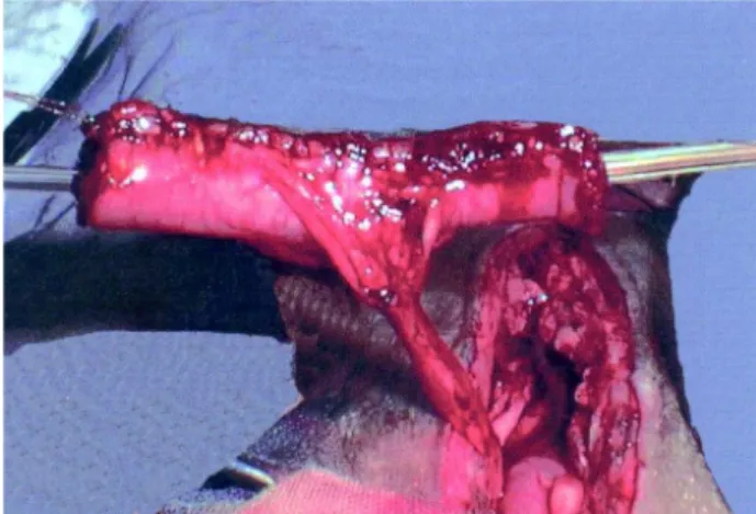
A 20-cm median incision was made in order to access the peritoneal cavity. Upon identifying the stomach and the jejunum, a jejunal segment mea-suring 2 cm in length was isolated, while preserving its vascular pedicle. The jejunal segment was then detubularized at the level of the anti-mesenteric edge and retubularized in the opposite direction through suture using separate stitches, thus creating a tube measuring 6 cm in length and 0.5 cm in diameter (Figure-1).
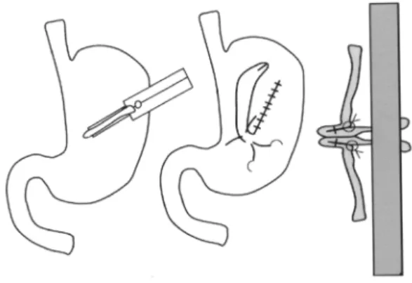
A longitudinal 8-cm seromuscular incision that preserved the mucosa was made in the middle third of the stomach between the lesser and the greater curvature, separating the seromuscular layer from the gastric mucosa in the 2 directions laterally to the in-cision. A small opening was made in the mucosa at the incision’s lower level where the lower margin of the jejunal tube was introduced and sutured to the orifice. The remainder of this tube was placed over the previously dissected gastric mucosa and then cov-ered by the seromuscular layer constituting the anti-reflux valvular mechanism. The upper margin of the
jejunal tube was exteriorized through the upper angle of the surgical incision (Figure-2).
Surgical Technique for the Control Group
We used the technique proposed by Webster (7), which gathers the Janeway principles (1) for the creation of a gastric tube using a linear stapler as pro-posed by Moss (6), and a valvular mechanism with placation stitches at the base of the tube as proposed by Spivack (1).
The gastrostomy tube was created at the level of the middle third of the stomach in the longitudinal direction. The stomach’s anterior wall was then pulled by 3 Allis forceps forming a plica measuring 2 cm in width and 6 cm in length. The TLC 55 cutting linear stapler was then inserted in the stomach’s wall under the forceps under traction, so that a circular tube would result after the stapling. Plication stitches were applied at the base to create the valvular system and the tube was then exteriorized and fixed to the skin through a passage between the fibers of the left rec-tus muscle of abdomen. At the end of the procedure, the surgical wound was closed as in the SG (Figure-3).
Evaluation
On the 90th postoperative day, all animals from both groups underwent tests for assessing
com-Figure 1 – Re-tubularized jejunal segment before being inserted
petency concerning leakage, initially with the live animal and then following its sacrifice.
Under general anesthesia, 2 orogastric tubes were introduced, one for infusion of saline solution with methylene blue up to a final volume of 1500 mL, and the other for measuring the gastric tension generated by saline infusion. A laparotomy was per-formed for clamping the pylorus. Tensions were mea-sured in linear cmof H2O, in a scale where the zero point corresponded to the level of the animal’s mid-axillary line.
Following sacrifice, the esophagus, stomach, gastrostomy segment and first duodenal portion were removed en-bloc and placed over a flat surface. Next, as in the previous test, 2 orogastric tubes were intro-duced into the esophagus, this time with ligation of the extremities in order to avoid gastroesophageal reflux and duodenal emptying. A saline solution drip with methylene blue was infused up to a final vol-ume of 1500 mL. The tensions were checked and measured at the same scale, establishing the table’s surface level as the zero point.
Macro and microscopic analyses of the gas-tric tubes and implanted intestinal segments were performed as well.
Results of continuous variables were ex-pressed by their mean and standard deviation.
Re-sults of categorical variables were expressed by their absolute and relative frequencies.
The Student’s “t” test for non-paired samples was used. The qui-square test was used for assessing a potential difference between frequencies. The Fisher’s exact test was used in 2x2 contingency tables. P value < 0.05 was considered statistically significant.
RESULTS
Study Group
Of the 12 animals that underwent continent gastrostomy using the Monti principle, 9 were as-sessed for the continence test. Two animals died in the immediate post-operative period due to anesthetic accident and were replaced. One died on the 63rd day due to peritonitis and gastric perforation caused by duodenal obstruction resulting from external compres-sion due to splenic volvulus.
On the third and fifth postoperative days, 2 animals presented dehiscence of the surgical wound that was restricted to the skin and repaired with no consequences.
The gastrostomy stomas maintained a good aspect with no signs of skin erosion at the
implanta-Figure 2 – Schematic drawing of surgical technique in the study
group.
Figure 3 – Schematic drawing of surgical technique in the
tion site. All allowed gastric catheterization with a Nelaton 8F catheter without any difficulty. In 2 ani-mals, we observed the formation of a pellicle over the stoma, which was easily removed during cath-eterization.
In all animals, the gastric mucosa and the je-junal segment had macroscopically normal aspects with no signs of ulceration or irritation.
Only 1 animal had leakage through the gas-trostomy when the saline infusion into the stomach reached 800 mL. The remaining animals endured a gastric volume of 1500 mL without leakage.
On the post-sacrifice continence test, leak-age was seen in 2 animals (Table-1).
Control Group
All animals survived the experiments, how-ever erosion of the skin surrounding the stomas was seen in 9 animals, and 5 of these presented tube re-traction with closure of the gastrostomies. Six ani-mals were re-operated upon, including 2 to correct stenosis of the gastrostomy stoma and 4 for laparo-tomy to reimplant the gastroslaparo-tomy tubes, which had subaponeurotic retractions (Table-2). The latter were
Under Anesthesia Following Sacrifice
N Volume (mL) Tension Leakage Volume (mL) Tension Leakage Obs.
(cm H2O) (cm H2O)
1 1500 15 - 1500 12
-2 Death
3 1500 10 - 1500 13
-4 1500 17 - 700 10 +
5 1500 17 - 1500 19
-6 800 14 + 400 8 +
7 1500 14 - 1500 14
-8 1500 30 - 1500 30
-9 Death
10 1500 10 - 1500 17
-11 1500 16 - 1500 12
-12 Death
Table 1 – Continence analysis in the study group. The mean infused volume under anesthesia (1422 mL +/- 426) was not
different from the infused volume following sacrifice (t = 0.824, p = 0.422).
tested for gastric continence 3 months after the cor-rections.
When tested for gastric continence under gen-eral anesthesia, only 2 animals had leakage. On the gastric continence test performed following sacrifice, only 1 of the 10 animals did not present leakage (Table-3).
When the SG and the CG were compared re-garding the leakage test under anesthesia, there was no statistically significant difference when the Fisher’s test was applied (p = 1.000). In relation to the post-sacrifice leakage test, there was a signifi-cantly higher frequency of leakage in the CG (p = 0.0005).
Histopathological analysis
Study Group
Table 2 – Complications in the control group.
N Skin Stomal Gastrostomy Re-operation Repair of Laparotomy Erosion Stenosis Retraction Stenosis
1 + - - - -
-2 + + + + +
-3 - - -
-4 + + + + + +
5 + - - - -
-6 + - - - -
-7 + + + + + +
8 + + - + +
-9 + + + + + +
10 + + + + + +
Under Anesthesia Following Sacrifice
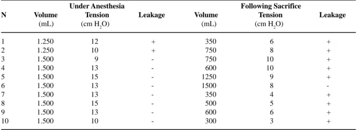
N Volume Tension Leakage Volume Tension Leakage (mL) (cm H2O) (mL) (cm H2O)
1 1.250 12 + 350 6 +
2 1.250 10 + 750 8 +
3 1.500 9 - 750 10 +
4 1.500 13 - 600 10 +
5 1.500 15 - 1250 9 +
6 1.500 13 - 1500 8
-7 1.500 13 - 350 4 +
8 1.500 15 - 500 5 +
9 1.500 13 - 600 6 +
10 1.500 10 - 300 3 +
Table 3 – Continence test in the control group. Mean volume of saline solution infused under anesthesia (1450 mL
+/-105) was significantly higher (t = 5.537, p < 0.001) than the volume infused following sacrifice (695 +/- 396 mL).
enlarged due to the presence of moderate mononuclear inflammatory infiltrate.
Around the anastomosis site between the skin and the jejunal tube, we observed mild inflammatory infiltrate, predominantly mononuclear, with no signs of ulceration. These findings were common to all the examined specimens.
Control Group
Skin ulceration around the gastrostomy tube stomas was constant, with a presence of reparative
changes in the gastric mucosa at this level. The tube mucosa had typical features of the mucosa found in the gastric body where parietal and principal cells were identified. In the chorion, there was mono- and polymorphonuclear infiltrate, less intense than the one detected in the SG.
COMMENTS
due to different causes, are unable to use the oral route for this purpose.
The rationale for using the Monti principle to construct a continent gastrostomy was based on the encouraging results obtained by different authors in the performance of vesicostomies and cecostomies that showed to be continent and easy to perform (8-11). The surgical technique does not present major difficulties and can be performed by anyone who is familiar with intestinal anastomoses.
The SG was the first to be performed, thus it was more exposed to the learning curve, especially concerning the anesthesia, which, due to technical reasons, resulted in the death of 2 animals.
The repetition of continence tests following sacrifice was designed to test the competence of the valvular mechanism without influence of the gastroe-sophageal reflux, a variable factor that could inter-fere with the results. The use of methylene blue in the test’s saline solution aimed to improve the visual-ization for identifying gastric leakage. The standard-ized maximum volume of 1500 mL of saline solution infused into the stomach was established after assess-ments performed on the pilot animal where the in-fused volume did not result in leakage.
When comparing both groups in relation to surgical technique, we observed that the technique employed in the control group with the aid of the lin-ear stapler while creating the gastric tube made the surgical procedure easier, reducing the surgical time. However, it did not seem to prevent the gastric leak-age observed in the post-sacrifice test, despite the appliance of plication stitches at its base in compli-ance with the anti-reflux Spivack technique (1). In the CG, the gastrostomy tube was exteriorized though the rectus muscle of the abdomen, which confers an additional valvular system. We believe that this ex-plains the different results observed between in vivo and post-sacrifice continence tests. The statistical analysis of leakage frequency following sacrifice gives mathematical support to the biological obser-vation. Moreover, when compared to the CG, the high-est volume of saline solution infused in the animals’ stomach in the SG gives additional support to the ef-ficiency of the valvular system projected for gastros-tomy continence.
The SG showed to be more effective concern-ing the control of gastric leakage, despite the gastros-tomy tube being exteriorized at the level of the linea alba; that is, without muscular influence. This allows us to suggest that the observed continence was de-pendent exclusively on the submucous valvular mechanism. Another important fact is that in the SG, the gastrostomy tube was created with a jejunal seg-ment whose mucosa does not have acid secretion, while in the CG a gastric tube was used, consequently creating an acid-secreting mucosa, which, in associa-tion with gastric leakage, may have contributed to the development of erosions and dehiscences around the gastrostomy stomas.
Similarly to the Janeway technique and also to other approaches that use part of the stomach as a gastrostomy device, another advantage of the gastros-tomy with the jejunal tube using the Monti principle is that this technique is difficult to apply to patients who undergo partial gastrectomy, while in the pro-posed technique, the jejunal tube added to the stom-ach can be applied to any segment (1,12).
The elaboration of catheterization tubes through transverse re-tubularization of small intesti-nal segments has provided its use in several segments of the digestive tract, allowing the performance of several functions through continent cecostomy and sigmoidostomy for antegrade intestinal enema used for treating neurogenic constipation (8-11). Other uses can be tested in the future, such as continent jejunos-tomy for feeding gastrecjejunos-tomy patients where a gas-trostomy is not possible.
At the present moment, 14 patients have un-dergone continent gastrostomy according to this tech-nique with quite satisfactory results.
REFERENCES
1. Partipilo AV: Surgical Technique and Principles of Operative Surgery. Philadelphia, Lea & Febiger. 1949, pp. 245-56.
2. Osborne RO, Toffler RB: Gastrostomy tube prolapse. Am J Gastroenterol. 1973; 60: 602-6.
3. Sherman ML, Cosgrove MJ, Dennis JM: Gastrostomy tube migration. Am Surg. 1973; 39: 122-3.
5. Monti PR, Lara RC, Dutra MA, de Carvalho JR: New techniques for construction of efferent conduits based on the Mitrofanoff princple. Urolology. 1997; 49: 112-15.
6. Moss G: A simple technique for permanent gastros-tomy. Surgery. 1972; 71: 369-70.
7. Webster MW Jr, Carey LC, Ravitch MM: The perma-nent gastrostomy: use of the gastrointestinal anasto-motic stapler. Arch Surg. 1975; 110: 658-60. 8. Gerharz EW, Tassadaq T, Pickard RS, Shah PJ,
Woodhouse CR, Ransley PG: Transverse re-tubularized ileum: early clinical experience with a new second line Mitrofanoff tube. J Urol. 1998; 159: 525-8.
9. Gosalbez R, Wei D, Gousse A, Castellan M, Labbie A: Refashioned short bowel segments for the construc-tion of catheterizable channels (the Monti procedure): early clinical experience. J Urol. 1998; 160: 1099-102. 10. Castellan MA, Gosalbez R Jr, Labbie A, Monti PR: Clinical applications of the Monti procedure as a con-tinent catheterizable stoma. Urology. 1999; 54: 152-6.
11. Yerkes EB, Rink RC, Cain MP, Casale AJ: Use of a Monti channel for administration of antegrade conti-nence enemas. J Urol. 2002; 168: 1883-5.
12. Bianchi A, Pearse B: The non-refluxing gastrostomy: an evolution. Pediatric Surg Int. 1997; 12: 494-6.
Received: October 17, 2004 Accepted after revision: January 3, 2005
Correspondence address:
Dr. Luiz Alberto P. de Araújo
Rua Edson Álvares, 211 / 601, Casa Forte Recife, PE, 52061-450, Brazil