ORIGIN
AL RESEAR
CH
Corresponding address: Francisco Fleury Uchoa Santos Júnior – Rua Eliseu Uchôa Beco, 600, Água Fria – Fortaleza (CE), Brazil. Zip Code: 60810-270 – Email: [email protected] – Finance source: CAPES and FUNCAP – Conlict of interests: Nothing to declare – Presentation: Jan. 2017– Accepted for publication: Apr. 2017–
Approved by the Ethics Committee on Animal Use on advice no. 3576780/2014.
Study developed in the Laboratório de Bioquímica e Expressão Gênica and Laboratório de Biofísica da Respiração of the Universidade Estadual do Ceará – Fortaleza, CE, Brazil.
1Professor at Centro Universitário Estácio do Ceará – Fortaleza (CE), Brazil. 2Doctoral student at Universidade Estadual do Ceará– Fortaleza (CE), Brazil. 3Doctoral student at Universidade Estadual do Ceará– Fortaleza (CE), Brazil. 4Professor at Universidade Estadual do Ceará – Fortaleza (CE), Brazil.
ABSTRACT | Immobilization is a condition that afects several segments and organic systems, including the respiratory system, leading to structural and functional alterations. The purpose of this study was to analyze pulmonary function and micromechanical structure after 14 days of movement restriction in rats. Fourteen female Wistar rats with body mass between 210±50 g were used, divided into two groups, composed of (n=7) each group: Control (C) and Immobilized (I). The immobilization procedure involved the abdomen (and last ribs), pelvis, hip and knee extension and the ankle in plantar lexion in the two week period. After the immobilization period, an analysis of the pulmonary function was performed using a mechanical ventilator for small animals, lexVent, and alveolar recruitment maneuvers. Subsequently, lung strips were removed from each animal for pulmonary micromechanics analysis. Statistical analysis was performed using the unpaired
t test with p<0.05, expressed as mean±standard error of the mean. Group I presented signiicant changes in the parameters of airway resistance (Raw) Pre RM (C=0.067±0.003 cmH2O.s/mL, I=0.095±0.004 cmH2O.s/mL, p<0.05) and Hysteresivity (η) Pre RM (C=0.203±0.004 cmH2O.s/mL, I=0.248±0.013 cmH2O.s/ mL, p<0.05), which returned to their normal values after RM. Raw Post RM (C=0.064±0.003 cmH2O.s/ mL, I=0.065±0.004 cmH2O.s/mL, p<0.05) and η
205
(C=0.209±0.005 cmH2O.s/mL, I=0.214±0.007 cmH2O.s/ mL, p<0.05). It is concluded that immobilization causes reversible functional changes in the respiratory system after 14 days of movement restriction evidenced by the reduction of RN and η after RM.
Keywords | Immobilization; Respiratory System; Lung.
RESUMO | A imobilização é uma condição que compromete diversos segmentos e sistemas orgânicos, inclusive o sistema respiratório, levando a alterações estruturais e funcionais. O objetivo deste estudo foi analisar a função pulmonar e estrutura micromecânica após 14 dias de restrição de movimento de ratas. Foram utilizados catorze ratas Wistar com massa corporal entre 210±50 g, distribuídas em dois grupos, compostos por (n=7) cada grupo: Controle (C) e Imobilizado (I). O procedimento de imobilização envolveu abdômen (e últimas costelas), pelve, quadril e joelho em extensão, além de tornozelo em lexão plantar, por duas semanas. Após esse período de imobilização, foi realizada a análise da função pulmonar por ventilador mecânico para pequenos animais (lexiVent) e manobras de recrutamento alveolar (MR). E, posteriormente, foram retiradas tiras do pulmão de cada animal para analisar a micromecânica pulmonar. Para a análise estatística, utilizou-se o teste t não pareado com signiicância estatística (p<0,05), expresso como média±erro padrão
Analysis of pulmonary function and micromechanics
structure after 14 days of movement restriction in
female rats
Análise da função pulmonar e estrutura micromecânica após 14 dias de restrição de
movimento em ratas
Análisis de la función pulmonar y estructura micromecanica después de 14 días de restricción
en el movimiento en ratas
Francisco Fleury Uchoa Santos Júnior1, Karla Camila Lima de Souza2, Daniel Silveira Serra3, Vânia
da média. O grupo I apresentou mudanças signiicantes nos parâmetros da resistência das vias aéreas (RN)
pré-MR(C=0,067±0,003 cmH2O.s/mL, I=0,095±0,004 cmH2O.s/mL,
p<0,05) e histerisividade (η) pré-MR(C=0,203±0,004 cmH2O.s/
mL, I=0,248±0,013 cmH2O.s/mL, p<0,05), que retornaram a
seus valores de normalidade pós-MR, considerando-se RN
pós-MR (C=0,064±0,003 cmH2O.s/mL, I=0,065±0,004 cmH2O.s/
mL, p<0,05) e η (C=0,209±0,005 cmH2O.s/mL, I=0,214±0,007
cmH2O.s/mL, p<0,05). Conclui-se que a imobilização acarreta
alterações funcionais reversíveis no sistema respiratório após 14 dias de restrição de movimento, o que é evidenciado pela redução de RN e η pós-MR.
Descritores | Imobilização; Sistema Respiratório; Pulmão.
RESUMEN | La inmobilización es una condición que compromete diversos segmentos y sistemas orgánicos incluso el sistema respiratorio, llevando a alteraciones estructurales y funcionales. El objectivo de este estudio fue analizar la función pulmonar y estructura micromecánica después de 14 días de restricción en el movimiento de ratas. Fueron utilizados catorce ratas Wistar con masa corporal entre 210±50 g, distribuidas en dos grupos, compuestos por (n=7) cada grupo: Control (C) y Inmobilizado (I). El procedimiento de inmobilización envolvió el abdomen (y
últimas costillas), la pelvis, la cadera y la rodilla en extensión y el tobillo en lexión plantar el periodo de dos semanas. Después del periodo de inmobilización fue realizado el análisis de la función pulmonar por medio del ventilador mecánico para pequeños animales lexVent y manobras de recrutamiento alveolar (MR). Posteriormente, fueron retirados pedazos del pulmón de cada animal para análisis de la micromecánica pulmonar. Para el análisis estadística se utilizó la prueba t
no pareada con signiicación estadística (p<0,05), expresa como media±error patrón de la media. El grupo I presentó cambios signiicativos en los parámetros de la resistencia de las vias aéreas (RN) pre-MR (C=0,067±0,003 cmH2O.s/mL,
I=0,095±0,004 cmH2O.s/mL, p<0,05) y histerisividade (η)
pre-MR(C=0,203±0,004 cmH2O.s/mL, I=0,248±0,013 cmH2O.s/
mL, p<0,05), que retornaron a sus valores de normalidad después de la MR. RN post-MR (C=0,064±0,003 cmH2O.s/mL,
I=0,065±0,004 cmH2O.s/mL, p<0,05) y η (C=0,209±0,005
cmH2O.s/mL, I=0,214±0,007 cmH2O.s/mL, p<0,05). Se concluye
que la inmobilización conlleva alteraciones funcionales reversibles en el sistema respiratorio, después de 14 días de restricción en el movimiento evidenciado por la reducción de la RN y η después de la MR.
Palabras clave | Inmobilización; Sistema Respiratorio; Pulmón.
INTRODUCTION
Immobilization is a clinical practice commonly used in situations of trauma and/or algic pathologies to functional reestablishment. Both situations may limit the patients’ full abilities such as their locomotion and some activities of daily life1,2.
Small periods of movement restriction, even short-term ones, can lead to several damages to the immobilized region3, including disorders in various body segments and organic systems, such as circulatory impairments1, ligament alterations4, increase of the connective issue5, edema6, articular rigidity4, muscle hypertrophy and atrophy7 and reduction in bone mineral density3,8.
In general, depending on the immobilized region and the restraint time, the respiratory system can be compromised. hese damages in the mechanical structure of the diaphragm and the consequent reduction of diaphragmatic movement and thoracic excursion can cause an increase in the mechanical
resistance and a decrease of the pulmonary ventilation, leading to atelectasies and pneumonias9.
Several studies approach the impact of immobilization on the locomotor system1-3,6,7; however, little is known about the respiratory system regarding the efects of devices that constrain the torso movement. In this context, the present study examined the impact that the restriction of abdominal movement from an experimental model containing multiple motor limitations can promote on the structure and function of the respiratory system and whether they are reversible with recruitment maneuver.
METHODOLOGY
between 210±50 g from the vivarium of the Instituto Superior de Ciências Biomédicas of UECE. During immobilization period, the animals were kept in light/dark cycle (12 h/12 h), in temperature-controlled environment between 22 and 25°C and with feed and water ad libitum.
Immobilization protocol
he animals were randomly divided in groups: Control and Immobilized, with seven animals in each group. he immobilization procedure was conducted with waterproof tape (brand Cremer® with 5 cm wide), which included the abdomen (and last ribs), pelvis, hip and knee in extension, besides the ankle in plantar lexion. he right paw of the animals was bandaged with commercial tape strips, 5 cm wide and 10 cm long. he adhesive strips structure was secured with extra strips on the torso over a bandage in the abdomen and pelvis of the animal. he strips were replaced or reinforced when damaged. he immobilization was kept for 14 days6.
Adequacy of the animals to the mechanical ventilator
he pulmonary function of the animals was analyzed in mechanical ventilator for small animals (FlexiVent, SCIREQ, Montreal/Canada). he animals were anesthetized with Pentorbarbital sodium 90 mg/ kg via intraperitoneal (i.p.) for subsequent tracheotomy and then connected to the mechanical ventilator. After 5 minutes of animal adaptation, the musculature was paralyzed through the injection of Pancuronium Bromide (2 mg/kg, i.p.) and then the experimental protocol for pulmonary function was started. he lungs were ventilated at a frequency of 90 respiratory incursions per minute; current volume of 10 mL/kg; with pressure limitation at 30 cm H2O; and a positive end-expiratory pressure (PEEP) of 3 cm H2O.
Mechanical measures protocol
To obtain the impedance measurement of the respiratory system (Zrs), it was used a disturbance called quick-prime. he pressure and low obtained from that disturbance were used to calculate the measure of Zrs, which was adjusted to the constant-phase model10. hen, the following parameters were determined: airway resistance (Raw), tissue resistance (G), tissue elastance (H) and hysteresivity (η). To obtain
the pressure-volume curve (PV), the tracheal pressure was elevated at 30 cm H2O in equally spaced pre-set pressure intervals, allowing to collect static compliance measures (Cst), estimation of inspiratory capacity (IC) and calculation of the PV curve area. he protocol was stipulated in 12 disruptions and a PV curve followed by two deep insulations (or recruitment maneuvers). he entire procedure lasted 15 minutes.
Pulmonary micromechanics
After the collection of ventilatory parameters, animals were euthanized with a lethal dose of Sodium Pentorbarbital (120 mg/kg, i.p.). horaco-abdominal region was open to exposure of internal organs. hen, heart and lungs were removed in bloc. From pulmonary parenchyma were taken strips of approximately 2x2 mm of transversal section and 6 mm in length, so that one tip of the strip was glued with Cyanoacrylate-based glue to the actuator (Model 300B-LR. Aurora Scientiic, Ontario/Canada) and the other tip was glued to a ixed base and immersed in a chamber for isolated organ with Krebs solution, aerated and with controlled temperature of 37°C. he length of the sample was adjusted until the basal force reach a value above the one generated by the weight of the sample, named Lo (resting length – mm).
Preconditioning occurred through sinusoidal oscillations for 10 minutes to an amplitude of 10% Lo and frequency of 1 Hz, until it reaches a stable loop11. After preconditioning, the sample was adjusted again and the reference length (Lr), measured with a caliper. he initial length (Li) was adjusted to 15% of Lr and the samples were oscillated to a range of 2.5% of Li in the frequencies of 0.1, 0.3, 1.3, and 10 Hz, with 20 cycles each. After, Li was adjusted to 25% of Lr, and the procedure repeated to obtain measures of elastance, resistance and hysteresivity.
Statistical analysis was performed using the unpaired t test and Two-way Anova with Sidak’s post test with statistical relevance (p<0.05). he parameters mentioned were expressed as mean±standard error.
RESULTS
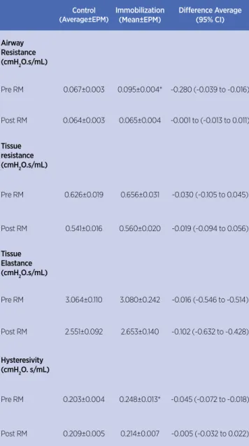
to obtain the values of Raw, G, H, η, Cst, IC and PV curve area (Table 1 and Table 2). Pulmonary mechanics showed that there was no statistically signiicant changes in the parameters of Raw and η (Table 02).
Table 1. Analysis of pulmonary function
Control Immobilization p-value
Static compliance
(mL/cmH2O) 0.763±0.043 0.731±0.034 0.5667 Inspiratory
Capacity (mL) 8.355±0.405 8.001±0.224 0.4514
Area of PV Curve (mL.cmH2O)
35.18±1.198 34.28±1.879 0.6901
PV: pressure-volume
Table 2. Recovery from the changes in pulmonary function after recruitment maneuver
Control (Average±EPM)
Immobilization (Mean±EPM)
Diference Average (95% CI)
Airway Resistance (cmH2O.s/mL)
Pre RM 0.067±0.003 0.095±0.004* -0.280 (-0.039 to -0.016)
Post RM 0.064±0.003 0.065±0.004 -0.001 to (-0.013 to 0.011)
Tissue resistance (cmH2O.s/mL)
Pre RM 0.626±0.019 0.656±0.031 -0.030 (-0.105 to 0.045)
Post RM 0.541±0.016 0.560±0.020 -0.019 (-0.094 to 0.056)
Tissue Elastance (cmH2O.s/mL)
Pre RM 3.064±0.110 3.080±0.242 -0.016 (-0.546 to -0.514)
Post RM 2.551±0.092 2.653±0.140 -0.102 (-0.632 to -0.428)
Hysteresivity (cmH2O. s/mL)
Pre RM 0.203±0.004 0.248±0.013* -0.045 (-0.072 to -0.018)
Post RM 0.209±0.005 0.214±0.007 -0.005 (-0.032 to 0.022)
SEM: standard error of the mean; CI: conidence interval; RM: recruitment maneuver. *Two-way Anova test with post multiple comparison Sidak test; p<0.05
he results concerning the micromechanics analysis of lung tissue are illustrated in Figure 1.
100000
10000
1000
100
10
0.1 0.3 1 3 10
100000
10000
1000
100
0.1 0.3 1 3 10
Resistance 15% L0 Resistance 25% L0
Elastance 15% L0 Elastance 25% L0
Frequency (Hz) Frequency (Hz)
Frequency (Hz) Frequency (Hz)
20
15
10
5
0
0.1 0.3 1 3 10
20
15
10
5
0
0.1 0.3 1 3 10
Control Immobilized
0.5
0.4
0.3
0.2
0.1
0
0.1 0.3 1 3 10 0.5
0.4
0.3
0.2
0.1
0
0.1 0.3 1 3 10
Hysteresivity 15% L0 Hysteresivity 25% L0
Frequency (Hz) Frequency (Hz)
Control Immobilized Control Immobilized Control
Immobilized Control Immobilized
Control Immobilized
Figure 1. Pulmonary Micromechanics Resistance, Elastance and Hysteresivity of the control (n=7) and immobilized (n=7) groups in function of the diferent frequencies (0.1, 0.3, 1, 3 and 10 Hz). Values are expressed as mean and standard deviation
DISCUSSION
A second peculiarity is related to an increase in the value of η on pulmonary mechanics. Hysteresivity is a parameter calculated from the relationship between the parameters G and H, and its value grows as the lung becomes mechanically heterogeneous, therefore, with irregular ventilatory distribution15,16. his fact can justify the increase identiied on parameter η, suggesting the presence of ventilatory heterogeneity related to the increase in Raw value.
A particularity of recruitment maneuver is its ability to normalize the values for Raw and η, as we can see in Table 2. To understand the standardization of the referred results, possibly there was a stretch of the smooth muscles after the administration of a deep insulation, since the smooth muscle of the central airways, once contrite, did not return to its normality without mechanical aid17.
According to the work of Bates et al.18 with mice, muscle stretch caused by an increase in lung volume afected muscle tone through neural pathways, causing relaxation and the return to normality patterns. Kapsali et al.19 reported a bronchoprotector efect in lung tissue of healthy individuals after an alveolar recruitment maneuver. he authors report that the bronchoprotection is an important pulmonary physiological function, and these facts may corroborate the indings of this study.
On the structural analysis of the pulmonary parenchyma, in Figure 1, it was observed that there were no structural changes in the components of the ibers network that make up the lung tissue, so probably changes do not occur in the amount of elastic and collagen ibers, which is in accordance with the results obtained for the G and H values.
Tissue resistance relects the energy loss generated by the viscosity concerning the lung movement and the pulmonary elastance, therefore, the elastic aspect of the tissue12. hese parameters are not independent, therefore, an increase in the G value is directly associated with an increase in the same proportion, in the H value, associated with the elastic characteristics of lung tissue, which can directly change the parameters of Cst, CI and PV curve area, as shown in Table 1.
In two previous studies with this experimental model of respiratory movement restriction and the same time of immobilization (14 days), there was a 14% diaphragmatic muscle hypertrophy, identiied by histology6 (Haematoxylin and Eosin stain) and a quantitative reduction of total proteins in the diaphragm1, facts that support the commitment of the
existence of skeletal respiratory musculature. Our study therefore suggests that the condition described earlier in this experimental model probably did not afect the function or structure of the pulmonary parenchyma deinitively. hese changes were probably caused by possible areas of alveolar collapse, with reduced respiratory capacity.
In this context, our study showed that restriction of the rib cage and abdominal wall in an animal model of 14 days of immobilization, which mimicked a condition of multiple restricted body segments, generated reversible changes in pulmonary function, without changing its structure.
CONCLUSION
Animals submitted to immobilization conditions presented functional and reversible changes in the respiratory system. hese changes showed increased airway resistance and hysteresivity, suggesting possible respiratory compromises due to movement restriction.
REFERENCES
1. Santos FFU Jr, Souza ALQ, Franco FGS, André NM, Ceccatto VM. Reabilitação diafragmática de ratos pós-imobilização com terapia aquática. Fisioter Bras. 2012;13(6):419-3.
2. Santos FFU Jr, Pires AF, Ribeiro NM, Mendonça VA, Alves JO, Soares PM, et al. Sensorial, structural and functional response of rats subjected to hind limb immobilization. Life Sci. 2015;137:158-63. doi: 10.1016/j.lfs.2015.07.020.
3. Vasconcelos APT, Santos FFU Jr. Alterações na densidade óssea pós-imobilização em ratos. Rev Saúde Diálogo. 2010;1(1):59-65.
4. Carvalho LC, Shimano AC, Picado CHF. Estimulação elétrica neuromuscular e o alongamento passivo manual na recuperação das propriedades mecânicas do músculo gastrocnêmio imobilizado. Acta Ortop Bras. 2008;16(3):161-4. doi: 10.1590/S1413-78522008000300007.
5. Abdalla DR, Bertoncello D, Carvalho LC. Avaliação das propriedades mecânicas do músculo gastrocnêmio de ratas imobilizado e submetido à corrente russa. Fisioter Pesqui. 2009;16(1):59-64. doi: 10.1590/S1809-29502009000100011. 6. Santos FFU Jr, Alves JSM, Machado AAN, Carlos PS,
Ferraz ASM, Barbosa R, et al. Alterações morfométricas em músculo respiratório de ratos submetidos à imobilização de pata. Rev Bras Med Esporte. 2010;16(3):215-8. doi: 10.1590/ S1517-86922010000300012.
8. Volpon JB, Cecim PES, Miyase CI, Gava NF. O alendronato de sódio na prevenção da osteopenia secundária á imobilização gessada, em ratas: avaliação histomorfométrica. Rev Bras Ortop. 2008;43(10):442-51. doi: 10.1590/ S0102-36162008001000004.
9. Cazeiro APM, Peres PT. A terapia ocupacional na prevenção e no tratamento de complicações decorrentes da imobilização no leito. Cad Ter Ocup. UFSCar. 2010;18(2):149-67.
10. Gomes RF, Shen X, Ramchandani R, Tepper RS Bates JH. Comparative respiratory system mechanics in rodents. J Appl Physiol. 2000;89(3):908-16.
11. Leite JH Jr, Rocco PR, Fafe DS, Romero PV, Zin WA. On the preparation of lung strip for tissue mechanics measurement. Respir Physiol Neurobiol. 2003;134(3):255-62. doi: 10.1016/ S1569-9048(02)00217-3.
12. Bates, JHT. Lung mechanics: an inverse modeling approach. Cambridge: Cambridge University Press, 2009.
13. Salerno DF, Werner RA, Albers JW, Becker MP, Armstrong TJ, Franzblau A. Reliability of nerve conduction studies among active workers. Muscle Nerve. 1999;22(10):1372-9.
14. Martin JG, Duguet A, Eidelman DH. The contribution of airway smooth muscle to airway narrowing and airway hyperresponsiveness in disease. Eur Respir J. 2000;16(2):349-54. 15. Bates JHT, Rincon M, Irvin CG. Animal models of asthma. Am J Physiol Lung Cell Mol Physiol. 2009;297(3):L401-10. doi: 10.1152/ajplung.00027.2009.
16. Fredberg JJ, Ingram RH Jr, Castile RG, Glass GM, Drazen JM. Nonhomogeneity of lung response to inhaled histamine assessed with alveolar capsules. J Appl Physiol. 1985;58(6):1914-22. 17. Wagers S, Lundblad LK, Ekman M, Irvin CG, Bates JH. The
allergic mouse model of asthma: normal smooth muscle in an abnormal lung? J Appl Physiol. 2004;96(6):2019-27. doi: 10.1152/japplphysiol.00924.2003.
18. Bates JH, Cojocaru A, Lundblad LK. Bronchodilatory efect of deep inspiration on the dynamics of bronchoconstriction in mice. J Appl Physiol. 2007;103(5):1696-705. doi: 10.1152/ japplphysiol.00698.2007.