Obstructive uropathy secondary to bilateral
ureteroinguinoscrotal herniation
_______________________________________________
Oladapo Feyisetan
1, Michael S. Floyd Jr.
1, Azi Samsudin
11 St Helens & Knowsley Teaching Hospitals NHS Trust – Urology Prescot, United Kingdom of Great
Britain and Northern Ireland
_______________________________________________________________________________________
621
RADIOLOGY PAGE
CASE PRESENTATION
A 55 year old man presented with acute renal failure. He was grossly overweight with a BMI of 48 and had a past history of sleep ap-noea, chronic lymphoedema and left ventricular dysfunction. Physical examination revealed a pendulous abdomen which extended to his knees and bilateral, irreducible inguinoscrotal hernias. Blood samples on admission revealed a serum creatinine of 187umol/l and an eGFR of 33ml/ min. CT urogram demonstrated bilateral hydro-nephroureter to the level of the vesico-ureteric junction. The ureters were found to be tortuous and appeared to extend below the bladder before
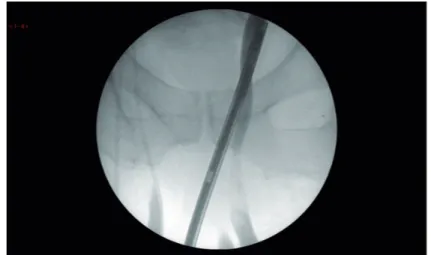
looping back up into the bladder. The absence of contrast within the ureters made the position of the lower ureter difficult to determine. Subse-quent MAG-3 renogram showed a split function of 39/61% with a right sided preponderance. Both kidneys were slow to peak and showed negligible drainage. Intraoperative retrograde pyelograghy showed the ureters to be grossly elongated, loop-ing down bilaterally through the hernial sacs within the scrotum before returning up to the kidneys (Figure-1). Conventional double pig tail ureteric stents were found to be not long enough to span the distance between the bladder and renal pelvis and 75cm-long ileal conduit stents were used successfully (Figure 2 and 3).
Figure 1 - Retrograde pyelograph showing the lower ends of both ureters looping below the pubic arch before ascending towards the kidneys.
IBJU | RADIOLOGY PAGE
622
DISCUSSION
Sliding inguinal hernias which contain the ureter have been reported in the literature (1, 2). Case reports also exist of associated ureteric ob-struction and with resultant ureterohydronephro-sis (3-5). Definitive management involves repair of the hernia with care taken to preserve the ure-ters. These patients are usually morbidly obese (6)
ARTICLE INFO
Int Braz J Urol. 2016; 42: 622-3
_____________________
Submitted for publication: March 19, 2015
_____________________
Accepted after revision: July 20, 2015
REFERENCES
1. Percival WL. Ureter within a sliding inguinal hernia. Can J Surg. 1983;26:283,286.
2. Giglio M, Medica M, Germinale F, Raggio M, Campodonico F, Stubinski R, et al. Scrotal extraperitoneal hernia of the ureter: case report and literature review. Urol Int. 2001;66:166-8. 3. Eilber KS, Freedland SJ, Rajfer J. Obstructive uropathy
secondary to ureteroinguinal herniation. Rev Urol. 2001;3:207-8.
4. Massoud W, Eschwege P, Hajj P, Awad A, Iaaza LA, Chabenne J, et al. Hydronephrosis secondary to sliding inguinal hérnia containing the ureter. Urol J. 2011;8:333-4.
5. Won AC, Testa G. Chronic obstructive uropathy due to uretero-inguinal hernia: A case report. Int J Surg Case Rep. 2012;3:379-81.
6. Akpinar E, Turkbey B, Ozcan O, Akdogan B, Karcaaltincaba M, Ozen H. Bilateral scrotal extraperitoneal herniation of ureters: computed tomography urographic findings and review of the literature. J Comput Assist Tomogr. 2005;29:790-2.
_______________________ Correspondence address: Oladapo Feyisetan, MD Department of Urology, Whiston Hospital, St Helens & Knowsley Teaching Hospitals NHS Trust, Merseyside, L35, 5DR, United Kingdom
Fax: +44 78 7040-5988 E-mail: [email protected]
Figure 2 - CT-KUB scout film. The stents outline the unusual anatomical passage of both ureters. The upper coil of the stents shows the renal pelvises have been pulled down to L4/L5vertebral level.
Figure 3 - In this sagittal section of the CT-KUB, the left ureter, as outlined by the stent, can be seen passing through the neck of the hernia sac.
and are likely to have co-morbidities that make surgery a high risk (3-5).
CONFLICT OF INTEREST
None declared.