Radiofrequency thermal ablation versus
conventional saphenectomy
Ablação térmica por radiofrequência versus safenectomia convencional
Jeferson Freitas Toregeani1
*
, Antônio Severino Trigo Rocha1, Claudio Jundi Kimura2,
Ricardo Adriano Gomes Araújo2, Américo Kazuo Kawai3, Larissa Sokol Rotta3, Andressa Midori Fusioka3
Abstract
Background: Varicose veins of the lower limbs have a high prevalence worldwide. New treatment techniques have been developed with the objectives of improving patients’ quality of life and reducing recovery times. Objective: To evaluate patients with incompetent saphenous veins treated using conventional saphenectomy or radiofrequency ablation (RF), in terms of postoperative status. Methods: From May 2012 to April 2013 146 varicose veins patients with saphenous insuiciency, 90 of whom were treated with conventional surgery (G1) and 56 with RF ablation (G2), were evaluated prospectively. Results: In G1, 88.61% of patients complained of postoperative pain and needed to take analgesics, compared with 28.85% in G2 (p<0.05). Mean pain rating on an analog scale from 0 to 10 was 3.91±2.13 points for G1 and 1.76±3.01 points for G2 (p<0.05). Recovery periods ranged from 26.63±13.3 days to 18.26±19.37 days, for G1 and G2 respectively. Mean time taken to become totally asymptomatic was 66.78±60.9 days for G1 and 38.38±46.8 days for G2 (p<0.05). Conclusions: he RF treatment method caused less postoperative pain and resulted in earlier recovery, when compared to conventional saphenectomy.
Keywords: varicose veins; radiofrequency; saphenectomy.
Resumo
Contexto: As varizes dos membros inferiores têm elevada prevalência mundial e as técnicas convencionais de tratamento têm seus resultados bem deinidos há décadas. O advento de novas tecnologias nos obriga a avaliar os resultados e compará-los com métodos tradicionais. Objetivo: Avaliar o tratamento de pacientes com varizes dos membros inferiores e insuiciência de safenas por safenectomia convencional (SF) ou ablação por radiofrequência (RF), quanto aos sintomas pós-operatórios. Materiais e Métodos: Entre maio/2011 e abril/2013, foram avaliados prospectivamente 146 pacientes com varizes dos membros inferiores e insuiciência de safenas, sendo 90 por SF (G1) e 56 por RF (G2). Resultados: Quanto aos quesitos avaliados, o G1 evidenciou 88,61% dos pacientes com queixa de dor pós-operatória com necessidade do uso de analgésicos e o G2, 28,85% (p<0,05). A média da graduação da dor através da escala analógica – de 0 a 10 – foi de 3,91±2,13 pontos no G1 e de 1,76±3,01 pontos no G2 (p<0,05). O período de recuperação variou de 26,63±13,3 dias para o G1 e 18,26±19,37 dias para o G2. O tempo médio até tornar-se assintomático foi 66,78±60,9 dias para G1 e 38,38±46,8 dias para G2. Conclusão: A RF propiciou menor dor pós-operatória e recuperação mais precoce quando comparada à SF.
Palavras-chave: varizes; radiofrequência; safenectomia.
1Universidade Estadual do Oeste do Paraná – UNIOESTE, Cascavel, PR, Brazil. 2Instituto Vascular, Cascavel, PR, Brazil.
3Faculdade Assis Gurgacz – FAG, Cascavel, PR, Brazil.
Financial support: None.
Conlicts of interest: No conlicts of interest declared concerning the publication of this article. Submitted: July 02, 2014. Accepted: August 04, 2014.
INTRODUCTION
Varicose veins of the lower limbs are dilated, elongated and tortuous veins and they have an elevated prevalence worldwide, making them one of the most important causes of discomfort and work incapacity.1 They affect 10 to 15% of men and 20 to
25% of women in the Western world.2 If multiple
points of relux are also included, in addition to the
saphenous veins, prevalence can reach 43%3 and if
cases of reticular veins and telangiectasia are also included, then more than 80% of the population has the disease.4
The etiology of primary varicose veins of the lower limbs is linked to changes to the veins walls and changes to the structure of collagen and/or elastin, to localized or segmental valve incompetence and/
or the presence of arteriovenous istulas at the level
of the microcirculation.2 Secondary varicose veins
are related to post-phlebitic syndrome, traumatic
arteriovenous istulas, angiodysplasias and causes of extrinsic compression.2
In Brazil, chronic venous insuficiency of the
lower limbs is the 14th most common disease for which the country’s social security system (INSS)
makes sickness beneit payments. According to the
Brazilian National Health Service (SUS - Sistema
Único de Saúde) statistical ofice, DATASUS, in
2004 the service spent 43 million Reais on surgical treatment for varicose veins of the lower limbs.5 In
2013, expenditure was 36.6 million Reais.6
New treatment techniques for varicose veins of the lower limbs have been developed with the objectives of reducing the length of hospital stays and recovery times,7 including thermal ablation using
lasers8 or radiofrequency,9 mechanical ablation10
and administration of sclerosant agents such as ultrasound-guided foam.11 It should be borne in mind
that the results and complications of conventional
surgery are well-deined, leading to an unpleasant situation in terms of cost and beneit, which are in constant conlict.
Radiofrequency (VNUS Medical Technologies Inc., San José, California, and, more recently, Covidien, Mansfield, Maryland) ablation is an intervention guided by catheter, in which energy is released in continuous or sinusoidal waves with frequencies of 200 to 3000 kHz causing tissues in contact with the catheter to heat up.12 The procedure’s
activity is concentrated against the vessel wall, destroying the endothelium and denaturing the
collagen, causing ibrosis of the lumen. The treatment
protocol includes a standardized rate of traction,
determined by the manufacturer of the catheter, so that it travels through a 7 cm segment of the vessel during each 20-second burst of energy, which makes it easier to reproduce the technique and offers more consistent results than treatment with laser, which focuses on transmission of energy into blood or water and has traction velocities that vary depending on the manufacturer or the service.12
One feature of minimally invasive techniques that can make them preferable to conventional
surgery is the possibility of using color Doppler
ultrasonography, which enables real-time evaluation of status in the immediate postoperative period and
identiication of tributaries or anomalous trajectories
that have not been correctly treated or which may
appear immediately after exclusion of the diseased
saphenous vein.13
It is understood that an ideal treatment for varicose veins of the lower limbs should be minimally invasive, possible to be repeated when necessary,
free from signiicant complications and inexpensive
and should also improve esthetics while effectively
eliminating points of relux and allowing a rapid
return to work.14 However, in practice this ideal
treatment does not exist because each of the available
techniques has its advantages and disadvantages, so it
is up to the vascular surgeon to consult the scientiic
evidence and choose the treatment that will achieve the best results for each patient.
OBJECTIVE
To evaluate postoperative results in a sample of patients with varicose veins of the lower limbs, with incompetent saphenous veins, who were treated either by conventional surgery or by radiofrequency ablation.
MATERIALS AND METHODS
This prospective study was conducted in the city of Cascavel, state of Paraná, Brazil, at the Instituto
Vascular, between May 2012 and April 2013. Since
this study constitutes research involving human beings, it was conducted in accordance with the provisions of National Health Council resolution
196/96 and was approved in advance by the Research Ethics Committee at the Faculdade Assis Gurgacz – FAG (Approval number: 085 / 2012).
the lower limbs; with ultrasonographic diagnosis
of insufficiency of at least one of the internal
saphenous veins along at least 50% of its course; with indications for surgery; CEAP classes 2 to 5;
free and spontaneous consent for the patient’s chosen procedure, plus signature of a form accepting responsibility for the costs incurred for use of the
VNUS Closure FAST radiofrequency equipment.
Patients with varicose veins of the lower limbs
were excluded from the study if they did not have a
saphenous vein that was incompetent along at least 50% of its length, if they had high surgical risk or
CEAP classes 1 or 6, or if they did not consent to
taking part in the study.
Patients were allocated to one of two groups: group 1 (G1) received conventional surgery and group 2 (G2) was treated with radiofrequency ablation. Allocation was based on the patients’ own
preferences, in view of the additional costs involved if RF is used.
Data were collected at initial consultations, from lower limb Doppler ultrasound reports, both
preoperative and postoperative, and during telephone calls made to ask about postoperative signs and symptoms.
The following data were acquired from the
Doppler ultrasound studies: diameter, in millimeters,
at the arch, thigh, leg and ankle, and presence of
signiicant relux, greater than 0.5 s, involving more than 50% of the total extent of an internal or external
saphenous vein.
Both groups underwent the same preoperative procedures, consisting of initial consultation,
evaluation using color Doppler ultrasonography,
preoperative evaluations including laboratory tests and cardiological and anesthetic assessments, verification that informed consent had been understood, trichotomy and demarcation of the legs using a percutaneous transilluminator. Perioperative procedures were similar for both groups, with spinal anesthesia, with the only difference between groups
being the open saphenectomy administered to G1 patients versus the RF ablation method used for G2
patients, followed by removal of reticular veins using mini-incisions and sclerotherapy and dressing with
micropore and 20 cm crepe bandages. During the
postoperative period all patients were given the same
prescription, consisting of 6 hours’ fasting; hydration with 500 mL of 0.9% saline solution; analgesia with tenoxicam and dipyrone when necessary; rest with legs raised and prophylaxis 4 hours after anesthesia with 20 mg of enoxaparin. Patients were discharged
after 12 to 24 hours. Both groups were instructed to
remove their bandages after 3 days and start wearing elastic stockings, if possible. Seven days after the operation, the micropore dressings were removed
for both groups and G1 patients’ saphenectomy
sutures were also removed. Patients’ progress was monitored at 30, 90 and 180 days and in the most recent assessment available.
Patients were contacted by telephone and asked
questions about the following: time in hospital (days); recovery time (days); time until asymptomatic; burns/
discoloration (and, if they occurred, the number of
days until they disappeared); pain and pain rating (on a scale from 0 to 10), use of medication (NSAIDS, Analgesics or opiates).
Statistical analysis was conducted using Epi Info 7
from the Centers for Disease Control and Prevention (CDC), Atlanta, USA.
RESULTS
The surgical group, G1, comprised 90 patients,
19 men (21.11%) and 71 women (78.89%), with mean age of 51.4 years (varying from 23 to 78).
The RF group, G2, comprised 56 patients, 14 men
(25.0%) and 42 women (75.0%), with mean age of 54.7 years (varying from 25 to 84).
The most common risk factors were arterial hypertension (28.77%), diabetes mellitus (8.22%)
and smoking (6.85%). There was a higher incidence of risk factors in G1 and this difference was statistically signiicant.
In G1, mean diameters at arch, thigh, leg and ankle
were 7.42 mm, 5.27 mm, 4.92 mm and 3.72 mm,
respectively, and in G2 the same measures were 7.06 mm, 5.30 mm, 4.94 mm and 3.69 mm, with no statistically signiicant difference between groups 1
and 2 (Table 1).
There were no major complications such as deaths, deep venous thrombosis or clinically detectable
infections in either group. In G2 there were two
cases of skin burns (3.57%), one of which left a hyperchromic discoloration.
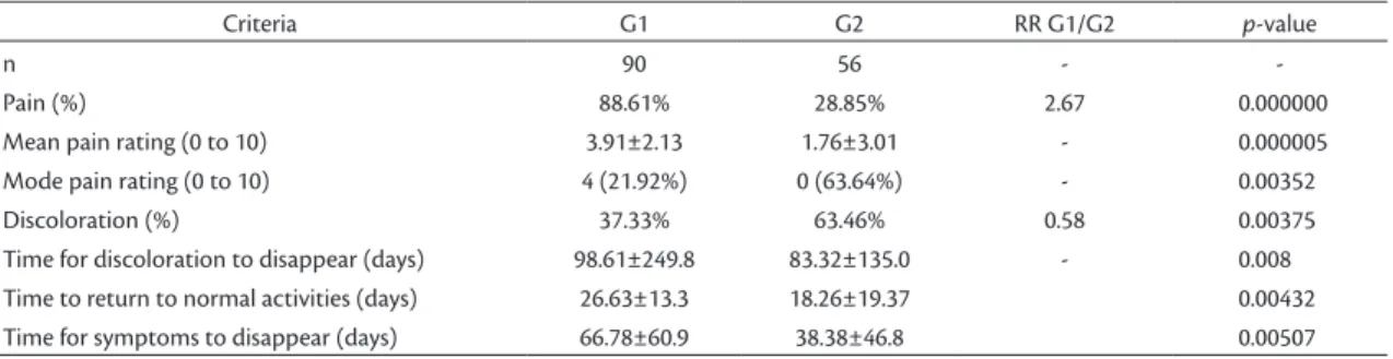
In G1, 88.61% of the patients complained of
postoperative pain and required analgesics and
Table 1. Mean diameters of saphenous veins in G1 and G2 (mm).
n G1 G2 p-value
90 56
-Arch 7.42±2.78 7.06±1.51
0.301
high 5.27±2.25 5.30±1.32
Leg 4.92±1.11 4.94±1.57
Ankle 3.72±0.87 3.69±0.63
anti-inlammatories, whereas in G2 this percentage
was 28.85% (p<0.05). The mean pain rating on the
analog scale was 3.91 points in G1 (varying from 0 to 8), with a mode of 4 (20.25%). In G2, mean pain rating was 1.76 (varying from 0 to 10), with a mode
of 0, chosen by 70.59% of the group (p<0.05). The relative risk of suffering pain during the postoperative
period was 3.07 times greater for patients in G1
(p<0.05) (Table 2).
Recovery time (return to normal physical
activities) was 26 days for G1 and 18 days for G2.
The mean time taken for patients to become entirely
asymptomatic was 66.78±60.9 days in G1 and 38.38±46.8 days in G2 (p<0.05) (Table 2).
Skin discoloration affected 37.33% of patients
in G1 during the postoperative period and 63.46% of the G2 patients (p<0.05). Furthermore, 45.10% of all patients with discoloration were in G1 and 54.10% were in G2 (p<0.05). Mean time
taken for discoloration to disappear varied from
98.61±249.8 days in G1 to 83.32±135.0 in G2. The
relative risk of discoloration on the lower limbs was
1.69 times greater for patients treated with RF (G2)
(p<0.05) (Table 2).
A combination of economic reasons and healthcare
providers’ regulations meant that the study protocol
only included serial Doppler ultrasonography for patients in G2. This showed that immediate and late
success rates of RF ablation were 99% and 92% respectively.
DISCUSSION
This study assessed a population seen at a private clinic and, therefore, did not offer any advantage with regard to the procedures proposed. The proportion of men to women was in line with the tendencies reported in several Brazilian publications,15,16 with
higher prevalence among the female population. The criteria for ‘incompetence’ of the saphenous
veins were based on the literature, and were deined as reflux time evidently greater than 0.5 in the
segments assessed combined with patients’ clinical complaints.17 The mean diameters of internal
saphenous veins observed in this study are similar to results published by Engelhorn et al.,18 who found
that veins larger than 8 mm at the arch, 6 mm at the
thigh and 4 mm at the leg had a greater than 90%
positive predictive value for relux, and are also
in line with data from our service that have been analyzed previously.19 In that study, assessment of
2,471 internal saphenous vein segments revealed a strong correlation between increased diameter and
the presence of relux and showed that more than 50% of cases in which veins were larger than 6.1 mm exhibited relux.19
The fact that patients were responsible for covering the cost of the radiofrequency catheter
constitutes a inancial limitation to formation of the groups, although it did not signiicantly affect the
populational characteristics of either, since statistical analysis showed that the groups had very similar data.
As has been described, the only differences
between the procedures were the method used
to treat internal saphenous vein relux. All of the
other elements involved in the operations were conducted in the same manner for both groups. In
general, countries in Europe and North America
tend to conduct ablation in outpatient settings,20,21
and do not attempt to resolve other aesthetic
venous problems which, to a certain extent, delayed
acceptance of endovascular techniques by Brazilian vascular surgeons because they felt that, initially, the procedure did not address all of the problems
presented by patients, i.e., saphenous vein relux
disease and the aesthetic problems caused by reticular and truncal veins and telangiectasias.
Introduction of a new technique is based on
the constant need for improvement. Although
the majority of vascular surgeons consider it to be well-defined, conventional surgery also has its prerequisites. Obese patients are always a
challenge, for example. Additionally, postoperative
complaints related to conventional saphenectomy
Table 2. Comparison of results for G1 and G2.
Criteria G1 G2 RR G1/G2 p-value
n 90 56 -
-Pain (%) 88.61% 28.85% 2.67 0.000000
Mean pain rating (0 to 10) 3.91±2.13 1.76±3.01 - 0.000005
Mode pain rating (0 to 10) 4 (21.92%) 0 (63.64%) - 0.00352
Discoloration (%) 37.33% 63.46% 0.58 0.00375
Time for discoloration to disappear (days) 98.61±249.8 83.32±135.0 - 0.008
Time to return to normal activities (days) 26.63±13.3 18.26±19.37 0.00432
have become a cause of growing concern among vascular surgeons. Subramonia and Lees22 state that
around 40% of patients who undergo conventional saphenectomy suffer neuropathic symptoms during the postoperative period. Notwithstanding, radiofrequency ablation is not entirely complication free, and cases of neuropathic pain have been described along the path of the saphenous nerve and the sural nerve12 and even neuropathy of the ibular
nerve with foot drop.23
Rasmussen et al.24 compared a number of
different methods for treatment of saphenous vein incompetence, including radiofrequency, intravenous laser, dense foam and conventional surgery.
According to these authors, all treatments were
effective, but radiofrequency and foam caused less pain during the postoperative period when compared
with laser or conventional surgery. According to a
randomized study conducted by Rautio et al.,20,25
patients subjected to conventional saphenectomy suffered paresthesia in 23% of cases, whereas 13% of patients treated with RF suffered neuropathic symptoms.
Patients subjected to radiofrequency thermal ablation had less pain during the postoperative period and many of these patients did not even take the analgesics they were offered. Several different studies have compared the incidence of postoperative symptoms in patients subjected to varicose vein treatments,20 with less pain and reduced consumption
of analgesics. According to Rautio et al.,20 patients
treated with conventional surgery took a mean of 1.6 600 mg ibuprofen tablets per day, whereas patients
treated with radiofrequency only took 0.4 tablets.
According to Shepherd et al.26 patients treated with RF had a mean score of 26.4 points (0-100) and took 8.8 ibuprofen tablets over the irst three days,
in contrast with patients treated with conventional
surgery who had a mean score of 36.8 points (0-100) and took a mean of 20.4 tablets over the irst three
days.
The time taken for recovery (return to normal
physical activities) varied from 26 days in G1 to 18 days in G2. The mean time to become entirely asymptomatic was 66.78±60.9 days in G1 and 38.38±46.8 days in G2 (p<0.05). It should be
remembered that if these patients had only been
treated for saphenous reflux they would have
recovered more rapidly, but regardless of this the RF group recovered more quickly, as had already
been demonstrated by some North-American studies, where, for example, the time taken to return to normal
activities varied from 4.7 to 6.5 days after RF ablation and 12.4 to 15.6 days after conventional treatment.20
Skin discolorations were more common in G2,
primarily at the sites of repeated punctures made
to conduct pre-ablative expansion, and consisted
of ecchymosis along the paths of the saphenous veins, in contrast with saphenectomy which initially caused a lower incidence of ecchymosis, because
the majority of the hematoma is conined to the
saphenous compartment. On the other hand, this may
also explain why the G2 patients, who exhibited more
discoloration during the postoperative period, also
exhibited faster disappearance of this discoloration, since it was more supericial. With regard to burns,
both occurred during the learning curve. One case was caused by overheating of the introducer kit
sheath and the other was caused by a very supericial path combined with excessive compression. Both cases were treated with local measures, but the irst
case resulted in a hyperchromic scar.
The results of this study are similar to data reported in the current literature, such as research by Lurie and
Highlife, 2006,10 conirming that the radiofrequency
technique has a greater impact in terms of improved
patient quality of life – with less postoperative pain and rapid return to daily activities– when compared
with conventional saphenectomy. The challenge that
remains is to develop a less expensive technology
so we can make it available to all patients with indications for saphenectomy.
CONCLUSIONS
This study has shown that radiofrequency thermal ablation caused less postoperative pain and offered faster recovery, when compared with conventional saphenectomy.
REFERENCES
1. Tisi P. Varicose veins. Clin Evid (Online). 2007;2007. PMid:19450366
2. Brake M, Lim CS, Shepherd AC, Shalhoub J, Davies AH. Pathogenesis and etiology of recurrent varicose veins. J Vasc Surg. 2013;57(3):860-8. http://dx.doi.org/10.1016/j.jvs.2012.10.102. PMid:23343668
3. Seidel AC, Miranda F Jr, Juliano Y, Novo NF, dos Santos JH, de Souza DF. Prevalence of varicose veins and venous anatomy in patients without truncal saphenous reflux. Eur J Vasc Endovasc Surg. 2004;28(4):387-90. http://dx.doi.org/10.1016/j.ejvs.2004.06.014. PMid:15350560
5. Figueiredo M, Araújo S, Barros N Jr, Miranda F Jr. Results of surgical treatment compared with ultrasound-guided foam sclerotherapy in patients with varicose veins: a prospective randomised study. Eur J Vasc Endovasc Surg. 2009;38(6):758-63. http://dx.doi. org/10.1016/j.ejvs.2009.07.015. PMid:19744867
6. DATASUS. Gastos com cirurgia de varizes bilaterais [site na
Internet]. 2014. [citado 2014 jul 02]. http://tabnet.datasus.gov. br/cgi/tabcgi.exe?sih/cnv/qiuf.def.
7. Puggioni A, Kalra M, Carmo M, Mozes G, Gloviczki P. Endovenous laser therapy and radiofrequency ablation of the great saphenous vein: analysis of early efficacy and complications. J Vasc Surg. 2005;42(3):488-93. http://dx.doi.org/10.1016/j.jvs.2005.05.014. PMid:16171593
8. Altin FH, Aydin S, Erkoc K, Gunes T, Eygi B, Kutas BH. Endovenous laser ablation for saphenous vein insufficiency: Short- and mid-term results of 230 procedures. Vascular. 2014. http://dx.doi. org/10.1177/1708538114522997. No prelo. PMid:24554352 9. Burihan MC. Endovenous ablation (radiofrequency and laser)
and foam sclerotherapy versus conventional surgery for great saphenous vein varices. Sao Paulo Med J. 2014;132(1):69. PMid:24474085
10. van Eekeren RR, Boersma D, Konijn V, de Vries JP, Reijnen MM. Postoperative pain and early quality of life after radiofrequency ablation and mechanochemical endovenous ablation of incompetent great saphenous veins. J Vasc Surg. 2013;57(2):445-50. http://dx.doi.org/10.1016/j.jvs.2012.07.049. PMid:23141679 11. Devereux N, Recke AL, Westermann L, Recke A, Kahle B.
Catheter-directed foam sclerotherapy of great saphenous veins in combination with pre-treatment reduction of the diameter employing the principals of perivenous tumescent local anesthesia. Eur J Vasc Endovasc Surg. 2014;47(2):187-95. http:// dx.doi.org/10.1016/j.ejvs.2013.10.017. PMid:24268395 12. Roth SM. Endovenous radiofrequency ablation of superficial and
perforator veins. Surg Clin North Am. 2007;87(5):1267-84, xii. http://dx.doi.org/10.1016/j.suc.2007.07.009. PMid:17936486 13. Winterborn RJ, Corbett CR. Treatment of varicose veins: the present
and the future—a questionnaire survey. Ann R Coll Surg Engl. 2008;90(7):561-4. http://dx.doi.org/10.1308/003588408X318228. PMid:18701012
14. Guex JJ, Isaacs MN. Comparison of surgery and ultrasound guided sclerotherapy for treatment of saphenous varicose veins: must the criteria for assessment be the same? Int Angiol. 2000;19(4):299-302. PMid:11305726.
15. Virgini-Magalhães CE, Salvadori RAM, Fagundes FB, et al. Cirurgia de varizes em regime de mutirão. J Vasc Bras. 2007;6(3):231-7. http://dx.doi.org/10.1590/S1677-54492007000300006. 16. Seidel AC, Mangolim AS, Rossetti LP, Gomes JR, Miranda
F Jr. Prevalência de insuficiência venosa superficial dos membros inferiores em pacientes obesos e não obesos. J Vasc Bras. 2011;10(2):124-30. http://dx.doi.org/10.1590/ S1677-54492011000200006.
17. Labropoulos N, Tiongson J, Pryor L, et al. Definition of venous reflux in lower-extremity veins. J Vasc Surg. 2003;38(4):793-8. http:// dx.doi.org/10.1016/S0741-5214(03)00424-5. PMid:14560232
18. Engelhorn CA, Engelhorn AL, Cassou MF, Salles-Cunha SX. Patterns of saphenous reflux in women with primary varicose veins. J Vasc Surg. 2005;41(4):645-51. http://dx.doi.org/10.1016/j. jvs.2004.12.051. PMid:15874929
19. Toregeani JF, Michelan G, Fenato RR, et al. Diâmetro versus refluxo – análise de 2471 segmentos de veia safena interna. J Vasc Bras. 2005;4(3, Supl. 1):S50.
20. Rautio T, Ohinmaa A, Perälä J, et al. Endovenous obliteration versus conventional stripping operation in the treatment of primary varicose veins: a randomized controlled trial with
comparison of the costs. J Vasc Surg. 2002;35(5):958-65. http:// dx.doi.org/10.1067/mva.2002.123096. PMid:12021712
21. Xenos ES, Bietz G, Minion DJ, et al. Endoluminal thermal ablation versus stripping of the saphenous vein: Meta-analysis of recurrence of reflux. Int J Angiol. 2009;18(2):75-8. http://dx.doi. org/10.1055/s-0031-1278330. PMid:22477498
22. Subramonia S, Lees T. Sensory abnormalities and bruising after long saphenous vein stripping: impact on short-term quality of life. J Vasc Surg. 2005;42(3):510-4. http://dx.doi.org/10.1016/j. jvs.2005.05.021. PMid:16171598
23. Kumar RS, Gopinath M. A rare cause of foot drop after radiofrequency ablation for varicose veins: case report and review of the literature. Neurol India. 2010;58(2):303-5. http://dx.doi. org/10.4103/0028-3886.63801. PMid:20508356
24. Rasmussen LH, Lawaetz M, Bjoern L, Vennits B, Blemings A, Eklof B. Randomized clinical trial comparing endovenous laser ablation, radiofrequency ablation, foam sclerotherapy and surgical stripping for great saphenous varicose veins. Br J Surg. 2011;98(8):1079-87. http://dx.doi.org/10.1002/bjs.7555. PMid:21725957
25. Lurie F, Creton D, Eklof B, et al. Prospective randomized study of endovenous radiofrequency obliteration (closure procedure) versus ligation and stripping in a selected patient population (EVOLVeS Study). J Vasc Surg. 2003;38(2):207-14. http://dx.doi. org/10.1016/S0741-5214(03)00228-3. PMid:12891099
26. Shepherd AC, Gohel MS, Brown LC, Metcalfe MJ, Hamish M, Davies AH. Randomized clinical trial of VNUS ClosureFAST radiofrequency ablation versus laser for varicose veins. Br J Surg. 2010;97(6):810-8. http://dx.doi.org/10.1002/bjs.7091. PMid:20473992
*
Correspondence
Jeferson Freitas Toregeani Rua Dom Pedro II, 2359 CEP 85812-120 - Cascavel (PR), Brazil Tel.: +55 (45) 32251288 E-mail: [email protected]
Author information
JFT - Vascular surgeon, Instituto Vascular; TSBACV (Vascular Ultrasound); Assistant professor of Vascular Surgery, Universidade Estadual do Oeste do Paraná (UNIOESTE) and Faculdade Assis Gurgacz (FAG); MSc in Biosciences and Health (UNIOESTE). ASTR - Vascular surgeon, Instituto Vascular; TSBACV (Angioradiology); Professor of Angiology and Vascular Surgery, Universidade Estadual do Oeste do Paraná (UNIOESTE). CJK - Vascular surgeon, Instituto Vascular (Vascular Ultrasound). RAGA - Vascular surgeon, Instituto Vascular. AKK - Vascular surgeon, Instituto Vascular; TSBACV (Angioradiology and Vascular Ultrasound). LSR - Medical student, Faculdade Assis Gurgacz (FAG). AMF - Medical student, Faculdade Assis Gurgacz (FAG).
Author contributions
Conception and design: JFT Analysis and interpretation: JFT, LSR, AMF Data collection: JFT, CJK, RAGA, AKK Writing the article: JFT, LSR, AMF Critical revision of the article: JFT, ASTR Final approval of the article*: JFT, CJK, ASTR, RAGA, AKK, LSR, AMF Statistical analysis: JFT, AMF, LSR Overall responsibility: JFT