Evaluation of pelvic varicose veins using color Doppler
ultrasound: comparison of results obtained with
ultrasound of the lower limbs, transvaginal ultrasound, and
phlebography
Avaliação de varizes pélvicas por Doppler colorido: comparação dos resultados obtidos com
ultrassom dos membros inferiores, ultrassom transvaginal e flebografia
Fanilda Souto Barros,1 José Maria Gomez Perez,2 Eliana Zandonade,3 Sérgio X. Salles-Cunha,4 Javier Leal Monedero,5
Ariadne Basseti Soares Hilel,6 Antônio Augusto Barbosa de Menezes,7 Daniela Souto Barros8
Abstract
Introduction: Pelvic varicose veins, one of the main causes of chronic pelvic pain and dyspareunia, are an important source of relux for lower limb varicose veins, especially in recurrent cases. Color Doppler ultrasound of the lower limbs and transvaginal ultrasound are the noninvasive diagnostic methods most commonly used to assess pelvic venous insuiciency, whereas phlebography is still considered as the gold standard.
Objectives: To determine the prevalence of lower limb varicose veins originating from the pelvis in a group of female patients and to determine the agreement between results obtained via color Doppler ultrasound of the lower limbs, transvaginal ultrasound, and phlebography.
Methods: he sample comprised female patients referred to a vascular laboratory for lower limb screening. Patients diagnosed with deep venous thrombosis were excluded. Data analysis included kappa coeicient of agreement, McNemar’s test, sensitivity and speciicity values.
Results: Of a total of 1,020 patients, 124 (12.2%) had indings compatible with relux of pelvic origin. Among these patients, 51 (41.2%) were recurrent cases. A total of 249 were submitted to transvaginal ultrasound. here was signiicant agreement between lower limb ultrasonographic indings and transvaginal indings. Phlebography was performed in 54 patients. he comparison between transvaginal ultrasound and phlebography was associated with a 96.2% sensitivity and 100% speciicity.
Conclusions: he authors draw attention to the relatively high prevalence of lower limb varicose veins originating from the pelvis, suggesting an important but underdiagnosed cause of recurrent varicose veins.
Keywords: Color Doppler ultrasound, pelvic varicose veins, transvaginal Doppler ultrasound, phlebography.
Resumo
Introdução: As varizes pélvicas, uma das principais causas de dor pélvica crônica e dispareunia, são uma importante fonte de reluxo para as varizes dos membros inferiores, especialmente em casos recorrentes. O Doppler colorido dos membros inferiores e o ultrassom transvaginal são os métodos diagnósticos não-invasivos mais comumente usados para avaliar a insuiciência venosa pélvica, enquanto a lebograia ainda é considerada como o padrão-ouro.
Objetivos: Determinar a prevalência de varizes dos membros inferiores originadas na pélvis em um grupo de pacientes do sexo feminino e determinar a concordância entre os resultados obtidos por Doppler colorido dos membros inferiores, ultrassom transvaginal e lebograia.
Métodos: A amostra incluiu pacientes do sexo feminino encaminhadas para o laboratório vascular para triagem dos membros inferiores. As pacientes diagnosticadas com trombose venosa profunda foram excluídas. A análise dos dados incluiu o coeiciente de concordância kappa, o teste de McNemar e os valores de sensibilidade e especiicidade.
his study was approved by the Research Ethics Committee of Universidade Federal do Espírito Santo (UFES), register no. CEP 151/08.
1 Especialista, Angiologia, Área de Atuação em Ecograia Vascular.
2 Doutor, Angiologia e Cirurgia Vascular. Professor, Universidade Federal do Espírito Santo (UFES), Vitória, ES, Brazil. 3 PhD. Professora e Chefe, Departamento de Estatística, UFES, Vitória, ES, Brazil.
4 PhD. Registered Vascular Technologist. Vascular ultrasound specialist, CompuDiagnostics, Phoenix, USA. 5 Cirurgião vascular. Chefe, Departamento de Cirurgia Vascular, Hospital Ruber International, Madrid, Espanha. 6 Especialista, Angiologia e Cirurgia Vascular.
7 Doutor, Angiologia e Cirurgia Vascular. Professor, UFES, Vitória, ES, Brazil.
8 Estudante de Medicina, Escola Superior de Ciências, Santa Casa de Misericórdia de Vitória, Vitória, ES, Brazil.
Resultados: De um total de 1.020 pacientes, 124 (12.2%) tiveram achados compatíveis com reluxo de origem pélvica. Entre essas pacientes, 51 (41.2%) eram casos recorrentes. Um total de 249 foram submetidas a ultrassom transvaginal. Houve concordância signiicativa entre os achados ultrassonográicos dos membros inferiores e os achados transvaginais. A lebograia foi realizada em 54 pacientes. A comparação entre o ultrassom transvaginal e a lebograia foi associada a 96.2% de sensibilidade e 100% de especiicidade.
Conclusões: Os autores chamam a atenção para a prevalência relativamente alta de varizes dos membros inferiores originadas na pélvis, sugerindo uma importante, embora subdiagnosticada, causa de varizes recorrentes.
Palavras-chave: Doppler colorido, varizes pélvicas, ultrassonograia Doppler transvaginal, lebograia.
Introduction
Varicose veins of pelvic origin are a major cause of relux that is not directly related with the saphenous vein system.1 hey can be restricted to the pelvic region itself or
extend to the perineum, vulvar region or lower limbs.2
Pelvic varicose veins can be identiied during physi-cal examination, indirectly via color Doppler ultrasound (CDU) of the lower limbs or directly via transvaginal
Doppler ultrasound or phlebography.3,4 he condition can
evolve asymptomatically or develop into pelvic conges-tion syndrome, with symptoms such as abdominal bloat-ing and dyspareunia or presence of varicose veins of the lower limbs with relux originating from subdiaphragmatic tributaries.5,6
Knowledge of diferent forms of drainage in the pelvic region is essential for a clear understanding of the patho-physiology and treatment of pelvic varicose veins. he ve-nous plexus located on the broad ligament of the uterus communicates with the uterine plexus, thus forming the gonadal or ovarian veins that usually converge directly into the inferior vena cava on the right side and into the renal vein on the let side. hese veins contain valves and are therefore extremely important for drainage; on the other hand, an insuicient number of these veins will result in pelvic varicose veins.7
CDU is the method of choice for the assessment of su-pericial venous insuiciency of the lower limbs. It success-fully identiies patterns of saphenous and nonsaphenous relux, including relux of pelvic origin.1,8 Transvaginal
CDU is used to assess organs and circulation in the pelvic region. Finally, selective phlebography is still considered the gold standard for the diagnosis of subdiaphragmatic varicose veins.4,5
he objective of the present study was to identify the prevalence of pelvic varicose veins in female patients re-ferred to a vascular laboratory for supericial venous system screening using three diagnostic methods: CDU of the low-er limbs, transvaginal CDU, and phlebography. he results obtained with the three methods were compared so as to determine inter-method agreement.
Material and methods
he sample included all female patients referred to the vascular laboratory at Angiolab-Vitória, located in the mu-nicipality of Vitória, southeast Brazil, for lower limb screen-ing usscreen-ing CDU from January 2006 to April 2008.
Sample size was calculated taking into consideration a total of 10,000 examinations per year, an expected preva-lence of pelvic varicose veins of 15%,4 a signiicance level of
5%, and a precision level of 2.5%. he minimum sample size was deined as 727 patients submitted to CDU of the lower limbs. In order to measure sensitivity between transvaginal CDU and phlebography, the same sample size was consid-ered, with an expected prevalence of 15%, an expected sen-sitivity of 95%, a signiicance level of 5%, and a precision level of 16%. he minimum number of patients necessary for submission to the two diagnostic tests (transvaginal CDU and phlebography) was found to be 54. Indication of transvaginal CDU and phlebography was based on clinical and symptomatic assessment of the patients.
he clinical classiication (CEAP) of the sample ranged
between 0 and 5.9 Patients with prior or recent deep venous
thrombosis in the iliac, femoropopliteal, and infrapopliteal segments were excluded from the study.
Patients were assessed by a physician specialized in angi-ology and experienced in vascular ultrasound, using an ATL-Philips® HDI 5000 ultrasound device with a 7.5 MHZ linear transducer for the assessment of lower limbs and a 4-8MHz endocavity probe for transvaginal ultrasound. he protocol used for lower limb venous mapping followed two stages: 1) patient in the supine position for assessment of the deep ve-nous system; and 2) patient standing for assessment of the
main sources of relux.10 Signiicant relux was deined as the
presence of retrograde low lasting for more than 0.5 s, moni-tored by placing the pulsed Doppler sample volume longitu-dinally in the center of the vessel (CDU longitudinal image) with adjustments in gain, ilter and pulse repetition settings.11
he protocol used for assessment of the pelvic region
(transvaginal ultrasound) was as follows12: examination
Transparietal abdominal investigation was carried out to evaluate the patency of the inferior vena cava and iliac vein system, as well as to identify the presence of extrinsic venous compression suggestive of pelvic vari-cose veins (May-hurner syndrome and nutcracker syn-drome). his exam was performed with patients in the supine position using a low-frequency convex transducer (2-5 MHz).
Transvaginal assessment was carried out with patients in the recumbent position using a 4-8 MHz endocavity probe with a sterile cover (condom). he transducer was introduced into the vaginal canal, allowing the identiica-tion of vessels in the bilateral adnexal region.
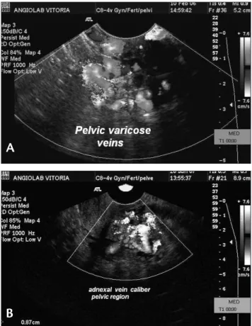
Pelvic varicose veins were deined as the presence of dilated (diameter ≥ 7 mm), tortuous vessels, with relux (presence of bidirectional low during Valsalva’s maneu
-ver) in the adnexal region11,13 (Figure 1).
Selective pelvic phlebography was performed using a Philips® device and the Seldinger technique. All operators were specialists in vascular and endovascular surgery. A right upper limb vein was used as access route, followed by selective catheterization with nonionic contrast of renal veins, iliac/gonadal veins and gonadal plexus. Vein diam-eter and the presence of venous relux in the pelvic region were evaluated, also trying to identify the direction of blood low and possible escapes to the lower limbs.
Statistical analysis
Prevalence rates of pelvic varicose veins were calcu-lated for the three examination methods. Sensitivity, speci-icity, positive predictive values (PPV) and negative predic-tive values (NPV) obtained for ultrasonographic indings were compared with those obtained with the gold standard method (phlebography).
Kappa coeicients and McNemar’s nonparametric test were used to measure agreement and disagreement between tests, respectively. he SPSS sotware version 15.0 was used, and the signiicance level was set at 5%.
he study was approved by the Research Ethics Committee at Universidade Federal do Espírito Santo (UFES), under the protocol no. 101/08.
Results
Prevalence
A total of 1,020 patients were analyzed; mean age was 48.1±14.2 years, and mean number of gestations was
3.3±2.3. CEAP classiication ranged from 1 to 2.
CDU of the lower limbs was performed in all 1,020 pa-tients; of these, 249 patients were submitted to transvaginal CDU, and 59 patients to selective phlebography.
he prevalence of pelvic varicose veins according to CDU of the lower limbs was 12.2% (124 positive cases out of 1,020), distributed as follows: 3% (31 cases) bilateral, 4.4% (45 cases) afecting the right limb only, and 4.7% (48 cases) afecting the let limb only. Among the positive cases, 51 patients (41.2%) were recurrent, i.e., had been previous-ly submitted to surgery (great saphenous vein stripping or high ligation of the saphenofemoral junction with preser-vation of the saphenous vein). hese patients included 14 bilateral cases (45.2%), 17 cases (37.8%) afecting the right side only, and 20 cases (41.7%) afecting the let side only.
Relux of pelvic origin in the lower limbs was as fol-lows: 48 cases (38.7%) of relux in the posterior aspect of the thigh, 35 (28.2%) converging into the great saphenous vein, 28 (22.6%) in the medial aspect of the thigh (parallel to the saphenous axis), 13 (10.5%) in perijunctional region, three (2.4%) converging into the small saphenous vein, and ive (4.0%) in other regions. More than one type of relux were detected in some patients.
Figure 1 - A) Dilated vessels, with relux, in the adnexal region identiied by transvaginal color Doppler ultrasound; B) measurement of adnexal vein caliber, identiied by transvaginal color Doppler ultrasound
A
According to transvaginal CDU, the prevalence of pel
-vic varicose veins was 60.2% (150 positive cases out of 249 examinations). The mean diameter of veins with relux was 8.5 mm (±1.7).With phlebography, the prevalence rate ob
-tained was 98.1% (53 positive cases out of 54 examinations).
here was a statistically signiicant association between tributaries of pelvic origin in the lower limbs and recurrent varicose veins (chi-square = 26.839; p = 0.001), as shown in Table 1.
Agreement between CDU of the lower limbs and transvaginal CDU
Table 2 shows the results obtained with the two tests. Comparison between CDU of the lower limbs and
transvaginal CDU (considered as the gold standard in this case) revealed a sensitivity of 41.3%, a speciicity of 93.9%, a positive predictive value of 48.5%, and a negative predictive value of 92.0% (predictive values were calculated based on the prevalence obtained with lower limb CDU, 12.2%).
Kappa coeicient for the comparison between the results obtained with CDU of the lower limbs and trans-vaginal CDU (in this case, no test was considered as gold standard) was 0.309 (p = 0.001), suggesting a statistically signiicant agreement between both methods. McNemar’s test indicated that transvaginal CDU yielded more positive results, i.e., had a higher sensitivity (p = 0.001).
Agreement between transvaginal CDU and phlebography
Table 3 shows the results obtained with the two tests. Comparison between transvaginal CDU and phlebography (gold standard) revealed a sensitivity of 96.2%, a speciicity of 100%, a positive predictive value of 100%, and a negative predictive value of 94.6% (predictive values were calculated based on the prevalence obtained with transvaginal CDU, 60.2%).
Kappa coeicient for the comparison between the re-sults obtained with transvaginal CDU and phlebography (in this case, no test was considered as gold standard) was 0.486 (p = 0.001), suggesting a statistically signiicant agree-ment between both methods. McNemar’s test indicated that both tests were equivalent (p = 0.500).
Table 4 summarizes the main results of the present study.
Discussion
he pelvic plexus is characterized by venovenous anas-tomoses connected to the lower limbs, involving or not the saphenous vein system. Although gonadal vein dilatation/
insuiciency is not rare among asymptomatic patients,14 a
correlation between pelvic varicose veins on the one hand
Lower limbs Recurrent, n (%) Total, n (%)
No Yes
Negative 707 (95.3) 189 (85.1) 896 (92.9) Positive 35 (4.7) 33 (14.9) 68 (7.1) Total 742 (100) 222 (100) 964 (100) Table 1 - Association between presence of tributaries of pelvic origin in
the lower limbs and recurrent varicose veins
Lower limbs Transvaginal Total Negative Positive
Negative 93 88 181
Positive 6 62 68
Total 99 150 249
Table 2 - Results obtained with CDU of the lower limbs and transvaginal CDU
Phlebography Transvaginal Total Negative Positive
Negative 1 0 1
Positive 2 51 53
Total 3 51 54
Table 3 - Results obtained with transvaginal CDU and phlebography
Variable CDU of the lower limbs Transvaginal CDU Phlebography
Sample size 1,020 249 54
Prevalence 12.2% 60.2% 98.1%
Gold standard used for comparisons Transvaginal CDU Phlebography
-Sensitivity 41.3%, 96.2%
-Speciicity 93.9%, 100%
-Positive predictive value 48.5%, 100%
-Negative predictive value 92.0% 94%
-Kappa 0.309 (p = 0.001) 0.486 (p = 0.001)
-McNemar’s test p = 0.001 p = 0.500
and chronic pelvic pain and recurrent varicose veins of the lower limbs on the other is already known.2,6,13,15,16
According to Labropoulos et al.,1 relux of
nonsaphe-nous origin accounts for 10% of the varicose veins of the lower limbs; of these, 34% originate from the pelvic region. Recurrent varicose veins of the lower limbs afect up to 52% of cases within 5 years; abdominal or pelvic origin account for 17%.17,18
According to Leal Monedero et al., the etiology of re-current varicose veins includes recanalizations through pel-vic “escape points” to the lower limbs via veins of the broad ligament, i.e., posterior branches that escape through the
internal pudendal, obturator and ischiatic veins.13
Geier et al.19 showed that 68% of female patients with
pelvic varicose veins conirmed by phlebography presented recurrent varicose veins of the lower limbs ater great sa-phenous vein stripping.
In our sample, the prevalence of relux of pelvic ori-gin among patients submitted to CDU of the lower limbs was 12.2%, a similar rate to that reported by Ashour et al.4 (15.8%), but lower when compared to the study by
Labropoulos et al. (34%).1 his discrepancy in prevalence
rates can be explained, at least in part, by the diferent de-grees of disease severity found in the populations assessed. Labropoulos et al.1 inform that 90% of the sample had a
CEAP classiication ranging from 1 to 3, compared to clas-siications 1 to 2 in 87% of our sample.
he prevalence of patients submitted to great saphe-nous vein stripping or to high ligation of the saphenofemo-ral junction with preservation of the great saphenous vein (recurrent cases) in this study was 41.2%. he association between recurrent varicose veins and tributaries of pelvic origin in the lower limbs was statistically signiicant (chi-square; p = 0.001), suggesting an important and so far un-derdiagnosed cause of recurrent varicose veins.
Predominant involvement of multiparous women (having had more than two children) and a higher number of cases afecting the let adnexal region and the let lower limb were similar to reports found in the literature.20,21
It is important to emphasize the presence of collateral-ization of tributaries into the posterior aspect of the thigh through recanalization of the ischiatic primitive system, as well as transference of the relux to the saphenous vein system in the presence of ostial competence of the saphe-nofemoral junction. hese indings are extremely relevant because they allow to focus treatment planning on the real source of relux.
here is no consensus in the literature with regard to the ideal cutof point for the correlation between ad-nexal vein diameter measured by transvaginal Doppler
ultrasound and presence of relux, and it is possible to ind values ranging from 5 to 8 mm across diferent studies.13,22,23
We considered a cutof of 7 mm, and the mean diameter of
veins with relux found in our sample was 8.5 mm (±1.7).
Agreement between CDU of the lower limbs and trans-vaginal CDU with regard to the identiication of pelvic re-lux was statistically signiicant. However, sensitivity was low (41.3%), which suggests that CDU of the lower limbs alone cannot be used as a criterion for the diagnosis of pel-vic varicose veins; rather, the performance of transvaginal CDU to conirm the diagnostic hypothesis is required. On the other hand, the speciicity 93.9%, and negative predic-tive value 92.0% associated with CDU of the lower limbs suggests that whenever this examination results negative for pelvic varicose veins, further investigation is not necessary.
Phlebography is currently the method of choice for the diagnosis of pelvic varicose veins; however, transvaginal CDU indings were equivalent to those obtained with the gold standard, with the advantage of being a noninvasive and risk-free diagnostic method.
he complex anatomical variations found in the pel-vis, associated with the rich network of anastomoses that is characteristic of the region, suggests that endovascular treatment with embolization should be considered as a therapeutic option in cases of pelvic varicose veins.6,13,23
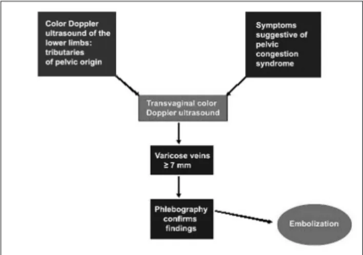
Based on the present indings, the authors propose an algorithm for the investigation of pelvic varicose veins (Figure 2). Patients with gynecological symptoms (pelvic congestion syndrome) or with clinical and ultrasonographic indings suggestive of varicose veins of pelvic origin should be referred for transvaginal assessment. If the presence of varicose veins of pelvic origin is conirmed (diameter ≥ 7
mm and presence of relux during Valsalva’s maneuver in adnexal vessels), then phlebography is recommended
Conclusions
The authors draw attention to the relevant preva-lence of varicose veins of pelvic origin found in a sample of patients referred to a vascular laboratory for venous screening of the lower limbs. This finding suggests an important and so far underdiagnosed cause of recurrent varicose veins, reinforcing the need to include venous screening of the lower limbs in the therapeutic plan-ning of the treatment of varicose veins. Moreover, the high agreement found between transvaginal CDU and phlebography findings for the diagnosis of pelvic vari-cose veins suggests that transvaginal CDU is very useful and should be considered as a less invasive diagnostic method prior to phlebography.
References
1. Labropoulos N, Tiongson J, Pryor L, et al. Nonsaphenous superi-cial vein relux. J Vasc Surg. 2001;34:872-7.
2. Francheschi C, Bahnini A. Treatment of lower extremity venous insuiciency due to pelvic leak points in women. Ann Vasc Surg. 2005;19:284-8.
3. Lasry JL, Coppé G, Balian E, Borie H. Pelvi-perineal venous insuf-iciency and varicose veins of the lower limbs: duplex Doppler di-agnosis and endoluminal treatment in thirty females. J Mal Vasc. 2007;32:23-31
4. Ashour MA, Soliman H, Khougeer GA. Role of discending venog-raphy and endovenous embolization in treatment of females with lower extremity varicose veins, vulvar and posterior thigh varices. Saudi Med J. 2007;28:206-12.
5. Greiner M, Gilling-Smith GL. Leg varices originating from the pel-vis: diagnosis and treatment. Vascular. 2007;15:70-8.
6. Creton D, Hennequin L, Kohler F, Allaert FA. Embolisation of symp-tomatic pelvic veins in women presenting with non-saphenous varicose veins of pelvic origin: three-year follow-up. Eur J Vasc Endovasc Surg. 2007;34:112-7.
7. Ulaker R. Atlas de anatomia vascular. Rio de Janeiro: Revinter; 2003. p. 635-729.
8. Engelhorn CA, Engelhorn AL, Cassou MF, Casagrande C, Gosalan CJ, Ribas E. Classiicação anatomofuncional da insuiciência das ve-ias safenas baseada no eco-Doppler colorido dirigida para o plane-jamento cirúrgico das varizes. J Vasc Bras. 2004;3:13-9.
9. Nicolaides AN, Allegra C, Bergan J, et al. Management of chronic venous disorders of the lower limbs: guidelines according to scien-tiic evidence. Int Angiol. 2008;27:1-59.
10. Coleridge-Smith P, Labropoulos N, Partsch H, Myers K, Nicolaides A, Cavezzi A. Duplex ultrasound investigation of the veins in chronic venous disease of the lower limbs: UIP consensus document. Part I: basic principles. Eur J Vasc Endovasc Surg. 2006;31:83-92
11. Labropoulos N, Tiongson J, Pryor L, et al. Deinition of venous re-lux in lower-extremity veins. J Vasc Surg. 2003;38:793-8.
12. Barros FS. Varizes pélvicas. In: Engelhorn CA, Morais Filho D, Barros FS, Coelho NA. Guia prático de ultra-sonograia vascular. Rio de Janeiro: Dilivros; 2006. p. 191-195.
13. Leal Monedero J, Ezpeleta SZ, Castro FC, Senosiain LDC. Recidiva varicosa de etiologia pélvica .In: homaz JB, Belczack CEQ. Tratado de lebologia e linfologia. Rio de Janeiro: Livraria Rubio; 2006. p. 301-22
14. Rozenblit AM, Ricci ZJ, Tuvia J, Amis ES Jr. Incompetent and di-lated ovarian veins: a common CT inding in asymptomatic parous women. Am J Roentgenol. 2001;176:119-22.
15. Liddle AD, Davies AH. Pelvic congestion syndrome: chron-ic pelvchron-ic pain caused by ovarian and internal iliac varchron-ices. Phlebolgy.2007;22:100-4.
16. Perrin M, Gillet JL. Management of recurrent varices at the popli-teal fossa after surgical treatment. Phlebology. 2008;23:64-8
17. Van Rij AM, Jiang P, Solomon C, Christie RA, Hill GB. Recurrence after varicose vein surgery (a prospective long-term clinical study with duplex ultrasound scanning and air plethysmography). J Vasc Surg. 2003;38:935-43.
18. Perrin MR, Labropoulos N, Leon LR Jr. Presentation of the pa-tient with recurrent varices after surgery (REVAS). J Vasc Surg. 2006;43:327-34.
19. Geier B, Barbera L, Mumme A, et al. Relux patterns in the ovarian and hypogastric veins in patients with varicose veins and signs of pelvic venous incompetence. Chir Ital. 2007;59:481-8.
20. Jacques NMP, Pinto MG. Vasos do tronco: Anatomia e aplicações médico-cirúrgicas. In: Mafei FHA, Lastória S, Yoshida WB, Rollo H, Giannini M, Moura R. Doenças Vasculares Periféricas. Rio de Janeiro: Guanabara Koogan; 2008. p. 17-69.
21. Scultetus AH, Villavicencio JL, Gillespie DL, Kao TC, Rich NM. he pelvic venous syndromes: analysis of our experience with 57 pa-tients. J Vasc Surg. 2002;36:881-8.
22. Kim HS, Malhotra AD, Rowe PC, Lee JM, Venbrux AC. Embolotherapy for pelvic congestion syndrome: long-term results. J Vasc Interv Radiol. 2006;17:289-97.
23. Ratnam LA, Marsh P, Holdstock JM, et al. Pelvic vein embolisation in the management of varicose veins. Cardiovasc Intervent Radiol. 2008;31:1159-64.
Correspondence: Fanilda Souto Barros R. Joaquim Lírio, 340/701, Praia do Canto CEP 29055-460 – Vitoria, ES, Brazil E-mail: [email protected]
NOTA EDITORIAL AO ARTIGO
Aldemar Araujo Castro*
* Mestre em Cirurgia Vascular, Universidade Federal de São Paulo (UNIFESP), São Paulo, SP. Professor assistente, Universidade Estadual de Ciências da Saúde de Alagoas, Maceió, AL.
O roteiro de avaliação da qualidade de um artigo so-bre teste diagnóstico1,2 é formado por três grandes questões,
que depois são subdivididas:
1. Os resultados são válidos?
1.1. Foi realizada uma comparação independente e mascara-da do teste diagnóstico com o padrão-ouro?
1.2. A amostra de pacientes utilizada no teste diagnóstico in-clui o espectro encontrado na prática clínica?
1.3. O resultado dos testes que está sendo avaliado inluen-ciou a decisão de realizar o padrão-ouro?
1.4. Foi realizada uma descrição do teste diagnóstico com de-talhes suicientes para permitir sua reprodução? 2. Quais são os resultados?
2.1 Os testes diagnósticos são apresentados com sensibilidade e especiicidade, valor preditivo positivo, valor preditivo negativo ou os dados estão disponíveis para calculá-los? 3. Os resultados irão ajudar no cuidado dos meus pacientes? 3.1. Os resultados dos testes são reprodutíveis e a
interpreta-ção é possível no local onde trabalho?
3.2. Os resultados são aplicáveis aos meus pacientes? 3.3. Os resultados poderão mudar minha conduta?
3.4. Os pacientes icaram melhores com os resultados do teste?
Ao utilizar o roteiro no artigo analisado3, são
encontra-das, segundo nossa avaliação, as seguintes respostas:
1.1 Foi realizada uma comparação independente e mascara-da do teste diagnóstico com o padrão-ouro?
Possivelmente não. A descrição no texto é incompleta para determinar se o novo ultrassom (US) transvaginal e, depois, a lebograia foram realizados de forma que o exa-minador não soubesse do resultado prévio, pois a indicação dos exames (US transvaginal e lebograia) foi feita de acor-do com os sinais e sintomas das pacientes.O melhor seria que todas fossem submetidas aos dois testes e ao padrão-ouro, independentemente dos resultados encontrados. Como o padrão-ouro é a lebograia, torna-se desnecessário o cálculo de sensibilidade ou especiicidade da comparação do US de membros inferiores com o US transvaginal. A ausência de um padrão-ouro adequado pode gerar resul-tados sem aplicabilidade clínica. A comparação entre o US transvaginal e o padrão-ouro em uma amostra com 98% da doença superestima a utilidade do teste diagnóstico.
1.2. A amostra de pacientes utilizada no teste diagnóstico in-clui o espectro encontrado na prática clínica?
Não. Foram selecionados os pacientes de um centro de referência que se submeteriam a US de membros inferio-res. O mais adequado seria incluir somente pacientes com sinais e sintomas da doença venosa pélvica crônica e sub-meter todos os indivíduos ao teste diagnóstico e ao padrão-ouro de forma independente e cega.
1.3. O resultado dos testes que está sendo avaliado inluen-ciou a decisão de realizar o padrão-ouro?
Sim. A indicação da realização da US transvaginal e da lebograia foi feita de acordo com sinais e sintomas das pacientes.
1.4. Foi realizada uma descrição do teste diagnóstico com de-talhes suicientes para permitir sua reprodução?
Sim. Foi detalhada toda a técnica.
Uma vez respondidas as quatro questões, devemos ter uma avaliação da validade da pesquisa. As respostas nega-tivas para a primeira e a segunda perguntas reduzem a ava-liação da qualidade do artigo. Cabe, então, a cada leitor a decisão pessoal de continuar a avaliação. A minha resposta é sim, apesar das limitações apontadas.
Respondendo à pergunta 2.1, sobre a importância: Os testes diagnósticos são apresentados com sensibilidade e es-peciicidade, valor preditivo positivo, valor preditivo nega-tivo ou os dados estão disponíveis para calculá-los?
Sim. Para a avaliação da qualidade em pesquisas sobre testes diagnósticos não interessam os testes estatísticos e os valores de p. Calcular a sensibilidade, a especiicidade, a prevalência e as razões de verossimilhança são suicien-tes. Nesse momento, a ajuda de calculadoras eletrônicas costuma ser útil. Realizados os cálculos, cabe a interpreta-ção. No entanto, por uma limitação da validade da pesquisa nas primeiras questões, os resultados encontrados são úteis em princípio para gerar boas hipóteses para serem testadas apropriadamente no futuro e, portanto, não seria apropria-do continuar com a avaliação de qualidade proposta por Jaeschke et al.1,2.
Assim, a avaliação da qualidade de um artigo é uma habilidade que deve ser desenvolvida e aprimorada por parte de angiologistas e cirurgiões vasculares. O roteiro apresentado é apenas um de uma série de roteiros que
existem para avaliação de cada tipo de estudo4,5. Ou seja,
Além do uso de roteiros de avaliação, é importante considerar o tipo de estudo. Na pesquisa publicada, existe a possibilidade de o estudo ser retrospectivo, transversal ou prospectivo, o que pode também inluenciar na qualidade da pesquisa. Os resultados são mais coniáveis nos estudos prospectivos do que nos retrospectivos por reduzir a possi-bilidade do viés de aferição.
Referências
1. Jaeschke R, Guyatt G, Sackett DL. Users’ guides to the medical li-terature. III. How to use an article about a diagnostic test. A. Are the results of the study valid? Evidence-Based Medicine Working Group. JAMA. 1994;271:389-91.
2. Jaeschke R, Guyatt GH, Sackett DL. Users’ guides to the medical literature. III. How to use an article about a diagnostic test. B. What are the results and will they help me in caring for my patients? he Evidence-Based Medicine Working Group. JAMA. 1994;271:703-7.
3. Barros FS, Perez JMG, Zandonade E, et al. Evaluation of pelvic vari-cose veins using color Doppler ultrasound: comparison of results obtained with ultrasound of the lower limbs, transvaginal ultra-sound, and phlebography. J Vasc Bras. 2010;9:xx-xx.
4. Oxman AD, Sackett DL, Guyatt GH. Users’ guides to the medi-cal literature. I. How to get started. he Evidence-Based Medicine Working Group. JAMA. 1993;270:2093-5.
5. Castro AA. Avaliação da qualidade da informação. In: Castro AA. Fiat lux. Maceió: Universidade Estadual de Ciências da Saúde de Alagoas; 2005. p. 1-14.
RÉPLICA DOS AUTORES
Fanilda Souto Barros1, Eliana Zandonade2
1 Especialista em Angiologia com Área de Atuação em Ecograia Vascular.
3 PhD. Professora e Chefe, Departamento de Estatística, Universidade Federal do Espírito
Santo (UFES), Vitória, ES, Brazil.
Em atenção ao comentário editorial sobre nosso arti-go, apresentamos uma relexão sobre medicina baseada em evidência (MBE), o tipo de estudo que realizamos, a forma como conduzimos a pesquisa e sua importância na prática clínica.
Ressaltamos que os comentários realizados foram de alta qualidade teórica. O autor da nota editorial classiicou apropriadamente o tipo de estudo, mas tem restrições me-todológicas quanto a pesquisas realizadas em clínica.
1. Entendendo sobre MBE
Segundo Fletcher & Fletcher1, MBE é um termo
mo-derno para a aplicação da epidemiologia clínica ao cuidado com os pacientes. A epidemiologia clínica é a ciência que faz predições sobre pacientes individuais utilizando eventos
clínicos em grupos de pacientes semelhantes. Utiliza-se do método cientíico sólido para garantir inferências corretas.
2. Estudos de testes diagnósticos
Fletcher & Fletcher1 airmam, no capítulo sobre
estu-do de testes diagnósticos, que a maioria das informações de um teste diagnóstico é obtida em ambientes clínicos, e não em ambientes de pesquisa. Nesses estudos, os médicos utilizam o teste no cuidado com os pacientes e, por ques-tões éticas, não conduzem uma avaliação mais aprofundada quando os testes preliminares são negativos. O problema metodológico advindo de estudos realizados dessa forma é a falta de dados suicientes para a análise.
No nosso estudo, dos três testes de diagnóstico realiza-dos, somente a lebograia (método invasivo e não isento de risco), sofreu parcialmente a falta de dados. Entretanto, o número de pacientes que foram submetidas a esse teste (le-bograia) por indicação clínica foi suiciente para a análise estatística de equivalência entre os testes.
3. Nossa pesquisa
A nossa pesquisa estudou pacientes encaminhadas ao laboratório vascular para o mapeamento venoso dos mem-bros inferiores para a avaliação de varizes. O objetivo foi correlacionar o achado de tributárias nos membros infe-riores sugestivas de origem pélvica com a real presença de varizes nesse território, visto que em alguns trabalhos2 essa
identiicação é feita somente com o estudo ultrassonográi-co dos membros inferiores. O ultrassom endovaginal é um exame considerado eicaz no estudo da região pélvica, e a lebograia é o exame considerado padrão-ouro para o diag-nóstico de varizes pélvicas.
Submeter todas as pacientes ao estudo lebográico, que não é isento de risco, no nosso entender não seria éti-co. Assim, coletamos os dados segundo os princípios da amostragem (cálculo de tamanho de amostra com nível de signiicância de 5%) e analisamos estatisticamente os re-sultados das pacientes que com a indicação clínica foram submetidas aos três exames. A análise estatística realiza-da mostra os resultados de sensibilirealiza-dade, especiicirealiza-dade, valor preditivo negativo e positivo e ainda a equivalência entre os testes.
4. O que aprendemos com nossos resultados
equivalência entre o eco-Doppler endovaginal e a lebogra-ia foi de cruclebogra-ial importânclebogra-ia na prática clínica, visto que a lebograia como diagnóstico de varizes pélvicas poderia ser abolida, sendo reservada apenas para quando o tratamento endovascular fosse indicado.
Referências
1. Fletcher RH, Fletcher SW. Epidemiologia clínica: elementos essen-ciais. Porto Alegre: ARTMED; 2006.