(Annals of the Brazilian Academy of Sciences)
Printed version ISSN 0001-3765 / Online version ISSN 1678-2690 http://dx.doi.org/10.1590/0001-3765201720150757
www.scielo.br/aabc
7-
epi
-grifonilide, a new lactone from
Bauhinia pentandra
:
complete
1H and
13C chemical shift assignments
MACIA C.S. DE ALMEIDA1
, LUCIANA G.S. SOUZA1
, DANIELE A. FERREIRA1
, FRANCISCO C.L. PINTO1
, DÉBORA R. DE OLIVEIRA2
, GILVANDETE M.P. SANTIAGO3
, FRANCISCO J.Q. MONTE1
,RAIMUNDO BRAZ-FILHO2,4 and TELMA L.G. DE LEMOS1
1
Departamento de Química Orgânica e Inorgânica, Centro de Ciências, Universidade Federal do Ceará, Av. Mister Hull, s/n, Pici, 60021-970 Fortaleza, CE, Brazil 2
Departamento de Química, Instituto de Ciências Exatas, Universidade Federal Rural do Rio de Janeiro, Rodovia BR 465, Km 07, s/n, Zona Rural, 23890-000 Seropédica, RJ, Brazil
3
Departamento de Farmácia, Universidade Federal do Ceará, Rua Capitão Francisco Pedro, 1210, Porangabuçu, 60451-970 Fortaleza, CE, Brazil
4
Setor de Química de Produtos Naturais, Universidade Estadual do Norte Fluminense Darcy Ribeiro, Av. Alberto Lamego, 2000, Parque Califórnia, 28013-600 Campos dos Goytacazes, RJ, Brazil
Manuscript received on October 30, 2015; accepted for publication on January 24, 2017
ABSTRACT
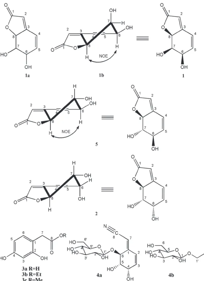
A new lactone, 7-epi-grifonilide (1), and six known compounds, 2, 3a – 3c, 4a and 4b, were isolated from the leaves of Bauhinia pentandra (Fabaceae). The structures elucidation of 1 and 2 were based on detailed 2D NMR techniques and spectral comparison with related compounds, leading to complete assignment of the 1H and 13C NMR spectra.
Key words:Bauhinia pentandra, Fabaceae, cyanoglucoside, lactones, RMN.
Correspondence to: Francisco José Queiroz Monte E-mail: [email protected]
INTRODUCTION
In the course of our continuing search for natural products from medicinal plants, we have
investigated the leaves of Bauhinia pentandra
(Bong.) D. Dietr. B. pentandra is widely distribute
in Northeast Brazil where is known as “mororó”
and used in folk medicine. The genus Bauhinia
contains many species of plants with medicinal
interest (Silva and Cechinel Filho 2002). It consists of about 300 species, distributed in most tropical
countries, including Africa, Asia and America
(Cechinel Filho 2009). Previous phytochemical studies with plants from Bauhinia genus report
the presence of lactones, lavonoids, terpenoids,
steroids, triterpenes, tannins, quinones and alkaloids (Silva and Cechinel Filho 2002, Maia Neto et al. 2008), while studies from B. pentandra reported the chemical composition of the essential and fatty oils (Duarte-Almeida et al. 2004, Almeida et al. 2015),
as well as the isolation of lavonoids (Salatino et
al. 1999) and cyanoglucosides (Silva et al. 2013). In this paper, we report the isolation and structure elucidation of lactones (1 and 2), phenylacetic
Compound 1 is new, and 2, 3a, 3b, 3c, 4a e 4b
has not been reported previously in the Bauhinia
genus. The structural assignments and relative stereochemistry of 1and 2 were based on detailed
2D NMR spectroscopy, while 3a, 3b, 3c, 4a e 4b
were identiied by comparison with NMR spectral
data from literature (Wu et al. 1979, Nahar et al. 2005, Kortesniemi et al. 2014).
MATERIALS AND METHODS
GENERAL EXPERIMENTAL PROCEDURES
Optical rotations were measured on a Jasco Polar-imeter, Model P-2000, operating at a wavelength of 589 nm and 20°C.NMR spectra were recorded in CD3OD solutions on Bruker DPX-500 spectrom-eter (equipped with the standard Bruker software)
with chemical shifts reported in δ units (ppm) rela
-tive to TMS as internal standard. HRMS were per-formed on Bruker (model mocrOTOF) mass spec-trometer equipped with a ESI ion source. HPLC analysis was performed on Shimadzu chromato-graph, model LC-20AT, equipped with two high pressure pumps and UV-Vis detector, model SPD-M20A. Semi-preparative reversed-phase chroma-tography was carried out on a Phenomenex column (C18, 250 x 10 mm, 10 uM). The solvents used were deionized water and methanol with spectral grade; detections in the range of 200 to 400 nm. CC was performed on Merck Silica gel 60 F254, and TLC on Merck Silica gel 60 plates (0.25 mm).
PLANT MATERIAL
Bauhinia pentandra leaves were collected in Medicinal Plant Garden, Universidade Federal do Ceará (UFC), Ceará, Brazil. A voucher specimen
No. 53444, idenied by Dr. Edson Paula Nunes,
was deposited at the Herbarium Prisco Bezerra, Departamento de Biologia (UFC).
EXTRACTION AND ISOLATION
The dried and pulverized leaves of B. pentandra
(507.0 g) were extracted at temperature room with
EtOH which gave a residue (80.7 g) after solvent evaporation under reduced pressure. A part of this extract (20.0 g) was prefractionated by CC on Silica gel under reduced pressure eluted successively with hexane, CH2Cl2, EtOAc and MeOH). The fraction eluted with EtOAc (1.97 g) was further chromatographed over a column of Silica gel with solvents of increasing polarity from hexane through EtOAc to MeOH. A total of 125 fractions were collected and combined based on their TLC patterns; 200 µL from combined fractions (F 60-69, 93.6 mg), was further subjected to reversed-phase chromatography, using H2O-MeOH (9 : 1) as mobile
phase in a isocratic system, low rate 4.0 mL/min
and monitored by HPLC semi-preparative column to yield 1 (15.5 mg, t
R 7.15 min) and 2 (10.7 mg,
tR 7.67 mn). Another portion of the EtOH extract (12.7 g) was prefractionated by CC on Silica gel eluted successively with hexane, CH2Cl2, EtOAc and MeOH. F-EtOAc fraction after evaporation of
the solvent aforded 448.0 mg. Part of this fraction
yield a resin dark brown consisting of the mixture of compounds 4a and 4b (9.3 mg, tR 16 min), soluble in MeOH.
RESULTS AND DISCUSSION
Compound 1 was obtained as an orange resin
soluble in methanol,
[ ]
α 22D - 3.11 (c 0.14, MeOH). The FT-IR spectrum showed absorption bandscharacteristic for OH (νmax 3419 cm-1), ester CO
(νmax 1733 cm-1) and C = C (ν
max 1633 cm -1). In
the HREIMS spectrum (positive mode), peaks at m/z 169.0498 ([M + H]+, calc 169.0501) and
m/z 191.0315 ([M + Na]+, calc 191.0320) were consistent with a molecular formula C8H8O4 for 1.
Comparative analysis of the {1H}- and DEPT 135o NMR spectra allowed to recognize signals corresponding six methines (three sp2 at δ
C 113.3,
121.0 and 143.0 and three sp3 oxygenated at δC 70.1,
72.8 and 83.5) and two no hydrogenated (oleinic
at δC 163.3 and carbonyl at δC 176.7) carbons atoms (Table I). The signals at δC 176.7 and 83.5
were used to characterize the presence of a
ive-membered lactone ring (Silverstein and Webster 2000). These assignments were consistent with the HREIMS empirical formula, supporting the
presence of two hydroxyl groups and ive degrees
of unsaturation/ring. Thus, NMR data suggested 1 as a bicyclic molecule.
The 1H NMR spectrum of compound 1 showed
virtually two sets of signals: δH 6.63 (dd, 10.0 and 2.5 Hz), 6.16 (d, 10.0 Hz) and 5.89 (s,) attributed
to the oleinic hydrogen atoms H-4, H-5 and H-2,
respectively, and δH 5.11 (br s, H-8), 4.52 (br s, H-6) and 4.49 (br s, H-7) corresponding to the methinic hydrogens attached to oxygenated carbons. The 2D COSY NMR experiment revealed spins interactions systems attributed to the presence of HO6
CH-5CH=4CH- and –O-8CH-7CH(OH)-6CHOH in 1, that were conirmed by 2D HSQC NMR spectral
data revealing heteronuclear correlation via one (1JCH) involving the CH-6 (δC 70.1/δH 4.52), CH-5
(δC 143.0/δH 6.16), CH-4 (δC 121.0/ δH 6.63), CH-8 (δC 83.5/ δH 5.11) and CH-7 (δC 72.8/ δH 4.49), along with additional transversal peak corresponding to CH-2 (δC 113.3/δH 5.89), shown in Table I. Thus, the chemical shifts δH 6.63, 6.16 and 5.89 indicate that the double bonds are conjugated with each other, and these in turn with the carbonyl group
(δC 176.7). The broad singlet signals at δH 5.11, 4.52 and 4.49 were correlated to the carbon atoms signals at δC 83.5, 70.1 and 72.8, respectively, in the HSQC NMR spectrum.
TABLE I 1
H- and 13
C NMR chemical shifts assignments 1 and 5 (CD3OD, 500 MHz). Chemical shifts in δ (ppm), coupling
constants (J) in Hz (in parenthesis)a.
Position
1 5 (Grifonilide)
δC δH (J in Hz) HMBC (JH-C) δC δH (J in Hz)
1 176.7 - 175.8
-2 113.3 5.89 (s) C1, C3, C4, C8 112.5 5.89 (d, ~2.0)
3 163.3 - 164.7
-4 121.0 6.63 (dd, 10.0, 2.5) C3, C6, C8 120.6 6.62 (dd, 9.5, 2.5)
5 143.0 6.16 (d, 10.0) C3, C7 144.2 6.27 (dd, 9.5, 1.9)
6 70.1 4.52 (br s) 73.6 4.33 (dt, 7.6, 2.5, 1.9)
7 72.8 4.49 (br s) C3, C5, C8 80.0 3.53 (dd, 10.8, 7.6)
8 83.5 5.11 (br s) C2, C3 85.1 4.90 (dd, 10.8, 1.9)
a
Number of hydrogens bound to carbon atoms deduced by comparative analysis of the {1H}- and DEPT- 13CNMR spectra. Chemical shifts and coupling constants (J) obtained from 1D 1
H NMR spectrum. The HSQC, HMBC and 1 H-1
The data were used to postulate the constitutional structure 1a, which was conirmed by correlations exhibited by the HMBC NMR
spectrum. Importantly, the oleinic hydrogen H-2
(δH 5.89) showed correlation with C-1 (δC 176.7,
2J
CH), C-3 (δC 163.3, 2J
CH) and CH-8 (δC 83.5, 3J
CH)
and the H-8 (δH 5.11) with δC C-3 (δC 163.3, 2JCH) and CH-2 (δC 113.3, 3J
CH), in agreement with the
presence of an α,β-unsaturated lactone. Further, the olefinic hydrogen H-4 (δH 6.63) revealed correlation with the carbons C-3 (δC 163.3, 2JCH) and CH-8 (δC 83.5, 3J
CH), while the other oleinic
H-5 (δH 6.16) showed heteronuclear coupling with C-3 (δC 163.3, 3JCH). The latter correlations, in turn, are in agreement with a lactone ring joint with a cyclohexene ring (1a).
The relative stereochemistry of 1a was determined from the coupling constants of relevant hydrogens and from the observed 1H-1H NOESY.
Thus, the signals corresponding to hydrogens H-6, H-7 and H-8 as broad singlets are consistent with the absence of diaxial interactions (1b). In agreement with these observations, the NOESY spectrum of 1
showed cross-peak assigned to dipolar interaction (spatial proximity) between H-6 and H-8 (1b).
These evidences, as well as the comparison with literature data reported for griffonilide [5, (6R,7S,7aS )-7,7a-dihydro-6,7-dihydroxybenzofu-ran-2(6H)-one] (Wu et al. 1979) led to elucidation of 1 [(6R,7R,7aS )-7,7a-dihydro-6,7-dihydroxyben-zofuran-2(6H)-one], a new 7-epi-grifonilide (Fig
-ure 1), which is being reported for the irst time in
the literature.
Complete and unambiguous 1H- and 13C-NMR
chemical shifts assignments of 1 based on 1H – 1H
COSY, 1H – 13C COSY-nJ
CH (n=1, HSQC; n=2 and
3, HMBC), and 1H – 1H NOESY were summarized in Table I.
Compound 2 was obtained as an orange resin
with solubility in methanol,
[ ]
α22D + 129.31 (c 0.1, MeOH). The FT-IR spectrum of 2 was very similarto 1, showing absorption bands characteristic for
OH (νmax 3395 cm-1), ester CO (νmax 1737 cm-1) and
C = C (νmax 1633 cm-1). In the HREIMS spectrum
(positive mode), peaks at m/z 169.0504 ([M + H]+, calc 169.0501) and m/z 191.0320 ([M + Na]+,
calc 191.0320) were consistent with a molecular formula C8H8O4 for 2, a isomer of 1.
Comparative analysis of the {1H}- and DEPT 135o NMR spectra of 2 allowed to recognize
signals corresponding six methines [three sp2 at δC
113.4 (CH-2), 123.4 (CH-4) and 139.8 (CH-5) and three sp3 oxygenated at δC 68.7 6), 74.5 (CH-7) and 83.5 (CH-8)] and two sp2 no hydrogenated
at δC 164.5 (C-3) and at δC 176.7 (C-1, conjugated carbonyl) carbon atoms (Table II). Thus, analysis of the 1H and 13C NMR spectra data of 2 conirmed
that it was closely relate to 1 (and to 5). In fact, the
1
H and 13C-NMR feature were virtually identical except for the small changes in chemical shifts (1H and 13C) and more pronounced changes in the
multiplicity and the coupling constants in the case of hydrogen atoms (Table II).
The main diference in the 1
H NMR spectrum of 1 and 2 consisted in the signal for H-7, which
had changed from a broad singlet at δH 4.49 of 1 to
a double doublets at δH 3.64 (J = 10.0 and 4.2 Hz) of 2, indicating diaxial (J = 10.0 Hz) interaction
with H-8. The 2D COSY NMR experiment also revealed spin systems compatible with the presence of the HO6CH-5CH=4CH- and –O-8CH-7
CH(OH)-6
CHOH in 2, that were conirmed by 2D HSQC NMR experiment by cross peaks corresponding to heteronuclear interactions involving CH-6 (δC
68.7/δH 4.40), CH-5 (δC 139.8/δH 6.42) and CH-4 (δC 123.4/δH 6.70) and CH-8 (δC 83.5/δH 5.23) and CH-7 (δC 74.5/δH 3.64), summarized in Table II.
The relative stereochemistry of 2 was deduced
from their mutual coupling constants of relevant hydrogens and from dipolar interaction revealed by 1H-1H NOESY. Unlike 1 was not observed
sig-nal nOe between H-6 (δH 4.40) and H-8 (δH 5.23) hydrogen of 2. The diaxial spin-spin interaction
duced from the large coupling constant (J = 10.4 Hz) covering these hydrogen atoms. The values of the coupling constants (J) observed in the signal at
δH 4.40 (H-6, dd, J6eq-5 = 5.3 and J6eq-7 = 4.2 Hz) sug-gested absence of diaxial coupling, as expected for the proposed stereochemistry.
These evidences, as well as the comparison with literature data reported for griffonilide [5, (6R,7S,7aS )-7,7a-dihydro-6,7-dihydroxybenzo-furan-2(6H)-one] (Wu et al. 1979) and dasycar-ponilide (Wu et al. 1979), led to elucidation of 2 [(6S,7S,7aS)-7,7a-dihydro-6,7-dihydroxybenzofu-ran-2(6H)-one], known as dasycarponilide (Figure
1), being irst reported in genus Bauhinia.
Complete and unambiguous 1H- and 13C-NMR
chemical shifts based on assignment of 2 based on
1
H – 1H COSY, 1H – 13C COSY-nJCH (n=1, HSQC; n=2 and 3, HMBC), and 1H – 1H NOESY were summarized in Table II.
The compounds 3 and 4 (Figure 1) were
identi-ied by analysis of the spectral data obtained main
by 1D and 2D NMR and mass spectra involving comparison with values described in the literature to 3 and 4 (Kortesnimi etal. 2014, Wuet al. 1979).
ACKNOWLEDGMENTS
The authors thank the institutions fostering research Conselho Nacional de Desenvolvimento
Científico e Tecnológico (CNPq), Coordenação de Aperfeiçoamento de Pessoal de Nível Superior (CAPES), Programa de Apoio aos Núcleos de Excelência (PRONEX) and Fundação Cearense de Apoio ao Desenvolvimento Científico e
Tecnológico (FUNCAP) for the inancial aid and
granted scholarship.
REFERENCES
ALMEIDA MCS, SOUZA LGS, FERREIRA DA, MONTE FJQ, BRAZ-FILHO R AND LEMOS TLG. 2015. Chemi-cal composition of the essential oil and fixed oil Bauhinia pentandra (Bong.) D. Dietr. Phcog Mag 11: 362-364. CECHINEL FILHO V. 2009. Chemical composition and
biological potential of plants from the genus Bauhinia. Phytother Res 23: 1347-1354.
DUARTE-ALMEIDA JM, NEGRI G AND SALATINO A. 2004. Volatile oils in leaves of Bauhinia (Fabaceae Caesalpinioideae). Biochem Syst Ecol 32: 747-753. KORTESNIEMI M, SINKKONEN J, YANG B AND
KALLIO H. 2014. 1
H NMR spectroscopy reveals the effect of genotype and growth conditions on composition of sea buckthorn (Hippophaë rhamnoides L.) berries. Food Chem 147: 138-146.
MAIA NETO M, ANDRADE NETO M, BRAZ-FILHO R, LIMA MAS AND SILVEIRA ER. 2008. Flavonoids and alkaloids from leaves of Bauhinia ungulata L. Biochem Syst Ecol 36: 227-229.
NAHAR L, RUSSELL WR, MIDDLETON M, SHOEB M AND SARKER SD. 2005. Antioxidant phenylacetic acid derivatives from the seeds of Ilex aquifolium. Acta Pharm 55: 187-193.
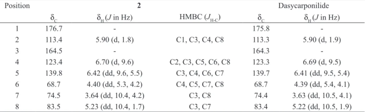
TABLE II 1
H- and 13
C NMR chemical shifts assignments 2 and Dasycarponilide (CD3OD, 500 MHz). Chemical shifts in δ
(ppm), coupling constants (J) in Hz (in parenthesis)a.
Position 2 Dasycarponilide
δC δH (J in Hz) HMBC (JH-C) δC δH (J in Hz)
1 176.7 - 175.8
-2 113.4 5.90 (d, 1.8) C1, C3, C4, C8 113.3 5.90 (d, 1.9)
3 164.5 - 164.3
-4 123.4 6.70 (d, 9.6) C2, C3, C5, C6, C8 123.3 6.69 (d, 9.5) 5 139.8 6.42 (dd, 9.6, 5.5) C3, C4, C6, C7 139.7 6.41 (dd, 9.5, 5.4) 6 68.7 4.40 (dd, 5.3, 4.2) C4, C5, C7, C8 68.7 4.39 (dd, 5.4, 4.1) 7 74.5 3.64 (dd, 10.4, 4.2) C3, C8 74.4 3.63 (dd, 10.5, 4.1) 8 83.5 5.23 (dd, 10.4, 1.7) C3, C7 83.4 5.22 (dd, 10.5, 1.9) a
Number of hydrogens bound to carbon atoms deduced by comparative analysis of the {1
H}-and DEPT-13 C NMR spectra. Chemical shifts and coupling constants (J) obtained from 1D 1H NMR spectrum. The HSQC, HMBC and 1
H-1
H-COSY spectra were also used to 1 H and 13
SALATINO A, BLATT CTT, SANTOS DYAC AND VAZ AMSF. 1999. Foliar flavonoids of nine species of Bauhinia. Rev Bras Bot 22: 17-20.
SILVA KL AND CECHINEL FILHO V. 2002. Plantas do gê-nero Bauhinia: composição química e potencial farmaco-lógico. Quím Nova 25: 449-454.
SILVA TMS, LINS ACS, SARMENTO-FILHA MJ, RAMOS CS, AGRA MF AND CAMARA CA. 2013. Riachin,
a new cyanoglucoside from Bauhinia pentandra and its antioxidant activity. Chem Nat Compd 49: 685-690. SILVERSTEIN RM AND WEBSTER FX. 2000. Identificação
espectrométrica de compostos orgânicos, 6ª ed., Rio de Janeiro: Livros Técnicos e Científicos, 460 p.