Dental anomalies in an orthodontic patient population
with maxillary lateral incisor agenesis
Mehmet Citak1,Elif Bahar Cakici2, Yasin Atakan Benkli3, Fatih Cakici2, Bircan Bektas4, Suleyman Kutalmış Buyuk3
1 Research Assistant, Department of Endodontics, Faculty of Dentistry, Ordu
University, Ordu, Turkey.
2 Assistant Professor, Department of Endodontics, Faculty of Dentistry, Ordu
University, Ordu, Turkey.
3 Assistant Professor, Department of Orthodontics, Faculty of Dentistry, Ordu
University, Ordu, Turkey.
4 Research Assistant, Department of Orthodontics, Faculty of Dentistry, Ordu
University, Ordu, Turkey.
Introduction: The purpose of this study was to evaluate the prevalence of dental anomalies in a subpopulation of orthodontic patients with agenesis of maxillary lateral incisors (MLI). Methods: The material of the present study included the records of the 1964 orthodontic patients. Panoramic radiographs and dental casts were used to analyze other associated eight dental anomalies, including agenesis of other teeth, dens invaginatus, dens evaginatus, peg shaped MLI, taurodontism, pulp stone, root dilaceration and maxillary canine impaction. Results: Out of the 1964 patients examined, 90 were found to have agenesis of MLI, representing a prevalence of 4.6%. The most commonly found associated-anomalies were agenesis of other teeth (23.3%), peg-shaped MLIs (15.6%), taurodontism (42.2%), and dilacerated teeth (18.9%). Conclusion: Permanent tooth agenesis, taurodontism, peg-shaped maxillary lateral incisor, and root dilacerations are frequently associated with maxillary lateral incisor agenesis.
Keywords: Dental anomalies. Hypodontia. Panoramic radiograph.
DOI: http://dx.doi.org/10.1590/2177-6709.21.6.098-102.oar
How to cite this article: Citak M, Cakici EB, Benkli YA, Cakici F, Bektas B, Buyuk SK. Dental anomalies in an orthodontic patient population with maxillary lateral incisor agenesis. Dental Press J Orthod. 2016 Nov-Dec;21(6):98-102. DOI: http://dx.doi.org/10.1590/2177-6709.21.6.098-102.oar
Submitted: March 18, 2016 - Revised and accepted: July 20, 2016
» The authors report no commercial, proprietary or financial interest in the products or companies described in this article.
» Patients displayed in this article previously approved the use of their facial and in-traoral photographs.
Contact address: Suleyman Kutalmış Buyuk E-mail: [email protected]
Objetivo: o objetivo do presente estudo foi avaliar a prevalência de anomalias dentárias em uma subpopulação de pacientes ortodônticos com agenesia de incisivos laterais superiores (ILS). Material e Métodos: o material do presente estudo incluiu os registros de 1964 pacientes ortodônticos. Radiografias panorâmicas e modelos de estudo foram usados para analisar outras anomalias dentárias associadas, incluindo a agenesia de outros dentes, dens invaginatus, dens evaginatus, ILS conoides, taurodon-tismo, calcificação pulpar, dilaceração radicular e impacção do canino superior. Resultados: dos 1964 pacientes examinados, constatou-se que 90 tinham agenesia do ILS, o que representa uma prevalência de 4,6%. As anomalias associadas mais comu-mente encontradas foram a agenesia de outros dentes (23,3%), ILS conoides (15,6%), taurodontismo (42,2%) e dentes com dilaceração (18,9%). Conclusão: a agenesia de dentes permanentes, o taurodontismo, incisivos laterais superiores conoides e dilaceração radicular estão frequentemente associados à agenesia de incisivos laterais superiores.
INTRODUCTION
Dental anomalies are typically caused by either
ge-netic or environmental stimuli.1,2 Mutations in AXIN2,
PAX9 and MSX1 have been determined in families
with dental agenesis.3,4 The most frequently observed
dental agenesis in children is deined as the absence of
one or more primary/permanent teeth.5 Data for
con-genital tooth agenesis prevalence vary between 0.3 and
11.3%6,7 for both males and females. However, the
prevalence of congenital tooth agenesis was shown to
be higher in females than in males, in some reports.5,8,9
Ater third molars, maxillary lateral incisors (MLI) are
the teeth that are the most frequently missing.5,10 Agenesis
of MLI has been documented for its higher prevalence than
of other permanent teeth.11 A correlation between MLI
agenesis and palatally displaced canines,12 tooth
transpo-sitions,13 and premolar rotations14 has also been reported.
However, reports on the prevalence of dental anomalies in a large MLI agenesis patient cohort have not been deter-mined. The purpose of the current study was to investigate the prevalence of MLI agenesis and other dental anomalies in an orthodontic subpopulation in Turkey.
MATERIAL AND METHODS
Panoramic radiographs of 1964 patients (1174 fe-males, 790 males) of the Department of Orthodontics of Ordu University, Turkey, between January 2013 and September 2015, were retrospectively analyzed. Patients, aged 12 to 25 years, with unilateral or bilat-eral agenesis of MLI and panoramic radiograph were included in the study. Patients with incomplete re-cords, permanent tooth extraction, and/or poor-quality
panoramic radiographs were excluded from the study. Care was used to ensure that all radiographs were taken by the same technician operating the same panoramic roentgen unit device (Kodak Cephalostat, Rochester NY, USA). In order to eliminate inter-examiner dif-ferences, all records were examined by one observer. All radiographs were evaluated by an orthodontist with more than 15 years of experience.
The following anomalies were determined in this study (Fig 1):
1) Agenesis: congenital developmental loss of one or more permanent teeth.
2) Dens invaginatus: caused by the invagination of
enamel into the dental papilla before the
mineraliza-tion phase.15
3) Dens evaginatus: malformation characterized by an
accessory cusp, composed of normal enamel and
den-tine, with or without pulp tissue.16
4) Microdontia (peg-shaped teeth): teeth that are substantially smaller than the average normal size. Mi-crodontia also refers to a tooth that does not ill its space in the dental arch or appears small due to absence of
ex-pected shape.17
5) Taurodontism: vertically extended, extremely oversized pulp cavities that are apically displaced at the
pulpal loor.18
6) Pulp stone: calciied masses on the pulp of healthy, diseased, and even unerupted teeth freely attached or embedded into the coronal rather than the root portion
of the pulp organ.19
7) Dilaceration: deviation in the linear relationship
of a crown of a tooth to its root.20
Figure 1 - A) Root dilacerations. B) Pulp stone. C) Taurodontism. D)Dens evaginatus. E)Dens invaginatus.
B D E
8) Impaction: a tooth that is predicted to remain un-erupted because of a physical barrier or delection along
its eruption path.21
Statistical analysis
Statistics were calculated with SPSS 15.0 statisti-cal sotware (SPSS Inc, Chicago, IL, USA). Anomaly prevalence was measured with respect to sex, side and dental location. Chi-square analysis and MLI prevalence were compared to previously published reports from the
Turkish population.22-28 A p-value of less than 0.05 was
considered signiicant.
To examine errors associated with digitizing and measurements, 10% of panoramic images were se-lected randomly and all dental anomalies were evalu-ated by the same author four weeks after the first ex-amination. Kappa coefficients were used to calculate the reliability of each dental anomaly determination from the two evaluation periods.
RESULTS
Kappa score of each dental anomaly was 1.00. This score indicated good agreement with the irst and second
evaluations and was observed for each dental anomaly. Out of the 1964 subjects (1174 females, 790 males) evalu-ated, 90 (62 females, 28 males) were determined to have MLI agenesis (prevalence = 4.58%; being 5.3% for females and 3.5% males. Diference between males and females
was statistically not signiicant [X2 = 3.26; p = 0.071]).
Bi-lateral MLI agenesis was found in 62 subjects (68.9%) and unilateral agenesis in 28 patients (31.1%).
The investigated dental anomalies in MLI
agen-esis patients were: dens invaginatus, dens evaginatus,
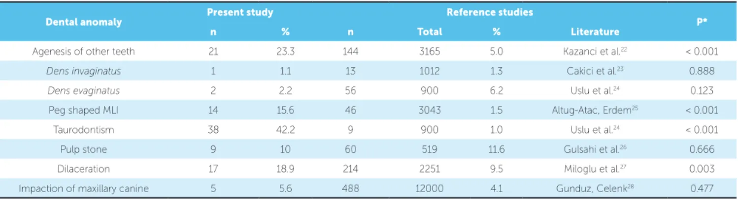
peg-shaped MLI, taurodontism, pulp stone, root dilacerations, impaction of maxillary canine, and missing teeth other than third molars (Table 1). The prevalence of MLI agenesis-associated dental-anom-alies was referenced to previous work, for consis-tency (Table 2). The prevalence of agenesis of other
teeth (p < 0.001), peg-shaped MLIs (p < 0.001),
taur-odontism (p < 0.001), and dilacerated teeth (p < 0.01)
were greater in our sample compared to the other studies. No statistical significant difference was shown
for the prevalence of dens invaginatus (p = 0.888), dens
evaginatus (p = 0.123), pulp stone (p = 0.666) and
im-paction of maxillary canines (p = 0.477).
Dental anomaly Male / Female Unilateral / Bilateral Total (%)
Agenesis of other teeth 5 / 16 7 / 14 21 (23.3)
Dens invaginatus 1 / 0 1 / 0 1 (1.1)
Dens evaginatus 1 / 1 1 / 1 2 (2.2)
Peg shaped MLI 7 / 7 14 / 0 14 (15.6)
Taurodontism 9 / 29 1 / 37 38 (42.2)
Pulp stone 0 / 9 5 / 4 9 (10)
Dilaceration 4 / 13 5 / 12 17 (18.9)
Impaction of maxillary canine 2 / 3 3 / 2 5 (5.6)
Table 1 - Distribution of MLI agenesis-associated dental-anomalies.
Table 2 - Comparison of the frequencies of dental anomalies subjects with maxillary lateral incisor agenesis and previous studies.
n = number of subjects, MLI; Maxillary lateral incisor, * p indicates results of chi-square test.
Dental anomaly Present study Reference studies P*
n % n Total % Literature
Agenesis of other teeth 21 23.3 144 3165 5.0 Kazanci et al.22 < 0.001
Dens invaginatus 1 1.1 13 1012 1.3 Cakici et al.23 0.888
Dens evaginatus 2 2.2 56 900 6.2 Uslu et al.24 0.123
Peg shaped MLI 14 15.6 46 3043 1.5 Altug-Atac, Erdem25 < 0.001
Taurodontism 38 42.2 9 900 1.0 Uslu et al.24 < 0.001
Pulp stone 9 10 60 519 11.6 Gulsahi et al.26 0.666
Dilaceration 17 18.9 214 2251 9.5 Miloglu et al.27 0.003
DISCUSSION
The prevalence of dental anomalies is variable among diferent populations. The aim of the present study was to determine the prevalence of MLI agenesis-associat-ed dental anomalies in orthodontic Turkish patients. We found that about 4.58% of patients had one or both maxillary incisors missing. These results are consistent with that reported by others, which ranges from 0.3%
and 11.3%.9,10,25 The prevalence of MLI agenesis varies
considerably between studies.7,29-32 Horowitz33 showed
a prevalence of 1.11% in an adolescent population (n = 1000; ages ranging from 7 to 16 years). Celikoglu
et al7 reported a prevalence of 2.4% from 3872 East
Ana-tolian adolescent patients in Turkey. The diferences may be related to sample size or selection, but may be difer-ent due to regional ethnic population, genetic variability, and/or environmental factors.
There was no statistically signiicant correlation between sex and MLI agenesis. Interestingly, we had more female subjects in our cohort. Some reports have
shown insigniicant diferences,34,35 while others have
determined signiicant sex-related changes.36,37
Previous studies24,38-41 have reported that tooth
agen-esis can be associated with other dental malformations, such as taurodontism, transposition, microdontia, ecto-pic eruption, supernumerary tooth or peg-shaped MLI.
Most of the papers8,42,43 published on MLI agenesis
demonstrated a reduction in crown size or a peg-shaped form of the contralateral MLI. MLI agenesis was detect-ed more commonly in females than males.
When we compared the prevalence rates of MLI
agen-esis-associated dental anomalies and reference values,22-28 it
was determined that the prevalence rates were signiicant-ly augmented for taurodontism, agenesis of other teeth, and peg-shaped MLIs. There have been only two studies
that have compared their work with7,38 reference values.
However, taurodontism, pulp stone, dens invaginatus, dens
evaginatus, and impaction of the maxillary canine were not
assessed in those studies. In this respect, our study is the irst one to show the diferent associated dental anomalies between subjects with MLI agenesis.
The most common MLI agenesis-associated dental anomaly was taurodontism; in our study, with a
preva-lence of 42.2%. Uslu et al24 showed 1% of taurodontism
prevalence in 900 orthodontic patients. Discrepancy in the results may be related to the location variations, and our taurodontism detection method. The diference
might arise from racial diferences or diferences in di-agnostic criteria.
Out of the 28 patients who had unilateral absence, 14 (50.0%) were found to have a peg-shaped lateral incisor
on the other side. Altug-Atac and Erdem25 reported that
1.51% of patients had peg-shaped MLI in an orthodontic
patient population. However, Albashaireh and Khaider42
demonstrated that peg-shaped and reduced size maxillary lateral incisors were found in 2.3% and 2.9% of patients, respectively. Similar to the prevalence of our results,
Albashaireh and Khader42 showed 50% microdontia or
peg-shaped MLIs on the other side in individuals with unilateral MLI agenesis. In this study, the prevalence of other teeth agenesis (23.3%) was very high, compared to
the reports by Celikoglu et al.7 The higher rate reported
may be attributed to orthodontic malocclusions, which
supports the indings by Garib et al.38
The prevalence of root dilacerations in our study (18.9%) was greater than that reported of a reference
study from the general population (9.5%).27 Diagnosing
dilacerations is mostly imperative for root canal
treat-ment, tooth extraction, and Orthodontics.34 The other
associated anomalies (dens invaginatus, dens evaginatus, pulp
stone and impaction of maxillary canine prevalence) were
similarly found with reference studies.23,24,26,28
Associations between tooth anomalies are clini-cally relevant, and early diagnosis may be helpful to
re-duce risk.44 Therefore, diagnosis and treatment options
should be precisely made. We found a higher prevalence of associated dental anomalies in MLI agenesis patients in Turkish orthodontic population. However, the orth-odontic literature shows diferent prevalence rates of
dental anomalies from the general population.7,9,22,25,34
This is mostly likely due to the greater variability of ra-cial factors, environmental stimuli and genetics.
CONCLUSIONS
1. Akcam MO, Evirgen S, Uslu O, Memikoğlu UT. Dental anomalies in individuals with cleft lip and/or palate. Eur J Orthod. 2010 Apr;32(2):207-13. 2. Kotsomitis N, Dunne MP, Freer TJ. A genetic aetiology for some common
dental anomalies: a pilot twin study. Aust Orthod J. 1996 Oct;14(3):172-8. 3. Nieminen P. Genetic basis of tooth agenesis. J Exp Zool B Mol Dev Evol.
2009 Jun 15;312B(4):320-42.
4. De Coster PJ, Marks LA, Martens LC, Huysseune A. Dental agenesis: genetic and clinical perspectives. J Oral Pathol Med. 2009 Jan;38(1):1-17. 5. Celikoglu M, Kazanci F, Miloglu O, Oztek O, Kamak H, Ceylan I. Frequency
and characteristics of tooth agenesis among an orthodontic patient population. Med Oral Patol Oral Cir Bucal. 2010 Sept 1;15(5):e797-801. 6. O’Dowling IB, McNamara TG. Congenital absence of permanent teeth
among Irish school-children. J Ir Dent Assoc. 1990;36(4):136-8. 7. Celikoglu M, Kamak H, Yildirim H, Ceylan I. Investigation of the
maxillary lateral incisor agenesis and associated dental anomalies in an orthodontic patient population. Med Oral Patol Oral Cir Bucal. 2012 Nov 1;17(6):e1068-73.
8. Pinho T, Tavares P, Maciel P, Pollmann C. Developmental absence of maxillary lateral incisors in the Portuguese population. Eur J Orthod. 2005 Oct;27(5):443-9. Epub 2005 Aug 31.
9. Silva Meza R. Radiographic assessment of congenitally missing teeth in orthodontic patients. Int J Paediatr Dent. 2003 Mar;13(2):112-6. 10. Fekonja A. Hypodontia in orthodontically treated children. Eur J Orthod.
2005 Oct;27(5):457-60.
11. Polder BJ, Van’t Hof MA, Van der Linden FP, Kuijpers-Jagtman AM. A meta-analysis of the prevalence of dental agenesis of permanent teeth. Community Dent Oral Epidemiol. 2004 June;32(3):217-26.
12. Sacerdoti R, Baccetti T. Dentoskeletal features associated with unilateral or bilateral palatal displacement of maxillary canines. Angle Orthod. 2004 Dec;74(6):725-32.
13. Peck S, Peck L, Kataja M. Concomitant occurrence of canine malposition and tooth agenesis: evidence of orofacial genetic ields. Am J Orthod Dentofacial Orthop. 2002 Dec;122(6):657-60.
14. Baccetti T. Tooth rotation associated with aplasia of nonadjacent teeth. Angle Orthod. 1998 Oct;68(5):471-4.
15. Oehlers FA. Dens invaginatus (dilated composite odontome). I. Variations of the invagination process and associated anterior crown forms. Oral Surg Oral Med Oral Pathol. 1957 Nov;10(11):1204-18.
16. Levitan ME, Himel VT. Dens evaginatus: literature review, pathophysiology, and comprehensive treatment regimen. J Endod. 2006 Jan;32(1):1-9.
17. Kocabalkan E, Ozyemişci N. Restoration of severe hypodontia associated with microdontia by using an overdenture: a clinical report. Chin Med J (Engl). 2005 Feb 20;118(4):350-2.
18. Shifman A, Chanannel I. Prevalence of taurodontism found in radiographic dental examination of 1,200 young adult Israeli patients. Community Dent Oral Epidemiol. 1978 July;6(4):200-3.
19. Al-Hadi Hamasha A, Darwazeh A. Prevalence of pulp stones in Jordanian adults. Oral Surg Oral Med Oral Pathol Oral Radiol Endod. 1998 Dec;86(6):730-2. 20. Hamasha AA, Al-Khateeb T, Darwazeh A. Prevalence of dilaceration in
Jordanian adults. Int Endod J. 2002 Nov;35(11):910-2.
21. Thilander B, Jakobsson SO. Local factors in impaction of maxillary canines. Acta Odontol Scand. 1968 May;26(2):145-68.
22. Kazanci F, Celikoglu M, Miloglu O, Ceylan I, Kamak H. Frequency and distribution of developmental anomalies in the permanent teeth of a Turkish orthodontic patient population. J Dent Sci. 2011 June;6(2):82-9.
23. Cakici F, Celikoglu M, Arslan H, Topcuoglu HS, Erdogan AS. Assessment of
REFERENCES
the prevalence and characteristics of dens invaginatus in a sample of Turkish Anatolian population. Med Oral Patol Oral Cir Bucal. 2010 Nov 1;15(6):e855-8. 24. Uslu O, Akcam MO, Evirgen S, Cebeci I. Prevalence of dental anomalies in various
malocclusions. Am J Orthod Dentofacial Orthop. 2009 Mar;135(3):328-35. 25. Altug-Atac AT, Erdem D. Prevalence and distribution of dental anomalies in
orthodontic patients. Am J Orthod Dentofacial Orthop. 2007 Apr;131(4):510-4. 26. Gulsahi A, Cebeci AI, Ozden S. A radiographic assessment of the prevalence
of pulp stones in a group of Turkish dental patients. Int Endod J. 2009 Aug;42(8):735-9.
27. Miloglu O, Cakici F, Caglayan F, Yilmaz AB, Demirkaya F. The prevalence of root dilacerations in a Turkish population. Med Oral Patol Oral Cir Bucal. 2010 May 1;15(3):e441-4.
28. Gunduz K, Celenk P. The incidence of impacted transmigrant canines: a retrospective study. Oral Radiol. 2010 Dec;26(2):77-81.
29. Baccetti T. A controlled study of associated dental anomalies. Angle Orthod. 1998 June;68(3):267-74.
30. Johannsdottir B, Wisth PJ, Magnusson TE. Prevalence of malocclusion in 6-year-old Icelandic children. Acta Odontol Scand. 1997 Dec;55(6):398-402. 31. Muller TP, Hill IN, Peterson AC, Blayney JR. A survey of congenitally missing
permanent teeth. J Am Dent Assoc. 1970 July;81(1):101-7.
32. Rolling S. Hypodontia of permanent teeth in Danish schoolchildren. Scand J Dent Res. 1980 Oct;88(5):365-9.
33. Horowitz JM. Aplasia and malocclusion: a survey and appraisal. Am J Orthod. 1966 June;52(6):440-53.
34. Thongudomporn U, Freer TJ. Prevalence of dental anomalies in orthodontic patients. Aust Dent J. 1998 Dec;43(6):395-8.
35. Endo T, Ozoe R, Kubota M, Akiyama M, Shimooka S. A survey of hypodontia in Japanese orthodontic patients. Am J Orthod Dentofacial Orthop. 2006 Jan;129(1):29-35.
36. Basdra EK, Kiokpasoglou M, Stellzig A. The Class II Division 2 craniofacial type is associated with numerous congenital tooth anomalies. Eur J Orthod. 2000 Oct;22(5):529-35.
37. Bergström K. An orthopantomographic study of hypodontia, supernumeraries and other anomalies in school children between the ages of 8-9 years. An epidemiological study. Swed Dent. 1977;1(4):145-57.
38. Garib DG, Alencar BM, Lauris JR, Baccetti T. Agenesis of maxillary lateral incisors and associated dental anomalies. Am J Orthod Dentofacial Orthop. 2010 June;137(6):732.e1-6.
39. Gomes RR, da Fonseca JA, Paula LM, Faber J, Acevedo AC. Prevalence of hypodontia in orthodontic patients in Brasilia, Brazil. Eur J Orthod. 2010 June;32(3):302-6.
40. Shapira Y, Kuftinec MM. Maxillary tooth transpositions: characteristic features and accompanying dental anomalies. Am J Orthod Detofacial Orthop. 2001 Feb;119(2):127-34.
41. Zhu JF, Marcushamer M, King DL, Henry RJ. Supernumerary and congenitally absent teeth: a literature review. J Clin Pediatr Dent. 1996 Winter;20(2):87-95. 42. Albashaireh ZS, Khader YS. The prevalence and pattern of hypodontia of the permanent teeth and crown size and shape deformity afecting upper lateral incisors in a sample of Jordanian dental patients. Community Dent Health. 2006 Dec;23(4):239-43.
43. Woolf CM. Missing maxillary lateral incisors: a genetic study. Am J Hum Genet. 1971 May;23(3):289-96.
44. Apajalahti S, Holtta P, Turtola L, Pirinen S. Prevalence of short-root anomaly in healthy young adults. Acta Odontol Scand. 2002 Jan;60(1):56-9.
Author contributions
Conception/design of the study: SKB, YAB, FC. Acquisition, analysis or interpretation: YAB, BB,