Class II malocclusion
nonextraction treatment with growth control*
Zilda Lúcia Valentim Assunção1
How to cite this article: Assunção ZLV. Class II malocclusion
nonextraction treatment with growth control. Dental Press J Orthod. 2014 Nov-Dec;19(6):113-22: DOI: http://dx.doi.org/10.1590/2176-9451.19.6.113-122.bbo
Submitted: August 26, 2014 - Revised and accepted: September 12, 2014
» Patients displayed in this article previously approved the use of their facial and intraoral photographs.
Contact address: Zilda Lúcia Valentim Assunção
Rua Prof. Almeida Cousin, 125 – Ed. Enseada Trade Center, sl. 1302 Enseada do Suá – CEP 29.050-565 - Vitória / ES — Brazil E-mail: [email protected]
» The author reports no commercial, proprietary or financial interest in the prod-ucts or companies described in this article.
1 Specialist in Orthodontics and Facial Orthopedics, State University of Rio de
Janeiro (UERJ). Certified by the Brazilian Board of Orthodontics and Facial Orthopedics (BBO).
*Case report, DI 34, approved by the Brazilian Board of Orthodontics and Dentofacial Orthopedics (BBO).
INTRODUCTION
The present study reports the case of a male pa-tient referred to orthodontic treatment at the age of 11 years and 8 months old. His chief complaint was being “too toothy”. The patient had a nasal breathing pattern, with history of allergy, onychophagia and ad-enoidectomy at the age of four. He also had good oral hygiene and was monitored by a pediatric dentist every 6 months. He reported having sufered dental trauma at the age of eight, which resulted in minor fracture on the incisal edge of the let maxillary central incisor.
DIAGNOSIS
As shown in Figure 1, facial analysis revealed a convex profile, with a well-defined mentolabial sul-cus and everted lower lip (Ul line: 1 mm; Ll S-line: 2 mm). Labial seal was strained and, at rest, maxillary incisors were prominent. At smiling, the patient had a wide buccal corridor despite satisfacto-ry smile arc. As shown in Figures 1 and 2, the patient was in mixed dentition, with Class II molar relation-ship and maxillary incisors bucally tipped, which fea-tures Angle Class II, division 1 malocclusion.
The present study reports a case of Angle Class II malocclusion treatment of a male growing patient with 10-mm overjet, excessive overbite and transverse maxillary deficiency. The case was presented to the Brazilian Board of Or-thodontics and Dentofacial Orthopedics (BBO), with DI equal to or greater than 10, as a requirement for the title of certified by the BBO.
Keywords: Angle Class III malocclusion. Palatal expansion technique. Growth and development.
DOI: http://dx.doi.org/10.1590/2176-9451.19.6.113-122.bbo
O presente relato de caso aborda o tratamento de uma má oclusão de Classe II de Angle, em paciente do sexo mascu-lino, em fase de crescimento, com sobressaliência de 10mm, sobremordida exagerada e deficiência maxilar transversa. Esse caso foi apresentado à Diretoria do Board Brasileiro de Ortodontia e Ortopedia Facial (BBO), representando a categoria com índice de grau de complexidade (IGC) igual ou acima de dez pontos, como parte dos requisitos para obtenção do título de Diplomada pelo BBO.
Figure 1 - Initial facial and intraoral photographs.
He had narrow maxillary arch with high palate; and mandibular arch with accentuated curve of Spee. Maxillary and mandibular midlines were coincident with each other and with the facial midline; severe overbite and overjet were evinced in 10 mm.
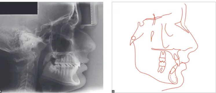
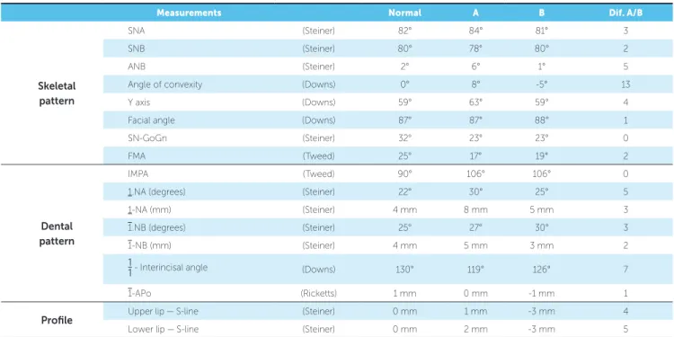
Periapical and panoramic radiographs (Figs 3 and 4) revealed the presence of all permanent teeth, in-cluding third molars; in addition to normal tooth as well as bone structures. Cephalometric examina-tion (Fig 5 and Tab 1) revealed that the patient had Class II skeletal pattern (ANB = 6o), protrusion of
the maxilla (SNA = 84o) and retrusion of the
man-dible (SNB = 78o) in relation to the base of the skull.
He had decreased mandibular plane with a tendency towards horizontal growth (SN-GoGn = 23o and
FMA = 17o). Maxillary incisors were severely
buc-cally tipped and protruded (1-NA = 30o and 8 mm),
while mandibular incisors were well positioned (1-NB = 27o and 5 mm).
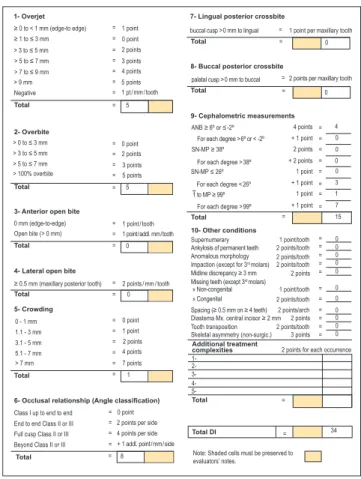
Discrepancy index (DI) was calculated and scored 34 points (Fig 6).
TREATMENT PLAN
Figure 2 - Initial casts.
Figure 5 - Initial lateral cephalogram (A) and cephalometric tracing (B).
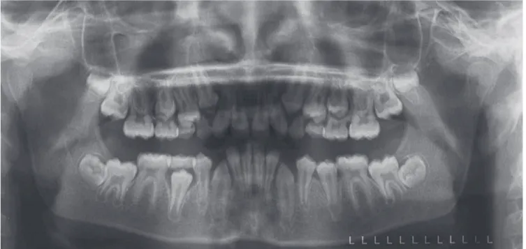
Figure 4 - Initial panoramic radiograph.
adenoidectomy, patient’s lateral cephalogram (Fig 5) revealed adenoid tissue on posterior pharyngeal wall. Thus, there was an urgent need to investigate wheth-er adenoid or othwheth-er type of tissue was obstructing patient’s upper airways and, therefore, causing na-sal breathing. Nevertheless, obstruction was absent; thereby rendering nasal breathing an acquired habit associated with hindered labial seal due to severe overjet. Should normal breathing not have been
restored after incisor retraction and achievement of potential passive labial seal, the patient would have been referred to speech therapy. Likewise, he would have been referred to psychological therapy to quit nail biting (onychophagia).
With a view to correcting skeletal and dental disharmonies, initial treatment planning consisted of using a Haas appliance to expand the maxilla in transverse direction. Additionally, with a view to
Figure 6 - Discrepancy index (DI) calculation.
correcting skeletal and dental disharmonies in the sagittal plane and to attain Class I molar relationship, treatment planning included the use of Kloehn-type headgear (cervical pull). Thus, treatment would take advantage of patient’s favorable growth pattern. Sub-sequently, to correct severe overbite, an interocclusal splint was installed. A preadjusted, fixed orthodontic Roth prescription appliance with 0.022 x 0.028-in slots was installed for alignment and leveling, incisors retraction and treatment finishing. After the active phase of treatment, a wraparound removable appli-ance, with anterior bite plate and mandibular inter-canine bar, was prescribed.
TREATMENT PROGRESS
Treatment began by adapting the orthodon-tic rings used to manufacture the Haas appliance.
Subsequently, the appliance was installed and the patient advised to turn the screw ¼ of a turn every 12 hours during 15 days in order to enhance maxil-lary shape. Once stability was attained, the expan-sion appliance remained in position for four months, acting as a retainer. During this period, the Kloehn-type headgear (cervical pull) was installed and used for 16 to 18 hours a day. After removing the expan-sion appliance, an acrylic plate was installed with a view to aiding severe overbite correction. The pa-tient and his family were informed about the need for compliance, particularly with regard to the headgear and the acrylic plate, necessary to achieve treatment objectives.
During the same phase of treatment, preadjusted brackets (Roth prescription, 0.022 x 0.028-in slots) were bonded to mandibular first molars and incisors for intrusion by means of Ricketts1 0.017 x 0.025-in
stainless steel utility arch. After intrusion and with premolars, maxillary canines and second molars erupted; maxillary and mandibular teeth were all bonded. Alignment and leveling were attained by means of stainless steel 0.016, 0.018 and 0.020-in archwire with mild step down bends at the region of mandibular incisors aimed at remaining intruded. Importantly, since treatment onset, both maxillary and mandibular archwires were often used in coor-dination. In the maxillary arch, retraction of incisors was carried out by means of a stainless steel 0.019 x 0.026-in archwire with a bull loop placed between lateral incisors and canines and used for space closure. After closing existing spaces, the finishing phase was carried out in both arches with the use of stainless steel 0.018 x 0.025-in straight archwires of individual shape, torque and coordination.
After confirming that all treatment objectives had been achieved, both maxillary and mandibular fixed appliances were removed and the retention phase started. To this end, a wraparound removable splint consisting of an stainless steel 0.032-in arch-wire and an anterior bite plate was installed in the maxillary arch with a view to preventing overbite re-lapse. As for the mandibular arch, a 0.032-in stain-less steel wire intercanine bar was installed. Impor-tantly, the patient proved highly compliant during the active phase of treatment as well as during the retention phase.
1- Overjet 7- Lingual posterior crossbite
8- Buccal posterior crossbite
9- Cephalometric measurements
10- Other conditions 2- Overbite
3- Anterior open bite
4- Lateral open bite
5- Crowding
6- Occlusal relationship (Angle classification) Total Total Total Total Total Total Total Total Total Total DI Total
≥ 0 to < 1 mm (edge-to edge) buccal cusp > 0 mm to lingual 1 point per maxillary tooth
2 points per maxillary tooth palatal cusp > 0 mm to buccal
SN-MP ≥ 38º
SN-MP ≤ 26º
1 to MP ≥ 99º
Skeletal asymmetry (non-surgic.) Additional treatment complexities Tooth transposition Diastema Mx. central incisor ≥ 2 mm
Note: Shaded cells must be preserved to evaluators’ notes.
Spacing (≥ 0.5 mm on ≥ 4 teeth) Missing teeth (except 3rd molars)
» Non-congenital » Congenital Midline discrepancy ≥ 3 mm Impaction (except for 3rd molars)
Anomalous morphology Ankylosis of permanent teeth Supernumerary
For each degree > 99º For each degree < 26º For each degree > 38º For each degree > 6º or < -2º ANB ≥ 6º or ≤ -2º
> 0 to ≤ 3 mm
1.1 - 3 mm 0 - 1 mm > 3 to ≤ 5 mm
3.1 - 5 mm > 5 to ≤ 7 mm
5.1 - 7 mm > 100% overbite
> 7 mm
1 point
4 points
+ 1 point
2 points/tooth 2 points/tooth 2 points/tooth 1 point/tooth 2 points/tooth 2 points/arch 2 points/tooth
2 points for each occurrence 2 points 2 points 3 points 1- 2- 3- 4- 5-1 point/tooth + 1 point
1 point 1 point 2 points + 1 point
+ 2 points
1 point 0 point 0 point 5 0 5 0 4 0 0 0 0 3 1 7 15 0 0 0 0 0 0 0 0 0 0 34 0 0 0 8 1 = = = = = = = = = = = = = = = = = = = = = = = = = = = = = = = = = = = = = = = = = = = = = = = = = = = = = = = ≥ 1 to ≤ 3 mm 0 point
0 point > 3 to ≤ 5 mm 2 points
2 points
+ 1 addl. point / mm / side 4 points per side 2 points per side
2 points > 5 to ≤ 7 mm 3 points
3 points > 7 to ≤ 9 mm 4 points
4 points 5 points
1 point / tooth 1 point / addl. mm / tooth
2 points / mm / tooth > 9 mm 5 points
1 pt / mm / tooth
7 points Negative
Open bite (> 0 mm) 0 mm (edge-to-edge)
≥ 0.5 mm (maxillary posterior tooth)
RESULTS
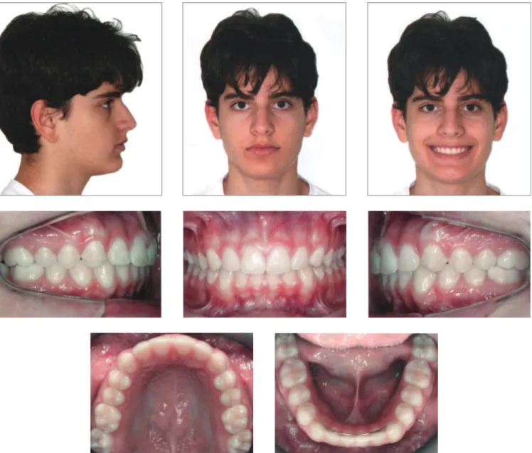
Patient’s final records (Figs 7-12 and Tab 1) as-sessment revealed that treatment objectives were achieved. Figure 7 shows that despite mild concave facial profile due to accentuated growth of the chin and nose, there was significant improvement in the relationship established between upper and lower lips, which resulted in passive lip sealing and, as a result, improved facial esthetics. Smile was more harmonious with reduction in buccal corridor width, thereby presenting satisfactory maxillary in-cisors exposure.
Dental assessment (Figs 7 and 8) revealed signifi-cant improvement in the shape of the maxillary arch due to correction of maxillary atresia. Alignment and leveling were successfully achieved. Class I molar and canine relationship was achieved on both sides. Overbite and overjet were corrected.
There was an increase in maxillary (from 51.5 mm to 58 mm) and mandibular intermolar width (from 47.5 mm to 51 mm). Since permanent mandibular incisors had not yet erupted at treatment onset, man-dibular intercanine width could not be assessed. Nev-ertheless, maxillary intercanine width increased from
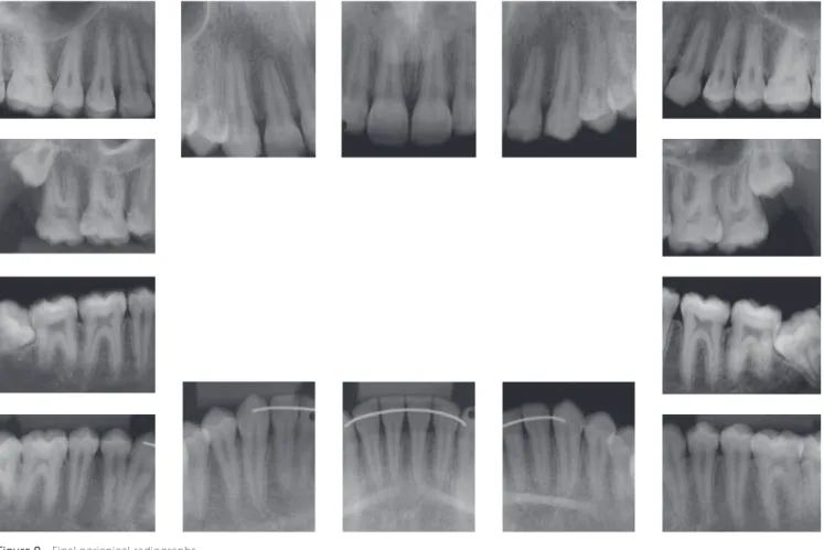
33.5 mm to 38 mm. Treatment finishing achieved balanced occlusion, with functional harmony in pro-trusive excursion as well as right and left lateral ex-cursion. Importantly, final results were achieved by means of mild apical remodeling of maxillary inci-sors despite significant repositioning of these teeth (Fig 9). Moreover, as shown by final panoramic ra-diograph (Fig 10), good root parallelism was attained in both maxillary and mandibular arches.
As expected, several skeletal changes were achieved (Fig 11 and Tab 1), with significant improvements in sagittal relationship between maxilla and mandible (SNA = 81o, SNB = 80o and ANB = 1o). The
head-gear appliance allowed not only the direction of max-illary growth to change, but also the expression of potential mandibular growth, even though orthope-dic effects were produced by the application of mild forces (300 g/side).2 There was remarkable reduction
in facial convexity and, despite cervical pull, opening
of the mandibular plane angle did not occur (GoGn-SN and FMA remained practically unaltered). Sig-nificant alteration was also found in the position of maxillary incisors (1-NA = 25o and 5 mm), which
notably contributed to overjet correction and im-provement of interincisal angle (1 / 1 = 126o).
Total cephalometric superimposition (Fig 12) revealed restricted anterior maxillary growth with downward displacement, only. Nevertheless, there was mild anterior displacement of the mandible, as well, which was responsible for patient’s mild con-cave profile. This fact was greatly reinforced by sig-nificant anterior growth of the chin. Overjet and overbite were corrected. Partial maxillary superim-position revealed expressive lingual movement of incisors, with altered tipping and posterior displace-ment of A Point. A large amount of vertical growth was observed in the mandible, followed by compen-satory alveolar growth in the region of molars.
Figure 9 - Final periapical radiographs.
Figure 11 - Final lateral cephalogram (A) and cephalometric tracing (B).
Figure 12 - Total (A) and partial (B) initial (black) and final (red) cephalometric tracings superimposition.
A B
A B
FINAL CONSIDERATIONS
Angle Class II malocclusion is occasionally asso-ciated with a narrow maxilla, which most of times creates the need to start orthodontic treatment by correcting maxillary transverse deficiency for sub-sequent correction of sagittal relationship.3 In the
case reported herein, the patient was at a fairly favor-able age; for this reason, rapid maxillary expansion
by means of Haas appliance was the technique of choice.4 After maxillary expansion, an increase in
Overbite correction was achieved by means of leveling the mandibular arch, which had accentuated curve of Spee, by means of intrusion of mandibu-lar incisors. In 1938, Hemley7 described treatment
carried out by means of anterior bite plate used to favor extrusion of posterior teeth, after which a cer-vical headgear associated with bite plate was used, yielding satisfactory clinical outcomes when treating Angle Class II malocclusion patients.
As previously reported, even though an acrylic ante-rior bite plate was used in association with cervical head-gear (Kloehn),8 patient’s mandibular plane remained
practically unaltered, probably due to the prevalence of horizontal growth pattern. Combined with accentuated growth of the chin and nose, this fact contributed to render patient’s proile slightly concave.9,10
Thus, reassessment of patient’s final records con-firms that treatment objectives were successfully achieved with Class I molar and canine relation-ship.11 Moreover, nasal breathing was reestablished,
thereby eliminating the need for speech therapy. It is worth noting that, despite patient’s and his family’s opposition, maxillary and mandibular third molars were eventually extracted.
Table 1 - Initial (A), intermediate (A1) and final (B) cephalometric values.
Measurements Normal A B Dif. A/B
Skeletal pattern
SNA (Steiner) 82° 84° 81° 3
SNB (Steiner) 80° 78° 80° 2
ANB (Steiner) 2° 6° 1° 5
Angle of convexity (Downs) 0° 8° -5° 13
Y axis (Downs) 59° 63° 59° 4
Facial angle (Downs) 87° 87° 88° 1
SN-GoGn (Steiner) 32° 23° 23° 0
FMA (Tweed) 25° 17° 19° 2
Dental pattern
IMPA (Tweed) 90° 106° 106° 0
1.NA (degrees) (Steiner) 22° 30° 25° 5
1-NA (mm) (Steiner) 4 mm 8 mm 5 mm 3
1.NB (degrees) (Steiner) 25° 27° 30° 3
1-NB (mm) (Steiner) 4 mm 5 mm 3 mm 2
1
1- Interincisal angle (Downs) 130° 119° 126° 7
1-APo (Ricketts) 1 mm 0 mm -1 mm 1
Profile Upper lip — S-line (Steiner) 0 mm 1 mm -3 mm 4
Lower lip — S-line (Steiner) 0 mm 2 mm -3 mm 5
1. Ricketts MR. Bioprogressive therapy as an answer to orthodontic needs.
Am J Orthod. 1976;70(4):359-97.
2. Bumrind S, Korn EL, Isaacson RJ, West EE, Molthen R. Quantitative
analysis of the orthodontic and orthopedic efects of maxillary traction. Am J Orthod. 1983;84(5):384-98.
3. Quaglio CL, Macedo A, Ferreira FAC. Idade ideal para correção
ortodôntica. Rev Assoc Paul Cir Dent. 2005;59(2):131-6.
4. Haas AJ. Rapid expansion of the maxillary dental arch and nasal cavity by opening the midpalatal suture. Angle Orthod.1961;31(2):73-90.
5. Haas AJ. Headgear therapy: the most eicient way to distalize molars.
Semin Orthod. 2000;6(2):79-90.
6. Lima AC, Lima AL, Lima Filho, RMA, Oyen OJ. Spontaneous mandibular
arch response after rapid palatal expansion: a long-term study on Class I malocclusion. Am J Orthod Dentofacial Orthop. 2004;126(5):576-82.
REFERENCES
7. Hemley S. Bite plates: their application and action. Am J Orthod. 1938;24:721-36.
8. Kloehn SJ. Evaluation of cervical anchorage force in treatment. Angle Orthod. 1961;31(2):91-104.
9. Harris EF, Dyer GS, Vaden JL. Age efects on orthodontic treatment:
skeletodental assessments from the Johnston analysis. Am J Orthod Dentofacial Orthop. 1991;100(6):531-6.
10. Moore AW. Orthodontic treatment factors in Class II malocclusion. Am J Orthod. 1959;45(5):323-52.