Angle Class II, division 2 malocclusion treated
with extraction of permanent teeth*
Sílvio Luís Dalagnol**
This study describes the orthodontic treatment of a woman with Angle Class II, division 2 malocclusion, impacted maxillary third molars, periodontal pocket, gingival recession and tooth wear. Treatment consisted of extraction of maxillary second premolars and anchorage control. This case was presented to the Committee of the Brazilian Board of Orthodontics and Facial Orthopedics (BBO) in the Free Case category as part of the requisites to obtain the BBO Diploma.
Abstract
Keywords: Angle Class II malocclusion. Adult. Impacted tooth. Periodontal pocket. Tooth extraction. Orthodontic anchorage.
** MSc in Orthodontics, Federal University of Rio de Janeiro State, Brazil. Diplomate, Brazilian Board of Orthodontics and Facial Orthopedics. * Case Report, Free Choice Case Category, approved by the Brazilian Board of Orthodontics and Facial Orthopedics (BBO).
HISTORY AND ETIOLOGY
The patient, encouraged by her periodontist, sought orthodontic treatment at 28 years of age. Her main complaint was unsatisfactory dental es-thetics. Her medical history was uneventful. Her dental history, however, reported by the periodon-tist, included a periodontal pocket in the mesial aspect of the right mandibular first molar (tooth #46), gingival recession in several teeth, tooth wear, and an indication for extraction of impacted maxillary third molars.
DIAGNOSIS
Facial evaluation revealed a harmonious, slightly concave profile, retruded lips, mild facial asymme-try, mandible shifted to the left and gingival display on the right side during smiling (Fig 1).
She presented an Angle Class II, division 2
malocclusion, with characteristic maxillary crowd-ing and less marked mandibular crowdcrowd-ing, mesial space in tooth #46 and a prosthesis, smaller when compared with its contralateral tooth. The maxil-lary gingival margins were uneven, there was dis-crete gingival recession in teeth #14, 22, 23 and 24, and the occlusal plane was uneven. Maxillary cen-tral incisors were retruded, inclined lingually and excessively worn, and lateral incisors were protrud-ing and malformed. Maxillary second premolars had restorations and their size was disproportion-ate in comparison with the other teeth. The maxil-lary and mandibular canines had an edge-to-edge relation, marked overbite, and a functional dis-placement from centric relation (CR) to maximal intercuspation (MI). The upper part of the midline was shifted to the right in relation to the mid sagit-tal plane, and the lower, to the left (Figs 1 and 2).
FIGURE 1 - Initial facial and intraoral photographs.
A B
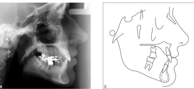
Radiographs showed a mesial periodontal pocket in tooth #46; teeth #38 and 48 were miss-ing, and teeth #18 and 28 were impacted (Fig 3). Cephalometric evaluation showed skeletal harmony: ANB was 3º, but the low values of the mandibular plane (SN-GoGn=29°and FMA=22°), the high value of the facial angle (89°) and the pogonion shape indicated a brachyfacial profile. Measurements to define dental pattern confirmed retrusion and lingual inclination of maxillary (1-NA=15° and 3 mm) and mandibular (1-NB=19° and 2.5 mm) in-cisors. Cephalometric measures are shown in Figure 4 and Table 1.
TREATMENT OBJECTIVES
As the main complaint was esthetical, the purpose of the treatment was to level maxillary gingival margins5 for esthetical and functional re-habilitation, and to extrude tooth #46 to reestab-lish normal periodontal space, as requested by the periodontist.
Specific objectives were to keep the harmoni-ous facial profile, to improve maxillary occlusal
plane by extrusion in the left side but not on the right side, and to obtain a Class II relationship be-tween molars and normal occlusion bebe-tween ca-nines according to Andrew’s keys of occlusion, all under maximal anchorage control, as well as to correct upper and lower midlines, overbite, incisor tipping and leveling of the curve of Spee.
Therefore, at the end of the treatment, facial harmony was expected to be preserved, smile esthetics improved, and centric relation (CR), maximal intercuspation (MI) and normal excur-sion corrected.
FIGURE 3 - Initial panoramic radiograph.
TREATMENT PLAN
To achieve the treatment objectives, we chose to extract maxillary second premolars2 because they were very small and had already been restored, although we were aware that this would complicate orthodontic mechan-ics. First, a fixed appliance would be placed in the upper arch, except for teeth #12 and 22 (Standard edgewise system, 0.018x 0.025-in slot) and a Kloehn extraoral appliance would be prepared for maximal anchorage. For maxil-lary leveling, 0.015-in round multistranded and 0.014 and 0.016-in stainless steel archwires would be used, but not for maxillary lateral in-cisors. After that, premolars and canines would be moved distally using chain elastics to create spaces for canines mesially. Then, lateral inci-sors would be bonded and leveled with nickel-titanium sectional archwires. The mandibular appliance would be mounted up to the second molars as soon as possible and according to the progression of maxillary incisor movement and creation of interocclusal spaces. The same sequence of archwires would be used to level the curve of Spee. Moreover, tooth #46 would be adjusted to enable its extrusion and the elimination of the periodontal pocket. After achieving normal canine occlusion according to Andrew’s keys, the incisors would undergo re-traction using a 0.017x 0.022-in stainless steel archwire with teardrop-loops. Finally, upper and lower continuous 0.017x 0.022-in stainless steel archwires would be used. According to gingival margin leveling and periodontal pock-ets in maxillary teeth, the appliance would be reassembled. After the removal of the fixed ap-pliance, a wraparound retainer (full time wear) would be prescribed to be worn until esthetic and functional restorations were made, an in-tercanine arch would be bonded for mandibu-lar retention, and the new maxilmandibu-lary retainer would be installed, in accordance with the new teeth shapes. The third molars would be
fol-lowed up to define whether they should be pre-served or extracted later on.
TREATMENT PROGRESSION
FIGURE 5 - Final facial and intraoral photographs.
A B TREATMENT RESULTS
The main treatment objectives were achieved, as confirmed by the patient’s final examinations (Figs 5-8).
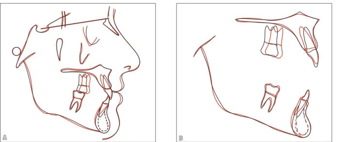
In the maxilla, the SNA angle was reduced in 1 degree because of bone remodeling result-ing from the correction of incisor tippresult-ing.1 In the mandible, the mandibular plane angles (SN-GoGn and FMA) and Y axis were reduced, and the facial angle increased, although a Kloehn extraoral appliance was used. The analysis of dental pattern confirmed that there were posi-tive changes in incisor position and tipping, as well as a better leveling of gingival margins. The evaluation of total cephalometric comparison confirmed profile, maxillary and dental changes. The partial comparison showed changes in A point1 and dental changes (Fig 9).
A Class II relation was achieved between molars, together with intentional maxillary pre-molar and pre-molar rotation to improve intercus-pation and normal canine occlusion according to Andrew’s keys. Dental midlines coincided with the facial midline. Overbite was corrected as the maxillary and mandibular incisors were
intruded and the curve of Spee was leveled. Gingival recessions did not change because tooth #22 recession did not allow for the defini-tion of a better contour for the gingival margins (Figs 5 and 6).
FIGURE 7 - Final panoramic radiograph.
A B
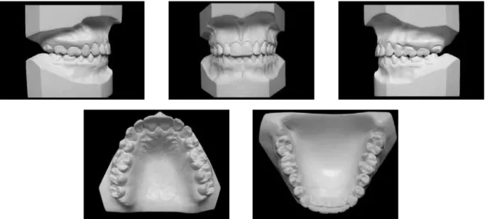
FIGURE 11 - Control dental casts four years and five months after treatment completion. Intercanine and intermolar distances
re-mained unaltered. Maxillary intercanine dis-tance increased 3.7 mm as a result of canine distal movement, and maxillary intermolar dis-tance decreased 4.1 mm due to the loss of an-chorage and the intentional mesial rotation of molars (Table 2).
The analysis of radiographs revealed bone leveling in tooth #46, and root dilacerations, mostly in the canines, which were visible only after rotations were corrected. Despite these di-lacerations, roots were parallel and root round-ing was compatible with the great amount of movement of maxillary central incisors. Root resorption, more marked in tooth #126, oc-curred primarily in the last phase of the treat-ment, and will be followed up. The progression of third molar eruption was small and will con-tinue under observation (Fig 7).
Facial profile remained harmonious despite slight lip retrusion,3 the smile was significantly
improved after the correction of the maxillary occlusal inclination and the adequate alignment and leveling of anterior teeth, and the esthetic and functional rehabilitation desired by the patient was achieved.
A B
A B
FIGURE 12 - Control panoramic radiograph four years and five months after treatment completion.
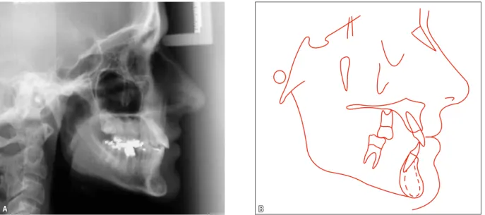
FIGURE 13 - Control cephalometric profile radiograph (A) and cephalometric tracing (B) four years and five months after treatment completion.
MEASUREMENTS Normal A B A/B
DIFFERENCE C
Skeletal Pattern
SNA(Steiner) 82° 81º 80° -1 80º
SNB(Steiner) 80° 78º 78° 0 78º
ANB(Steiner) 2° 3° 2° -1 2º
Convexity angle (Downs) 0° 0.5° 0° -0.5 0.5º
Y axis (Downs) 59° 59° 58° -1 58º
Facial angle (Downs) 87° 89° 90° 1 90º
SN–GoGn (Steiner) 32° 29° 28° -1 28º
FMA(Tweed) 25° 22° 20° -2 20º
Dental Pattern
IMPA(Tweed) 90° 93° 99° 6 99º
–1 – NA (degrees)(Steiner) 22° 15° 23° 8 22º
–1 – NA (mm)(Steiner) 4 mm 3 mm 4 mm 1 4 mm
–
1 – NB (degrees)(Steiner) 25° 19° 25° 6 26º
–
1 – NB (mm)(Steiner) 4 mm 2.5 mm 3 mm 0.5 3 mm
–11 – Interincisal angle (Downs) 130° 142º 128° -14 130º
–
1 – APo (mm)(Ricketts) 1 mm -1.5 mm -0.5 mm 1 -0.5 mm
Facial Proile
Upper lip – S line(Steiner) 0 mm -3 mm -4.5 mm -1.5 -4.5 mm
Lower lip – S line (Steiner) 0 mm -2 mm -3.5 mm -1.5 -3.5 mm
TABLE 1 - Summary of cephalometric measurements.
TABLE 2 - Transverse distances of dental arches.
MEASUREMENTS A B A/B
DIFF. C
Mandibular
intercanine distance 25.6 mm 25.6 mm 0 25.6 mm
Mandibular
intermolar distance 43.6 mm 43.6 mm 0 43.1 mm
Maxillary
intercanine distance 33 mm 35.6 mm +2.6 35.6 mm
Maxillary
intermolar distance 48.8 mm 44.7 mm -4.1 44.7 mm
FINAL CONSIDERATIONS
As the patient’s main complaint was about dental esthetics, the main treatment objectives have been achieved. Correct incisor alignment and leveling resulted in the desired esthetic and functional rehabilitation.
the second premolars, we chose to extract them, although we were aware that this would make an-chorage more difficult.
The evaluation of total cephalometric com-parison (Fig 14) confirmed the preservation of the skeletal pattern and the changes in dental pattern and facial profile. Bone remodeling due to the correction of maxillary incisor tipping and associated with the marked retraction of maxil-lary lateral incisors was confirmed in the partial
comparison of the maxilla. As maxillary lateral incisors worked as support for the lips in the be-ginning of the treatment, those changes might have resulted in a slight lip retrusion and made the profile more concave.
Control examinations (Figs 10-14) showed that the smile improved, the profile remained harmonious and occlusion remained stable, which confirms that the treatment objectives have been achieved.
1. Tien An TL, Cuoghi OA, Mendonça MR, Bertoz FA. O efeito da retração dos dentes anteriores sobre o ponto A em pacientes submetidos ao tratamento ortodôntico corretivo. Rev Dental Press Ortod Ortop Facial. 2008 mar-abr;13(2):115-23.
2. Brandt S, Sairstein R. Different extractions for different malocclusions. Am J Orthod. 1975 Jul;68(1):15-41. 3. Hershey HG. Incisor tooth retraction and subsequent proile
change in postadolescent female patients. Am J Orthod. 1972 Jan;61(1):45-54.
4. Kloehn SJ. Evaluation of cervical anchorage force in treatment. Angle Orthod. 1961 Apr; 31(2):91-104.
5. Kokich VG. Esthetics: the orthodontic-periodontic restorative connection. Semin Orthod. 1996;2(1):21-30.
6. Mirabella AD, Artun J. Risk factors for apical root resorption of maxillary anterior teeth in adult orthodontic patients. Am J Orthod Dentofacial Orthop. 1995 Jul;108(1):48-55.
REFERENCES
Contact address Sílvio Luís Dalagnol Av. Batel, 1230, Cj. 706, Batel CEP: 80.420-906 - Curitiba / PR, Brazil E-mail: [email protected] Submitted: April 2011