Fatal pulmonary embolism in hospitalized patients: a
large autopsy-based matched case-control study
Solange Aparecida Petilo Carvalho Bricola,I Edison Ferreira Paiva,I Arnaldo Lichtenstein,I Reinaldo Jose´ Gianini,IIJurandir Godoy Duarte,ISamuel Katsuyuki Shinjo,III Jose Eluf-Neto,IIMilton Arruda MartinsI
IFaculdade de Medicina da Universidade de Sa˜o Paulo, Department of Medicine, Sa˜o Paulo/SP, Brazil.IIFaculdade de Medicina da Universidade de Sa˜o
Paulo, Preventive Medicine, Sa˜o Paulo/SP, Brazil.IIIFaculdade de Medicina da Universidade de Sa˜o Paulo, Rheumatology, Sa˜o Paulo/SP, Brazil.
OBJECTIVE: Pulmonary embolism is an underdiagnosed major cause of death for hospitalized patients. The objective of this study was to identify the conditions associated with fatal pulmonary embolism in this population.
METHODS:A total of 13,074 autopsy records were evaluated in a case-control study. Patients were matched by age, sex, and year of death, and factors potentially associated with fatal pulmonary embolism were analyzed using univariate and multivariate conditional logistic regression.
RESULTS: Pulmonary embolism was considered fatal in 328 (2.5%) patients. In the multivariate analysis, conditions that were more common in patients who died of pulmonary embolism were atherosclerosis, congestive heart failure, and neurological surgery. Some conditions were negatively associated with fatal pulmonary embolism, including hemorrhagic stroke, aortic aneurism, cirrhosis, acquired immune deficiency syndrome, and pneumonia. In the control group, patients with hemorrhagic stroke and aortic aneurism had short hospital stays (8.5 and 8.8 days, respectively), and the hemorrhage itself was the main cause of death in most of them (90.6% and 68.4%, respectively), which may have prevented the development of pulmonary embolism. Cirrhotic patients in the control group also had short hospital stays (7 days), and 50% died from bleeding complications.
CONCLUSIONS:In this large autopsy study, atherosclerosis, congestive heart failure, and neurological surgery were diagnoses associated with fatal pulmonary embolism.
KEYWORDS: Atherosclerosis; Heart Failure; Neurosurgery; Pulmonary Embolism; Venous Thromboembolism.
Bricola SA, Paiva EF, Lichtenstein A, Gianini RJ, Duarte JG, Shinjo SK, et al. Fatal pulmonary embolism in hospitalized patients: a large autopsy-based matched case-control study. Clinics. 2013;68(5):679-685.
Received for publication onNovember 23, 2012;First review completed onJanuary 6, 2013;Accepted for publication onFebruary 6, 2013 E-mail: [email protected]
Tel.: 55 11 2661-7690
& INTRODUCTION
Venous thromboembolism (VTE) is a major cause of morbidity and mortality (10), and pulmonary embolism (PE) is responsible for up to 10% of hospitalized patient deaths (2). The estimates of PE prevalence in patients at risk for VTE have been derived from the results of randomized clinical trials, and prospective population-based analyses have provided additional insight into the risk of the population as a whole (1–4). However, the literature contains few studies that describe the incidence or the clinical characteristics of hospitalized patients who suffer from fatal VTE events.
Risk stratification and preventive strategies for both surgical and medical patients are presented in the American College of Chest Physicians Guidelines for VTE prophylaxis (3). Risk in surgical patients is based on age, the type of surgery, and the presence of other VTE risk factors. In medical patients, prophylaxis is recommended in cases of congestive heart failure (CHF) or severe respiratory disease and for bedridden patients with active cancer, previous VTE, sepsis, acute neurologic disease, or inflammatory bowel disease. Other guidelines recommend thrombopro-phylaxis for medical patients aged over 40 years who demonstrate reduced mobility associated with additional risk factors (4).
While the indications for thromboprophylaxis are well established, the diagnosis of PE remains a significant clinical challenge (5–8). Autopsy remains the gold standard for diagnosing fatal PE, but the number of autopsies performed has decreased substantially in many countries (9–11). In addition, recent studies have recognized PE as one of the most frequently missed diagnoses in living patients (12–15).
Copyrightß2013CLINICS– This is an Open Access article distributed under the terms of the Creative Commons Attribution Non-Commercial License (http:// creativecommons.org/licenses/by-nc/3.0/) which permits unrestricted non-commercial use, distribution, and reproduction in any medium, provided the original work is properly cited.
No potential conflict of interest was reported.
In this retrospective matched case-control study, we analyzed the autopsy records of 13,074 patients with the primary objective of identifying conditions associated with fatal PE in hospitalized clinical and surgical patients. Our results may aid in identifying patients who could benefit from VTE prophylaxis and whose risks should be empha-sized. The clinical suspicion of PE prior to death was also investigated.
& MATERIALS AND METHODS
Study population
This study was performed in a 1,100-bed university hospital at the Faculdade de Medicina da Universidade de Sa˜o Paulo in Brazil, which admits adult medical and general surgery patients. Autopsies are routinely performed if requested by physicians and when considered necessary. This study was approved by the local ethical committee (protocol number 901/03).
Study design
All autopsy reports from 1995 to 2004 were screened, and autopsies in which PE was a finding (as determined by pathologists) were selected for review. We chose the years 1995 to 2004 because autopsies were much more common during this period compared to the following years (70% of total deaths), thereby avoiding selection bias. The autopsy protocol included a systematic search for pulmonary thrombi in all pulmonary lobes and a microscopic analysis of all suspect areas of the lungs. For each confirmed PE case, two additional patients matched by sex, age (¡5 years), and
year of death were selected as controls. Patients without PE but with deep vein thrombosis (DVT), as noted on the autopsy reports, were not eligible for use as controls. Pediatric and orthopedic patients were typically admitted to other institutes within the hospital complex and were not included in this study. All autopsy reports and discharge summaries were independently reviewed by two physicians from the research group (EFP and AL) to classify PE as the primary cause or a contributing factor of death (‘‘fatal’’ and ‘‘non-fatal’’ PE), define the main reason for hospital admission, to identify whether a PE diagnosis was suspected before the autopsy. PE was considered fatal when the pathologists classified it as the only or main cause of death. The consensus reached between the two classify-ing physicians was 97%. When the two physicians dis-agreed, the case was reevaluated by both physicians together to reach a consensus.
All medical records were screened to determine whether VTE prophylaxis had been prescribed during hospitaliza-tion. Only unfractionated heparin (UFH) and enoxaparin were used for prophylaxis. UFH administered every 8 to 12 hours at a dose of 5,000 IU and enoxaparin administered once daily at a dose of 40 mg (or 20 mg if the creatinine clearance was below 30 mL/min/1.73 m2) were considered the proper treatment. For the prophylaxis to be considered adequate, the patients needed to have received the correct dose and could not have been subjected to 72 or more consecutive hours without prophylaxis during any period of hospitalization.
The entire patient group was considered for the analysis of PE incidence and the clinical accuracy of PE diagnosis before death. Only fatal cases and the appropriate controls
were included in the analysis of conditions associated with PE.
Statistical analysis
A database was created using Microsoft Access Version 2003. Student’s t-test, Fisher’s exact test, and the chi-squared test were employed to compare characteristics between the cases and controls. Univariate and multi-variate conditional logistic regressions were employed to estimate the association with fatal PE (OR and 95% CI). These regressions were performed by matching for sex, age, and year of death, and two controls were evaluated per case. The multivariate regression model included every variable that presented ap-value ,0.20 in the univariate
regression. Variables with p-values $0.05 were excluded
sequentially according to decreasing p-values. The final
multivariate model contained only variables withp-values
,0.05. The analyses were performed using the Stata 10.1TM software.
& RESULTS
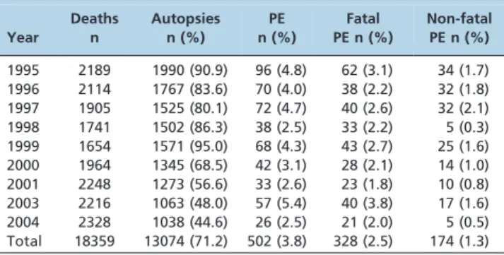
Autopsies were performed for 13,074 (71.2%) of the 18,359 deaths. Autopsies from 2002 were excluded because the hospital’s computer system changed that year, and the new system was unable to transfer data from the old system. Table 1 shows how the autopsy rate decreased over the course of the study period, and a significant difference was found after 1999. The average percentage of deaths for which autopsies were performed between 1995 and 1999 was 87%, and it decreased to 53.9% between 2000 and 2004 (p= 0.005).
PE was detected in 502 autopsies (3.8%) and was considered the main cause of death (fatal PE) in 328 of these (2.5%) cases. There were no significant differences in the percentages of autopsies with PE or the percentage of PE cases classified as fatal or non-fatal over the course of the study period (Table 1).
The sensitivity of the clinical PE diagnosis was only 48.2% for the entire group, although it was higher in cases of fatal PE compared to non-fatal PE (61.9%vs. 22.4%, respectively, p,0.001). The specificity of the clinical diagnosis of PE was
greater than 98% for all of the subgroups.
For the analysis of conditions associated with PE, 984 patients (328 fatal PE cases and 656 non-PE controls) were selected. The baseline characteristics are shown in Table 2. There were no significant differences in gender or age between the groups, which indicated successful matching. Arterial insufficiency, atherosclerosis, cancer, CHF, and chronic obstructive pulmonary disease (COPD) were more common in the fatal PE group, whereas acquired immuno-deficiency syndrome (AIDS), aortic aneurism, cirrhosis, infections, pneumonia, and hemorrhagic stroke were more common in the control group.
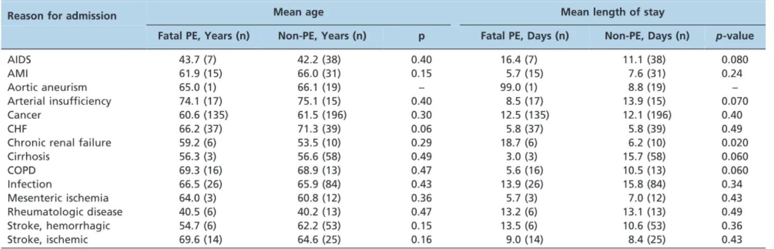
Age and length of stay were analyzed according to the main disease groups to test the hypothesis that older patients and those with longer hospital stays would have greater risks for VTE (Table 3). The only significant difference was related to the mean hospital stay of patients with chronic renal failure, which was longer for those in the fatal PE group (18.7 daysvs. 6.2 days in the non-PE group; p= 0.020).
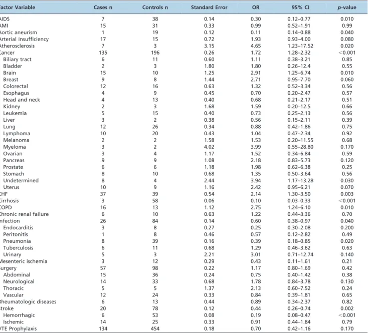
related to fatal PE autopsy findings and adjusted for sex, age and admission time. Atherosclerosis (OR = 4.65, 95% CI, 1.23–17.52, p= 0.020), cancer (OR = 1.72, 95% CI, 1.28–2.32, p,0.001), CHF (OR = 2.14, 95% CI, 1.30–3.50,p= 0.003), and
COPD (OR = 2.75, 95% CI, 1.24–6.10, p= 0.010) were
asso-ciated with fatal PE. Of the types of cancers documented, brain cancer (OR = 2.91, 95% CI, 1.25–6.74, p= 0.010) and
cancer of undetermined origin (OR = 3.94, 95% CI, 1.17– 13.28,p= 0.030) were most significantly associated with fatal
PE. Fatal PE was negatively associated with AIDS (OR = 0.30, 95% CI, 0.12–0.77, p= 0.010), aortic aneurism
(OR = 0.11, 95% CI, 0.14–0.88,p= 0.040), cirrhosis (OR = 0.10,
95% CI, 0.03–0.33,p,0.001), hemorrhagic stroke (OR = 0.19,
95% CI, 0.08–0.47, p,0.001), infection (OR = 0.60, 95% CI,
0.38–0.97, p= 0.040), and pneumonia (OR = 0.39, 95% CI,
0.18–0.85, p= 0.020). In the entire group (n = 1,506), VTE
prophylaxis was considered adequate in only 20.4% of cases. Lack of prophylaxis was not shown to be associated with fatal PE in the univariate analysis (Table 4).
The conditional multivariate logistic regression indicated that fatal PE was associated with atherosclerosis (OR = 4.03, 95% CI, 1.06–15.01,p= 0.040), CHF (OR = 1.77, 95% CI, 1.04–
2.97,p= 0.030), and neurological surgery (OR = 3.96, 95% CI,
1.48–10.65, p= 0.006). A negative association with fatal PE
was found in patients with AIDS (OR = 0.22, 95% CI, 0.08– 0.60, p= 0.003), aortic aneurism (OR = 0.12, 95% CI, 0.01–
0.90, p= 0.040), cirrhosis (OR = 0.08, 95% CI, 0.03–0.27, p,0.001), pneumonia (OR = 0.35, 95% CI, 0.15–0.79, p= 0.010), and hemorrhagic stroke (OR = 0.09, 95% CI,
0.03–0.25,p,0.001) (Table 5).
& DISCUSSION
VTE prevention in hospitalized patients has improved in recent years and now includes recommendations for improving the diagnosis of VTE and new algorithms for VTE prophylaxis (3,4,16). However, the percentage of patients whose deaths are the result of PE has not decreased, and this fact may be related to many factors, such as the increase in patient age or the large number of VTE risk factors (3,17). In addition, most doctors do not follow the appropriate thromboprophylaxis protocols (18,19). Thus, VTE remains a major cause of death for hospitalized patients, and a substantial number of VTEs discovered at autopsy are not diagnosed during hospitaliza-tion (14,20–23).
Autopsies remain invaluable for determining the effects of prophylaxis and diagnosing and treating VTE (9); unfortunately, the number of autopsies performed has decreased substantially in many countries (10,24). Although we observed a statistically significant decrease in the number of autopsies over the study period, the autopsy rate at our hospital was greater (71.2% of deaths) than that reported in other studies. Data from the United States National Center for Health Statistics over a similar time period showed that the autopsy rate was low in 1993 (9.7% of all deaths in the US) and further decreased to only 7.1% in 2003 (24).
In a classic study by Lindblad, a systematic search for VTE identified PE in 26.4% of all autopsies (9.4% were considered fatal PE) (9). The percentage of PE patients in our patient sample (3.8% for all PE and 2.5% for fatal PE) was much lower. Differences in the percentages of autopsies found with PE in the different studies may be caused by Table 1 -Number and percentage of autopsies performed
and PEs found during the study.
Year
Deaths n
Autopsies n (%)
PE n (%)
Fatal PE n (%)
Non-fatal PE n (%)
1995 2189 1990 (90.9) 96 (4.8) 62 (3.1) 34 (1.7) 1996 2114 1767 (83.6) 70 (4.0) 38 (2.2) 32 (1.8) 1997 1905 1525 (80.1) 72 (4.7) 40 (2.6) 32 (2.1) 1998 1741 1502 (86.3) 38 (2.5) 33 (2.2) 5 (0.3) 1999 1654 1571 (95.0) 68 (4.3) 43 (2.7) 25 (1.6) 2000 1964 1345 (68.5) 42 (3.1) 28 (2.1) 14 (1.0) 2001 2248 1273 (56.6) 33 (2.6) 23 (1.8) 10 (0.8) 2003 2216 1063 (48.0) 57 (5.4) 40 (3.8) 17 (1.6) 2004 2328 1038 (44.6) 26 (2.5) 21 (2.0) 5 (0.5) Total 18359 13074 (71.2) 502 (3.8) 328 (2.5) 174 (1.3)
PE: pulmonary embolism.
Table 2 -Baseline characteristics in fatal PE and non-PE groups.
Characteristic Fatal PE Non-PE p-value
n = 328 n = 656
Age in years, mean (SD) 62.2 (16.4) 61.5 (16.5) 0.26
Male, n (%) 163 (49.7) 326 (49.7) 1.00
Length of stay in days, mean (SD) 10.8 (15.1) 11.7 (16.0) 0.200
AIDS, n (%) 7 (2.1) 38 (5.8) 0.009
AMI, n (%) 15 (4.6) 31 (4.7) 1.00
Aortic aneurism, n (%) 1 (0.3) 19 (2.9) 0.006 Arterial insufficiency, n (%) 17 (5.2) 15 (2.3) 0.020
Atherosclerosis, n (%) 7 (2.1) 4 (0.6) 0.049
Cancer, n (%) 135 (41.2) 196 (29.9) 0.001
Biliary tract, n (%) 6 (4.4) 11 (5.6) 0.80
Bladder, n (%) 2 (1.5) 3 (1.5) 1.00
Brain, n (%) 15 (11.1) 10 (5.1) 0.060
Breast, n (%) 9 (6.7) 8 (4.1) 0.32
Colorectal, n (%) 12 (8.9) 16 (8.2) 0.84
Esophagus, n (%) 4 (3.0) 9 (4.6) 0.57
Head and neck, n (%) 4 (3.0) 13 (6.6) 0.200
Kidney, n (%) 2 (1.5) 3 (1.5) 1.00
Leukemia, n (%) 5 (3.7) 15 (7.7) 0.160
Liver, n (%) 3 (2.2) 10 (5.1) 0.26
Lung, n (%) 12 (8.9) 26 (13.3) 0.29
Lymphoma, n (%) 10 (7.4) 20 (10.2) 0.44
Melanoma, n (%) 2 (1.5) 2 (1.0) 1.00
Myeloma, n (%) 3 (2.2) 2 (1.0) 1.00
Ovarian, n (%) 3 (2.2) 4 (2.0) 1.00
Pancreas, n (%) 9 (6.7) 9 (4.6) 1.00
Uterus, n (%) 10 (7.4) 9 (4.6) 0.40
Chronic renal failure, n (%) 6 (1.8) 10 (1.5) 0.80
CHF, n (%) 37 (11.3) 39 (5.9) 0.005
Cirrhosis, n (%) 3 (0.9) 58 (8.8) ,0.001
COPD, n (%) 16 (4.9) 13 (2.0) 0.020
Infections, n (%) 26 (7.9) 84 (12.8) 0.020
Pneumonia 8 (2.4) 39 (5.9) 0.020
Tuberculosis 6 (1.8) 11 (1.7) 1.00
Urinary 5 (1.5) 3 (0.5) 0.130
Endocarditis 3 (0.9) 8 (1.2) 0.76
Peritonitis 1 (0.3) 8 (1.2) 0.29
Others 4 (1.2) 15 (2.3) 0.33
Mesenteric ischemia, n (%) 3 (0.9) 12 (1.8) 0.41
Rheumatologic, n (%) 6 (1.8) 13 (2.0) 1.00
Stroke (hemorrhagic), n (%) 6 (1.8) 53 (8.1) ,0.001 Stroke (ischemic), n (%) 14 (4.3) 25 (3.8) 0.73
Others, n (%) 29 (8.8) 46 (7.0) 0.31
different autopsy protocols, different hospital populations and the percentage of autopsies performed. We avoided selection bias by studying a period in which the percentage of autopsies was particularly high in our hospital, approxi-mately 70% of the total number of deaths. The small difference between the number of total PE and fatal PE cases found in our series may be due to the autopsy protocol used to search for visible thrombi and perform microscopic analysis only of the areas with macroscopic findings. Because the main objective of this study was to identify conditions associated with fatal PE, this factor was not considered a major limitation.
Clinical suspicion of PE has a high specificity, but the reported sensitivity was approximately 30% (20–23). Sensitivity can also vary from less than 10% to more than 65%, depending on the related conditions. Diagnosis is typically more difficult in older patients and for those with respiratory diseases, specifically COPD and pneumonia (21). The severity of the PE episode may be another possible explanation for this variation, and this possibility was demonstrated in the present study, in which the sensitivity of antemortem diagnoses was much higher in patients with fatal PE (61.9%) compared to patients with non-fatal PE (22.4%).
Cancer, CHF, and COPD have long been considered important risk factors for VTE (3,4,25–27). In the present study, the association of these factors with fatal PE was noted in the univariate analysis, but only the association with CHF was confirmed in the multivariate analysis. The association between brain cancer and fatal PE was also lost in the multivariate analysis; however, brain cancer is considered one of the cancers most frequently associated with VTE complications. In one study, it was identified as the second most frequent VTE-related cancer (OR = 2.37, 95% CI, 2.04–274) (28). Furthermore, neurological surgery has been shown to be associated with VTE (29), which was confirmed in our study.
Hemorrhagic stroke is considered to be highly associated with DVT and PE (3,4), but this was not the case in our study. In fact, we found a negative association with fatal PE in stroke patients, which may be explained by the severity of their conditions. In 48 of the 53 patients in the control group (90.6%), the hemorrhage itself was the main cause of death.
These patients also had short hospital stays prior to their deaths, which may have prevented the development of VTE. After excluding 1 patient who was in the hospital for 122 days, the average hospital stay of these patients was 8.48 days.
Aortic aneurism and cirrhosis were also found to have a negative association with fatal PE. Only 1 patient in the fatal PE group had an aortic aneurism, and his hospital stay was 99 days. Of the 19 patients in the control group, 13 (68.4%) died from the dissection or rupture of the aneurism, and 10 died prior to the third day of admission. Of the cirrhotic patients in the control group, 29 (50%) died from bleeding complications, and the median hospital stay prior to death for these patients was only 7 days.
One interesting finding of our study was the strong association between atherosclerosis and fatal PE (OR = 4.03, 95% CI, 1.06–15.71,p= 0.040). Although
athero-sclerosis is not typically thought to be an independent risk factor for VTE (30,31), intriguing evidence from recent studies has suggested that statins may prevent VTE in healthy older adults (32,33). In a randomized, double-blind, placebo-controlled trial, 20 mg of rosuvastatin daily significantly reduced the occurrence of symptomatic VTE in outpatients with no cardiovascular disease histories. The PE rates were 0.09 in the rosuvastatin group and 0.12 in the placebo group (HR = 0.77, 95% CI, 0.41–1.45,p= 0.42)
(32). The anti-inflammatory effect of statins on the microvascular endothelium may be the possible mechan-ism of action (33). Another interesting finding was the negative association between pneumonia and fatal PE. Pneumonia has classically been considered a risk factor for VTE (3,4), and it is unclear why our data did not confirm this association.
AIDS is considered to present many risk factors for VTE, including frequent hospitalization, the use of central venous catheters, an age older than 45 years, and the activation of endothelial cells from infections with cytomegalovirus, herpes, and HIV itself (34,35). The mean age of the AIDS patients in our study was 43.7 years, and the average hospital stay of these patients was only 11.1 days. These findings may indicate that these patients presented to the hospital with serious conditions that did not allow enough time for PE to develop.
Table 3 -Age and length of hospital stay according to reason for admission.
Reason for admission Mean age Mean length of stay
Fatal PE, Years (n) Non-PE, Years (n) p Fatal PE, Days (n) Non-PE, Days (n) p-value
AIDS 43.7 (7) 42.2 (38) 0.40 16.4 (7) 11.1 (38) 0.080
AMI 61.9 (15) 66.0 (31) 0.15 5.7 (15) 7.6 (31) 0.24
Aortic aneurism 65.0 (1) 66.1 (19) – 99.0 (1) 8.8 (19) –
Arterial insufficiency 74.1 (17) 75.1 (15) 0.40 8.5 (17) 13.9 (15) 0.070
Cancer 60.6 (135) 61.5 (196) 0.30 12.5 (135) 12.1 (196) 0.40
CHF 66.2 (37) 71.3 (39) 0.06 5.8 (37) 5.8 (39) 0.49
Chronic renal failure 59.2 (6) 53.5 (10) 0.29 18.7 (6) 6.2 (10) 0.020
Cirrhosis 56.3 (3) 56.6 (58) 0.49 3.0 (3) 15.7 (58) 0.060
COPD 69.3 (16) 68.9 (13) 0.47 5.6 (16) 10.5 (13) 0.060
Infection 66.5 (26) 65.9 (84) 0.43 13.9 (26) 15.8 (84) 0.34
Mesenteric ischemia 64.0 (3) 60.8 (12) 0.36 5.7 (3) 7.0 (12) 0.43
Rheumatologic disease 40.5 (6) 40.2 (13) 0.47 13.2 (6) 13.1 (13) 0.49
Stroke, hemorrhagic 54.7 (6) 62.2 (53) 0.15 13.5 (6) 10.6 (53) 0.36
Stroke, ischemic 69.6 (14) 64.6 (25) 0.16 9.0 (14) 8.4 (25) 0.43
Table 4 -Conditional univariate logistic regression of association between risk factors and fatal PE adjusted for sex, age and time of admission.
Factor Variable Cases n Controls n Standard Error OR 95% CI p-value
AIDS 7 38 0.14 0.30 0.12–0.77 0.010
AMI 15 31 0.33 0.99 0.52–1.91 0.99
Aortic aneurism 1 19 0.12 0.11 0.14–0.88 0.040
Arterial insufficiency 17 15 0.72 1.93 0.93–4.00 0.080
Atherosclerosis 7 3 3.15 4.65 1.23–17.52 0.020
Cancer 135 196 0.26 1.72 1.28–2.32 ,0.001
Biliary tract 6 11 0.60 1.11 0.38–3.21 0.85
Bladder 2 3 1.80 1.80 0.26–12.4 0.55
Brain 15 10 1.25 2.91 1.25–6.74 0.010
Breast 9 8 1.44 2.71 0.95–7.70 0.060
Colorectal 12 16 0.63 1.32 0.52–3.34 0.56
Esophagus 4 9 0.45 0.70 0.20–2.47 0.57
Head and neck 4 13 0.40 0.68 0.21–2.17 0.51
Kidney 2 3 1.68 1.59 0.20–12.5 0.66
Leukemia 5 15 0.40 0.73 0.25–2.13 0.56
Liver 3 2 0.38 0.56 0.15–2.11 0.39
Lung 12 26 0.34 0.88 0.42–1.86 0.75
Lymphoma 10 20 0.43 1.04 0.47–2.34 0.92
Melanoma 2 2 1.58 1.53 0.20–11.55 0.68
Myeloma 3 2 4.02 3.99 0.55–28.80 0.170
Ovarian 3 4 1.17 1.52 0.34–6.84 0.59
Pancreas 9 9 1.08 2.18 0.83–5.73 0.120
Prostate 6 6 1.18 1.98 0.62–6.38 0.25
Stomach 8 10 0.68 1.35 0.50–3.64 0.56
Undetermined 8 4 2.44 3.94 1.17–13.28 0.030
Uterus 10 9 1.16 2.42 0.95–6.21 0.070
CHF 37 39 0.54 2.14 1.30–3.50 0.003
Cirrhosis 3 58 0.06 0.10 0.03–0.33 ,0.001
COPD 16 13 1.12 2.75 1.24–6.10 0.010
Chronic renal failure 6 10 0.63 1.22 0.44–3.36 0.70
Infection 26 84 0.14 0.60 0.38–0.97 0.040
Endocarditis 3 8 0.27 0.25 0.30–2.08 0.200
Peritonitis 1 8 0.46 0.57 0.12–2.82 0.49
Pneumonia 8 39 0.16 0.39 0.18–0.85 0.020
Tuberculosis 6 11 0.68 1.29 0.46–3.62 0.63
Urinary 5 3 2.21 3.01 0.71–12.74 0.140
Mesenteric ischemia 3 12 0.29 0.43 0.11–1.61 0.21
Surgery 57 98 0.22 1.17 0.80–1.69 0.42
Abdominal 15 36 0.24 0.75 0.40–1.42 0.38
Neurological 14 33 0.68 1.78 0.84–3.78 0.130
Thoracic 5 5 1.37 2.13 0.60–7.52 0.24
Vascular 12 24 0.33 0.84 0.39–1.81 0.65
Rheumatologic diseases 6 13 0.44 0.89 0.34–2.37 0.82
Stroke 20 78 0.12 0.44 0.26–0.74 0.002
Hemorrhagic 6 53 0.08 0.19 0.08–0.47 ,0.001
Ischemic 14 25 0.33 0.91 0.44–1.84 0.79
VTE Prophylaxis 134 454 0.18 0.70 0.42–1.16 0.170
OR: odds ratio; CI: confidence interval; AIDS: acquired immunodeficiency syndrome; AMI: acute myocardial infarction; CHF: congestive heart failure and COPD: chronic obstructive pulmonary disease.
Table 5 -Conditional multivariate logistic regression of the association between risk factors and fatal PE, adjusted for age and time of admission.
Factor Variable Cases n Controls n Standard Error OR 95% CI p-value
AIDS 7 38 0.11 0.22 0.08–0.60 0.003
Aortic aneurism 1 19 0.12 0.12 0.01–0.90 0.040
Atherosclerosis 7 3 2,80 4.03 1.06–15.71 0.040
CHF 37 39 0.47 1.77 1.04–2.97 0.030
Cirrhosis 3 58 0.05 0.08 0.03–0.27 ,0.001
Pneumonia 8 39 0.15 0.35 0.15–0.79 0.010
Neurological surgery 14 33 2.00 3.96 1.48–10.65 0.006
Stroke, hemorrhagic 6 53 0.05 0.09 0.03–0.25 ,0.001
One recent publication that evaluated the adequacy of VTE prophylaxis in 37,356 hospitalized patients across 32 countries reported that thromboprophylaxis was prescribed for only 50% of these patients (18,19). In our hospital, the use of VTE prophylaxis has only increased during recent years, which suggests that we would expect to observe low rates of prophylaxis during the period studied. This finding proved accurate, as only 20.4% of patients in this study received adequate prophylaxis. This low prophylaxis rate may explain why no association between prophylaxis and fatal PE was found.
This study evaluated a large number of autopsies performed on hospital patients who had been admitted for general medical care or surgery; atherosclerosis, CHF, and neurological surgery were found to be associated with fatal PE. AIDS, aortic aneurism, cirrhosis, and hemorrhagic stroke were negatively associated with fatal PE, and this finding may be explained by the severity of these condi-tions, which often led to patient death after a short hospital stay and before PE could develop.
& ACKNOWLEDGMENTS
We are greatly indebted to Paulo Hila´rio Saldiva, MD, PhD, Marisa Dolhnikoff, MD, PhD, and Thais Mauad, MD, PhD, from the Department of Pathology of the Faculdade de Medicina da Universidade de Sa˜o Paulo for their valuable suggestions during the study.
& AUTHOR CONTRIBUTIONS
Bricola SA and Lichtenstein A were responsible for the study conception and design, data acquisition, critical review and final approval of the manuscript. Paiva EF contributed to the study conception and design, data acquisition, analysis and interpretation, drafting and final approval of the manuscript. Gianini RJ and Neto JE were responsible for the data analysis and interpretation, critical review and final approval of the manuscript. Duarte JG and Shinjo SK were responsible for the data acquisition, critical review and final approval of the manuscript. Martins MA contributed to the study conception and design, data analysis and interpretation, critical review and final approval of the manuscript.
& REFERENCES
1. Alpert JS, Dalen JE. Epidemiology and natural history of venous thromboembolism. Prog Cardiovasc Dis. 1994;36(6):417-22, http://dx. doi.org/10.1016/S0033-0620(94)80050-2.
2. Sandler DA, Martin JF. Autopsy proven pulmonary embolism in hospital patients: are we detecting enough deep vein thrombosis? J R Soc Med. 1989;82(4):203-5.
3. Geerts WH, Bergqvist D, Pineo GF, Heit JA, Samama CM, Lassen MR, et al. Prevention of venous thromboembolism: American College of Chest Physicians Evidence-Based Clinical Practice Guidelines (8th edition). Chest. 2008;133(6 Suppl):381S-453S, http://dx.doi.org/10. 1378/chest.08-0656.
4. Rocha AT, Paiva EF, Lichtenstein A, Milani R Jr, Cavalheiro CF, Maffei FH. Risk-assessment algorithm and recommendations for venous thromboembolism prophylaxis in medical patients. Vasc Health Risk Manag. 2007;3(4):533-53.
5. Perrier A, Roy PM, Sanchez O, Le Gal G, Meyer G, Gourdier AL, et al. Multidetector-row computed tomography in suspected pulmonary embolism. N Engl J Med. 2005;352(17):1760-8.
6. Goodman LR, Curtin JJ, Mewissen MW, Foley WD, Lipchik RJ, Crain MR, et al. Detection of pulmonary embolism in patients with unresolved clinical and scintigraphic diagnosis: helical CT versus angiography. AJR Am J Roentgenol. 1995;164(6):1369-74, http://dx.doi.org/10.2214/ajr. 164.6.7754875.
7. Michiels JJ, Gadisseur A, van der Planken M, Schroyens W, De Maeseneer M, Hermsen JT, et al. Screening for deep vein thrombosis and pulmonary embolism in outpatients with suspected DVT or PE by the sequential use of clinical score: a sensitive quantitative D-dimer test and noninvasive diagnostic tools. Semin Vasc Med. 2005;5(4):351-64, http://dx.doi.org/10.1055/s-2005-922480.
8. Michiels JJ, Gadisseur A, Van Der Planken M, Schroyens W, De Maeseneer M, Hermsen JT, et al. A critical appraisal of non-invasive
diagnosis and exclusion of deep vein thrombosis and pulmonary embolism in outpatients with suspected deep vein thrombosis or pulmonary embolism: how many tests do we need? Int Angiol. 2005;24(1):27-39.
9. Lindblad B, Sternby NH, Bergqvist D. Incidence of venous thromboem-bolism verified by necropsy over 30 years. BMJ. 1991;302(6778):709-11, http://dx.doi.org/10.1136/bmj.302.6778.709.
10. Rosenbaum GE, Burns J, Johnson J, Mitchell C, Robinson M, Truog RD. Autopsy consent practice at US teaching hospitals: results of a national survey. Arch Intern Med. 2000;160(3):374-80, http://dx.doi.org/10.1001/ archinte.160.3.374.
11. Goldman L, Sayson R, Robbins S, Cohn LH, Bettmann M, Weisberg M. The value of the autopsy in three medical eras. N Engl J Med. 1983;308(17):1000-5.
12. Ayoub T, Chow J. The conventional autopsy in modern medicine. J R Soc Med. 2008;101(4):177-81.
13. Shojania KG, Burton EC, McDonald KM, Goldman L. Changes in rates of autopsy-detected diagnostic errors over time: a systematic review. JAMA. 2003;289(21):2849-56, http://dx.doi.org/10.1001/jama.289.21.2849. 14. Coradazzi AL, Morganti AL, Montenegro MR. Discrepancies between
clinical diagnoses and autopsy findings. Braz J Med Biol Res. 2003;36(3):385-91, http://dx.doi.org/10.1590/S0100-879X2003000300014. 15. Perkins GD, McAuley DF, Davies S, Gao F. Discrepancies between clinical and postmortem diagnoses in critically ill patients: an observational study. Crit Care. 2003;7(6):R129-32, http://dx.doi.org/10.1186/cc2359. 16. Nicolaides AN, Breddin HK, Fareed J, Goldhaber S, Haas S, Hull R, et al.
Prevention of venous thromboembolism. International Consensus Statement. Guidelines compiled in accordance with the scientific evidence. Int Angiol. 2001;20(10):1-37.
17. Goldhaber SZ. Pulmonary embolism. Lancet. 2004;363(9417):1295-305, http://dx.doi.org/10.1016/S0140-6736(04)16004-2.
18. Cohen AT, Tapson VF, Bergmann JF, Goldhaber SZ, Kakkar AK, Deslandes B, et al. Venous thromboembolism risk and prophylaxis in the acute hospital care setting (ENDORSE study): a multinational cross-sectional study. Lancet. 2008;371(9610):387-94. Erratum in: Lancet. 2008;371(9628):1914, http://dx.doi.org/10.1016/S0140-6736(08)60202-0. 19. Deheinzelin D, Braga AL, Martins LC, Martins MA, Hernandez A,
Yoshida WB, et al. Incorrect use of thromboprophylaxis for venous thromboembolism in medical and surgical patients: results of a multi-centric, observational and cross-sectional study in Brazil. J Thromb Haemost. 2006;4(6):1266-70, http://dx.doi.org/10.1111/j.1538-7836.2006. 01981.x.
20. Pineda LA, Hathwar VS, Grant BJ. Clinical suspicion of fatal pulmonary embolism. Chest. 2001;120(3):791-5, http://dx.doi.org/10.1378/chest. 120.3.791.
21. Goldhaber SZ, Hennekens CH, Evans DA, Newton EC, Godleski JJ. Factors associated with correct antemortem diagnosis of major pulmon-ary embolism. Am J Med. 1982;73(6):822-6.
22. Kotovicz F, Mauad T, Saldiva PH. Clinico-pathological discrepancies in a general university hospital in Sa˜o Paulo, Brazil. Clinics (Sao Paulo). 2008;63(5):581-8, http://dx.doi.org/10.1590/S1807-59322008000500003. 23. Morpurgo M, Schmid C, Mandelli V. Factors influencing the clinical
diagnosis of pulmonary embolism: analysis of 229 postmortem cases. Int J Cardiol. 1998;65(Suppl 1):S79-82.
24. Hoyert DL, Kung HC, Xu J. Autopsy patterns in 2003. National Center for Health Statistics. Vital Health Stat. 2007;20(32):1-21.
25. Stein PD, Beemath A, Meyers FA, Skaf E, Sanchez J, Olson RE. Incidence of venous thromboembolism in patients hospitalized with cancer. Am J Med. 2006;119(1):60-8.
26. Beemath A, Stein PD, Skaf E, Al Sibae MR, Alesh I. Risk of venous thromboembolism in patients hospitalized with heart failure. Am J Cardiol. 2006;98(6):793-5, http://dx.doi.org/10.1016/j.amjcard. 2006.03.064.
27. Rizkallah J, Man SF, Sin DD. Prevalence of pulmonary embolism in acute exacerbations of COPD: a systematic review and metaanalysis. Chest. 2009;135(3):786-93, http://dx.doi.org/10.1378/chest.08-1516.
28. Thodiyil PA, Kakkar AK. Variation in relative risk of venous thromboembolism in different cancers. Thromb Haemost. 2002; 87(6):1076-7.
29. Khaldi A, Helo N, Schneck MJ, Origitano TC. Venous thromboembolism: deep venous thrombosis and pulmonary embolism in a neurosurgical population. J Neurosurg. 2011;114(1):40-6, http://dx.doi.org/10.3171/ 2010.8.JNS10332.
30. Prandoni P, Bilora F, Marchiori A, Bernardi E, Petrobelli F, Lensing AW, et al. An association between atherosclerosis and venous thrombosis. N Engl J Med. 2003;348(15):1435-41.
31. Prandoni P. Venous thromboembolism and atherosclerosis: is there a link? Thromb Haemost. 2007;5(Suppl 1):270-5, http://dx.doi.org/10. 1111/j.1538-7836.2007.02467.x.
32. Glynn RJ, Danielson E, Fonseca FA, Genest J, Gotto AM Jr, Kastelein JJ, et al. A randomized trial of rosuvastatin in the prevention of venous thromboembolism. N Engl J Med. 2009;360(18):1851-61.
observational studies. Thromb Res. 2011;128(5):422-30, http://dx.doi. org/10.1016/j.thromres.2011.05.012.
34. Ahonkhai AA, Gebo KA, Streiff MB, Moore RD, Segal JB. Venous thromboembolism in patients with HIV/AIDS: a case-control study. J Acquir Immune Defic Syndr. 2008;48(3):310-4.