Early osteoarthritis and reduced quality of life after
retirement in former professional soccer players
Gustavo Gonc¸alves Arliani, Diego Costa Astur, Ricardo Kim Fukunishi Yamada, Andre´ Fukunishi Yamada, Gustavo Kenzo Miyashita, Bert Mandelbaum, Moise´s Cohen
Universidade Federal de Sa˜o Paulo (UNIFESP/EPM-DOT), Departamento de Ortopedia e Traumatologia, Centro de Traumatologia do Esporte (CETE), Sa˜o Paulo/SP, Brazil.
OBJECTIVES:This study aims to compare the prevalence of osteoarthritis in two groups: one comprising former professional soccer players and the other comprising non-professional-athlete participants.
METHODS:Twenty-seven male former professional soccer players and 30 male volunteers from different non-sports professional areas participated in the study. All participants underwent bilateral knee radiography and magnetic resonance imaging. In addition, the quality of life, knee pain and joint function were evaluated and compared using questionnaires given to all participants in both groups. Specific knee evaluations, with regard to osteoarthritis and quality of life, were performed in both groups using the Knee Injury and Osteoarthritis Outcome Score subjective questionnaires and the Short-form 36. The chi-squared test, Fisher’s exact test, the Mann-Whitney U test and Student’s t-test were used for group comparisons.
RESULTS:The between-groups comparison revealed significant differences in the following: pain, symptoms and quality of life related to the knee in the Knee Injury and Osteoarthritis Outcome Score subscales; the physical aspects subscale of the SF-36; total whole-organ magnetic resonance imaging scores with regard to the dominant and non-dominant knees. Former soccer players had worse scores than the controls in all comparisons.
CONCLUSIONS:Both the clinical and magnetic resonance evaluations and the group comparisons performed in this study revealed that former soccer players have a worse quality of life than that of a control group with regard to physical aspects related to the knee; these aspects include greater pain, increased symptoms and substantial changes in radiographic and magnetic resonance images of the knee.
KEYWORDS: Osteoarthritis; Knee; Soccer; Football; MRI; Retirement.
Arliani GG, Astur DC, Yamada RK, Yamada AF, Miyashita GK, Mandelbaum B, et al. Early osteoarthritis and reduced quality of life after retirement in former professional soccer players. Clinics. 2014;69(9):589-594.
Received for publication onFebruary 18, 2014;First review completed onApril 10, 2014;Accepted for publication onApril 10, 2014 E-mail: [email protected]
Tel.: 55 11 5571-6621
& INTRODUCTION
Soccer is undoubtedly the most popular sport in the world. Approximately 200,000 professional and 240 million amateur athletes, of whom approximately 80% are male, play soccer around the world (1,2). Currently, participation in sports is viewed as favorable for healthy development. As a sport, soccer has undergone many changes in recent years, primarily due to increasing physical demands that force athletes to work close to their maximum capacity, predis-posing them to injury and to overloaded joints (3,4).
Previous studies have shown a higher incidence of osteoarthritis in the knees and ankles of former professional soccer, volleyball and basketball players than in those of the normal population (5-7).
However, no studies have evaluated the knees of retired players using magnetic resonance imaging (MRI) and linked past injuries with quality of life after retirement.
This study sought to compare the prevalence of osteoar-thritis in two homogeneous groups (matched by gender, age and presence of previous knee surgery); one group was composed of former professional soccer players and the other group was composed of non-professional-athlete participants. These groups were evaluated and compared regarding quality of life, knee pain and joint function, using questionnaires administered to all participants.
& MATERIALS AND METHODS
The Ethics and Research Committee of the Universidade Federal de Sa˜o Paulo (UNIFESP) approved this study (CEP 0629/10) and all participants signed informed consent forms.
Copyrightß2014CLINICS– This is an Open Access article distributed under the terms of the Creative Commons Attribution Non-Commercial License (http:// creativecommons.org/licenses/by-nc/3.0/) which permits unrestricted non-commercial use, distribution, and reproduction in any medium, provided the original work is properly cited.
No potential conflict of interest was reported.
Sample calculation
The calculation to determine the necessary sample size of each group was performed based on the results of a pilot study. This pilot study, performed with 10 former profes-sional soccer players, was conducted to statistically determine the minimum number of individuals who were required to participate in the main analysis. The sample size calculation was performed considering 80% power, 95% confidence intervals and a between-group difference of 40 points in the whole-organ magnetic resonance imaging score (WORMS). The control group score was taken from a previous WORMS study. This pilot study determined that the minimum number of participants in each group was 27 individuals.
Participants
This controlled, cross-sectional study was conducted between January 2011 and January 2013. The study participants were 27 former professional soccer players and 30 volunteers from different non-sports professional areas. All of the participants were male. The former athletes were recruited from a group of 32 former professional soccer players. The group consisted of former athletes from an association of former soccer players who met the inclusion criteria. From this total sample, five former players were not included in the final sample; three were currently living in distant regions and two declined to participate in the study. The 30 non-professional-athlete volunteers were recruited from various sectors of the university campus. These volunteers had different occupations and were matched with the former players by age and the presence of previous knee surgery. At most, the volunteers played soccer and other sports recreationally.
Inclusion and exclusion criteria
Former players group. The former professional soccer players included in the study, (a) had played first division soccer, (b) were between 30 and 55 years old at the time of the study, (c) had a professional sports career that lasted at least five years and (d) had participated in both training and matches during that period.
Individuals with (a) congenital diseases associated with knee deformity, (b) prior histories of lower limb fractures, (c) histories of inflammatory, metabolic, or infectious arthropathy in one or both knees, (d) hip arthroplasty or arthroplasty in one or both knees, (e) any contraindication to MRI, or (f) acute orthopedic lesions (in the last three months) were excluded from both groups.
Control group
The control group was composed of individuals who were matched by gender, age and the presence of previous knee surgery with the former players. In the control group, one subject had undergone ACL reconstruction, four individuals had undergone ACL reconstruction plus meniscectomy and the other four subjects had undergone meniscectomy. In the group of former players, one individual had undergone ACL reconstruction, three had undergone ACL reconstruction plus meniscectomy and seven former players had undergone isolated meniscectomy.
Evaluation
To characterize the clinical sample, the following vari-ables were evaluated: age, current weight, weight during
professional athletic career, height, body mass index (BMI), position that former athletes played during career, lower limb dominance, duration of professional career, reason for retirement, injuries and surgeries during career, current participation in recreational soccer, history of drug injec-tions in the knee during career and current occupation.
Current bilateral knee pain was quantified with a Visual Analogue Scale (VAS) with a 100-mm ruler on one side. After explaining that the line represented their feeling with regard to pain, individuals were instructed to score the pain in their knees from 0 mm (no pain) to 100 mm (maximum pain). Thus, their score was recorded on a millimeter ruler from 0 to 100 to quantify the pain sensitivity of each participant (8).
All participants underwent bilateral knee radiography using anteroposterior and lateral incidences (AP) with load, axial patella with 45 degrees of flexion and posteroanterior (PA) with load and 45 degrees of knee flexion. The following conditions were used: 66 KV, a bulb-to-film distance of 100 cm and high-resolution 35-43-cm radio-graphic (Kodak, Rochester, NY, USA) film. The radiographs taken with the knee in extension were obtained after placing the patella in the central portion of the frame. The radio-graphs were classified based on signs of osteoarthritis; classification was performed by a radiologist with ten years of experience evaluating musculoskeletal images and by a senior orthopedic surgeon. Image analyses were performed following a blind protocol; in the event of a disagreement, the findings were discussed until a consensus was reached. Kellgren and Lawrence’s (KL) classification was used with the radiographs (9). Radiographic osteoarthritis was defined as present when the KL classification was greater than or equal to 2.
The alignment of the lower limbs was measured via the medial angle formed between the femur and tibia (FT angle) using the method described by Moreland et al. (10). Straight lines were drawn through the center of the femoral and tibial diaphyses. The medial angle, formed by the meeting of these two lines in the center of the tibial spines was taken as the anatomical angle (OsiriX Imaging Software). Based on the measurement of this angle, the knees were divided into three groups: normal alignment, varus and valgus. The alignment was defined as normal when the FT angle was between 182
˚
and 184˚
, as valgus when the angle was greater than 184˚
, and as varus when the angle was less than 182˚
, based on the values defined by Moreland et al. (10).Specific knee evaluations with regard to osteoarthritis and quality of life were performed using the Knee Injury and Osteoarthritis Outcome Score (KOOS) subjective question-naire and the SF-36, respectively (11,12).
Blinded MRI evaluation was conducted by a radiologist with ten years of experience in assessing musculoskeletal imaging. The radiologist used a global evaluation method for osteoarthritic knees (Whole-Organ Magnetic Resonance Imaging Score; WORMS), which is applicable to traditional MRI examinations. This method is used to determine the direct prevalence of injury in all knee structures and quantifies the injuries using objective scores ranging from 0 to 332 (13).
suppression, proton density-weighted FSE and T1-weighted FSE on the coronal plane. The image parameters were as follows: 2566128 array, slice thickness between 3.5 mm and
4.0 mm, 0.4 mm spacing and a field of view (FOV) of 18 cm to 20 cm.
Data analyses
Quantitative variables were reported as means, medians, and standard deviations, and qualitative variables were displayed as percentages and absolute frequencies. All scales and quantitative variables were tested for normality (Kolmogorov-Smirnov test); the Mann-Whitney test was used for non-normally distributed data. The chi-squared test, Fisher’s exact test, the Mann-Whitney U test, and Student’s t-test were used for between-group comparisons. Correlation analysis between the results of the SF-36 and the WORMS was performed using Spearman’s correlation test. Values were considered significant at p,0.05 for all analyses. Data analyses were performed using SPSS 18.0.
& RESULTS
The BMI of the former soccer players at the time they played professionally was, on average, 23.13 kg/m2 (SD = 1.7), increasing to 25.73 kg/m2 (SD = 3.15) at the time of the study (p,0.001). During their career, the former players played in the following positions: goalkeeper (3;
11.1%), defender (7; 25.9%), midfielder (11; 40.7%) and forward (6; 22, 2%). During their careers, 44.4% (12) of the former soccer players underwent injection of drugs in the knee, with a mean of 5.33 injections per athlete (median: 3.5). In the group of former players, 81.5% were currently practicing the sport non-professionally and 85.2% currently worked, with the vast majority (95.6%) performing jobs related to football (coach, commentator, entrepreneur). On average, the careers of the former players lasted 14.89 years (median: 15 years).
The former players and the control group had similar demographic characteristics. The mean ages of the former players and the control group were 45.67¡5.91 years and
43.70¡6.15 years (p= 0.225), respectively; the body mass index (BMI) was higher in the control group than in the group of former players (mean = 28.35¡3.66vs. 25.73¡3.15,
respectively; p= 0.006). Other analyzed demographic char-acteristics are shown in Table 1.
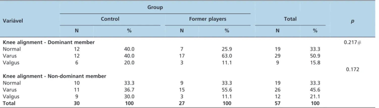
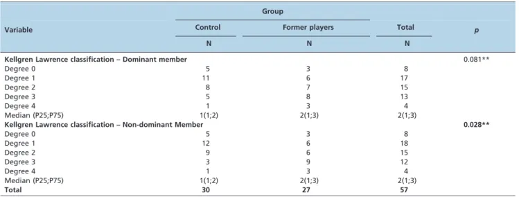
Group comparison of the lower limb alignment did not reveal significant differences (Table 2). The distribution of the radiograph analyses for the classification of knee osteoarthritis is shown in Table 3. The prevalence of osteoarthritis in the former players and in the control group in dominant knees was 66.6% and 46.7%, respectively (p= 0.081). However, the prevalence of OA in non-dominant knees in the two groups was 66.6% and 43.3%, respectively (p= 0.028).
A significant difference was revealed in the physical aspects subscale of the SF-36 (p= 0.005): the former players had lower scores than the control group (Table 4).
However, the between-group comparisons based on the VAS and on the KOOS revealed significant differences in the subscales of pain, symptoms and quality of life related to the knee: the former players had lower scores than the control group on the KOOS and higher scores on the VAS (Table 5).
According to the knee MRI analyses, the comparison of total WORMS scores revealed a significant difference with regard to both knees: the former players showed higher (worse) scores than the control group. When the three compartments of the knee (the medial tibiofemoral, lateral tibiofemoral and patellofemoral compartments) were ana-lyzed separately, significant differences were found between the groups. Again, the former players showed higher (worse) scores than the control group with regard to all compartments of both knees (Table 6).
Table 1 -Demographic characteristics of the groups.
Group
Variable Control
Former
Players Total p
N % N % N %
Dominance 0.722*
Right 26 86.7 22 81.5 48 84.2
Left 4 13.3 5 18.5 9 15.8
Knee Surgery 0.396
No 21 70.0 16 59.3 37 64.9
Yes 9 30.0 11 40.7 20 35.1
Currently Playing Soccer 0.044
No 13 43.3 5 18.5 18 31.6
Yes 17 56.7 22 81.5 39 68.4
Total 30 100 27 100 57 100
Chi-squared test
*Fisher’s exact test.
Table 2 -Knee Alignment Comparison between groups.
Group
Varia´vel Control Former players Total p
N % N % N %
Knee alignment - Dominant member 0.217#
Normal 12 40.0 7 25.9 19 33.3
Varus 12 40.0 17 63.0 29 50.9
Valgus 6 20.0 3 11.1 9 15.8
Knee alignment - Non-dominant member
0.172
Normal 10 33.3 9 33.3 19 33.3
Varus 11 36.7 15 55.6 26 45.6
Valgus 9 30.0 3 11.1 12 21.1
Total 30 100 27 100 57 100
The correlation between the SF-36 scores and the total score obtained from the MRIs of the dominant knee of the former athletes using Spearman correlation was inverted on the functional capacity subscale (r = -0.608;p= 0.001). Thus, the former players with the highest WORMS scores had less functional capacity when evaluated by the SF-36.
& DISCUSSION
The results of clinical and MRI evaluations of a group of former soccer players and those of a control group indicated that the former players had a worse quality of life with regard to physical aspects related to their knees, including greater pain symptoms and greater changes in their knee MRIs.
The results of the present study were similar to those of previous studies, with signs of osteoarthritis shown in the knee radiographs of 66% of the former players. Most of these patients (55.5%) presented with mild to moderate osteoarthritis. Previous studies have reported high rates of
radiographic osteoarthritis in the knees of former profes-sional soccer players, ranging from 43% to 69% (14). Soccer can increase the risk of osteoarthritis in the knee joint in two ways: the first is because of the increased risk of knee injury during the player’s career and the second is because of the overload placed on this joint while playing the sport (15-17). Appel et al. suggested that merely participating in the sport is a risk factor for osteoarthritis of the knees, regardless of the occurrence of knee injuries (18). In this study, the former players and the control group did not significantly differ with regard to the number of knee surgeries that the participants underwent, although the former players had undergone a greater number of meniscectomies than the control group.
Other risk factors related to developing osteoarthritis of the knee include high BMI and lower limb malalignment. Reijman et al. reported that a high BMI increases the risk of developing symptomatic and radiographic osteoarthritis of the knee (19). In this study, the control group had a significantly higher mean BMI, but they had a lower Table 3 -Osteoarthritis classification distribution comparison in dominant and non-dominant members between groups.
Group
Variable Control Former players Total p
N N N
Kellgren Lawrence classification – Dominant member 0.081**
Degree 0 5 3 8
Degree 1 11 6 17
Degree 2 8 7 15
Degree 3 5 8 13
Degree 4 1 3 4
Median (P25;P75) 1(1;2) 2(1;3) 2(1;3)
Kellgren Lawrence classification – Non-dominant Member 0.028**
Degree 0 5 3 8
Degree 1 12 6 18
Degree 2 9 6 15
Degree 3 3 9 12
Degree 4 1 3 4
Median (P25;P75) 1(1;2) 2(1;3) 2(1;3)
Total 30 27 57
**Mann-Whitney test.
Table 4 -SF-36 subscales comparison between groups.
Variable Group Median P25 P75 N p
Physical Functioning Control 95 0 3 30 0.131
Former players 90 2.0 5 27
Role-Physical Control 100 88.75 100 30 0.005
Former players 50 65.0 100 27
Bodily Pain Control 84 75 100 30 0.095
Former players 62 25.0 100 27
General Health Control 89.5 84.4 100 30 0.878
Former players 87 62.5 100 27
Vitality Control 82.5 72 97 30 0.172
Former players 75 72.0 97 27
Social Functioning Control 100 67.5 95 30 0.123
Former players 87.5 60.0 90 27
Role-Emotional Control 100 100 100 30 0.100
Former players 100 33.3 100 27
Mental Health Control 84 71 93 30 0.917
Former players 84 68.0 96 27
prevalence of radiographic osteoarthritis than the former players. Perhaps in this context, BMI may be less relevant than the more intense articulate overload to which the athletes were exposed during their careers.
A study of former soccer players in the UK showed that former athletes diagnosed with osteoarthritis of the knee had a worse quality of life related to health than did former players without osteoarthritis. The effect was more pro-nounced with regard to physical aspects and many former athletes required treatment for knee injuries after retirement (5). The present study found similar results: the former players had poorer physical aspect indices and worse quality of life scores related to the knee compared with those of the control group.
The knee MRI comparisons revealed significant differ-ences in both the dominant and the non-dominant knees. Specifically, the former players showed higher (worse) scores than the control group. However, these degenerative changes were not significant in comparisons of dominant knee radiographs between the groups. The fact that MRI is a more sensitive and specific imaging technique than radio-graphy may explain these differences. Krajnc et al. found an increased risk of developing early osteoarthritis in the non-dominant knees of former professional soccer players with or without previous injury to that joint. The authors claimed that this result might be explained by the use of techniques
that require the non-dominant leg during participation in the sport, which exposed the knee to an increased risk of injury (14). Most previous studies only used radiographic images to correlate soccer participation with degenerative changes in the knee. However, the association between radiographic osteoarthritis and the symptoms reported by patients is weak (10). Because of this weak relationship, we also used quality of life scores specifically concerning the knee to measure and compare the two groups of partici-pants. A study of young asymptomatic soccer players who were evaluated using knee MRIs revealed certain types of specific bone abnormalities, but in the absence of symptoms, the isolated presence of the injuries revealed by these exams might not be of clinical importance (20).
The results of this study indicate probable specific adverse effects associated with playing professional soccer. We believe that it is extremely important to define and discuss the correlations between soccer participation and these negative effects to develop plans that solve or reduce these problems.
The small sample size in this study is a weakness with regard to conducting subgroup analysis. Another limitation of this study is the retrospective data collection of the prior injuries and knee surgeries of the participants. A further limitation is the impossibility of using long standing radiographs to measure the mechanical axis of the lower Table 5 -KOOS subscales and VAS comparison between groups.
Variable Group Median P25 P75 N p
Pain Control 94.4 85.42 100 30 0.005*
Former players 88.9 66.7 97.222 27
Symptoms Control 94.64 89.29 100 30 0.002*
Former players 85.71 71.4 92.860 27
Function in daily living Control 100 95.22 100 30 0.060
Former players 97.06 88.2 100 27
Function in sport and recreation Control 100 78.75 100 30 0.193
Former players 85 75.0 100 27
Knee related Quality of life Control 93.75 75 100 30 0.027*
Former players 75 50.0 93.75 27
Mann-Whitney test
VAS Control 0 0 3 30 0.001*
Former players 3 2 5 27
Mann-Whitney test; *p,0.05.
Table 6 -Whole-Organ Magnetic Resonance Imaging Score findings comparison between groups.
Variable Group Median P25 P75 N p
MFTC Dominant Control 0 0.0 0.0 30 0.005
Former Players 2 0.0 16.0 27
LFTC Dominant Control 0 0.0 0.0 30 ,0.001
Former Players 2 0.0 9.0 27
PFC Dominant Control 4.25 1.8 8.3 30 0.031
Former Players 9 3.0 20.0 27
TOTAL Dominant Control 7 2.8 14.8 30 0.007
Former Players 12 7.0 49.5 27
MFTC Non-dominant Control 0 0.0 0.0 30 ,0.001
Former Players 3 0.0 8.0 27
LFTC Non-dominant Control 0 0.0 0.0 30 ,0.001
Former Players 1 0.0 6.0 27
PFC Non-dominant Control 2.75 1.0 7.6 30 0.011
Former Players 8.5 2.0 12.0 27
TOTAL Non-dominant Control 6 1.0 13.1 30 0.003
Former Players 14 5.0 43.0 27
Mann-Whitney test
limbs. However, the alignment of the lower limbs was measured using a validated method described by Moreland et al. (10).
The clinical and MRI evaluations as well as the group comparisons performed in this study revealed that former soccer players have a worse quality of life with regard to physical aspects related to the knee, including greater pain, more symptoms and greater changes in radiographic and MRI images of the knee, compared with a control group. In this study, the prevalence of radiographic osteoarthritis in the group of former soccer players was 66.6%.
& ACKNOWLEDGMENTS
We would like to thank FAPESP (Fundo de Amparo a` Pesquisa do Estado de Sa˜o Paulo) for financial support.
& AUTHOR CONTRIBUTIONS
Arliani GG conceived and designed the study, was responsible for analysis and interpretation of the data, manuscript drafting, critical revision of the manuscript for important intellectual content, final approval of the manuscript, provision of study materials and patients, and obtaining funding. Astur DG conceived and designed the study, was responsible for critical revision of the manuscript for important intellectual content, final approval of the manuscript, data collection and assembly, and adminis-trative, technical and logistic support. Yamada RK was responsible for critical revision of the manuscript for important intellectual content, final approval of the manuscript, provision of study materials and patients, statistical expertise, data collection and assembly, and administrative, technical and logistic support. Yamada AF conceived and designed the study, was responsible for manuscript drafting, final approval of the manuscript, provision of study materials and patients, collection and assembly of data, and administrative, technical, and logistic support. Miyashita GK conceived and designed the study, final approved the manuscript and was also responsible for provision of study materials and patients, obtaining funding, data collection and assembly, and adminis-trative, technical and logistic support. Mandelbaum B conceived and designed the study, and was responsible for critical revision of the manuscript for important intellectual content and final approved the manuscript. Cohen M conceived and designed the study, was responsible for data analysis and interpretation, critical revision of the manuscript for important intellectual content, final approval of the manuscript, provision of study materials and patients, obtaining funding, data collection and assembly, and administrative, technical and logistic support.
& REFERENCES
1. Junge A, Dvorak J. Soccer injuries: a review on incidence and prevention. Sports Med. 2004;34(13):929-38, http://dx.doi.org/10.2165/00007256-200434130-00004.
2. Timpka T, Risto O, Bjormsjo M. Boys soccer league injuries: a community-based study of time-loss from sports participation and long-term sequelae. Eur J Public Health. 2008;18(1):19-24.
3. Maffulli N, Longo UG, Gougoulias N, Caine D, Denaro V. Sport injuries: a review of outcomes. Br Med Bull. 2011;97:47-80, http://dx.doi.org/10. 1093/bmb/ldq026.
4. Molloy MG, Molloy CB. Contact sport and osteoarthritis. Br J Sports Med. 2011;45(4):275-7.
5. Turner AP, Barlow JH, Heathcote-Elliott C. Long term health impact of playing professional football in the United Kingdom. Br J Sports Med. 2000;34(5):332-6.
6. Drawer S, Fuller CW. Propensity for osteoarthritis and lower limb joint pain in retired professional soccer players. Br J Sports Med. 2001; 35(6):402-8.
7. Conaghan PG. Update on osteoarthritis part 1: current concepts and the relation to exercise. Br J Sports Med. 2002;36(5):330-3.
8. Huskisson EC. Measurement of pain. J Rheumatol. 1982;9(5):768-9. 9. Kellgren JH, Lawrence JS. Radiological assessment of osteo-arthrosis.
Ann Rheum Dis. 1957;16(4):494-502, http://dx.doi.org/10.1136/ard.16.4. 494.
10. Moreland JR, Bassett LW, Hanker GJ. Radiographic analysis of the axial alignment of the lower extremity. J Bone Joint Surg Am. 1987;69(5):745-9. 11. Ciconelli R, Ferraz M, Bosi M, Santos W, Meina˜o I, Quaresma MR. Traduc¸a˜o para a lı´ngua portuguesa e validac¸a˜o do questiona´rio gene´rico de avaliac¸a˜o de qualidade de vida SF-36 (Brasil SF-36). Rev Bras Reumatol. 1999;39(3):143-50.
12. Goncalves RS, Cabri J, Pinheiro JP, Ferreira PL. Cross-cultural adaptation and validation of the Portuguese version of the Knee injury and Osteoarthritis Outcome Score (KOOS). Osteoarthritis Cartilage. 2009;17(9):1156-62, http://dx.doi.org/10.1016/j.joca.2009.01.009. 13. Peterfy CG, Guermazi A, Zaim S, Tirman PF, Miaux Y, White D, et al.
Whole-Organ Magnetic Resonance Imaging Score (WORMS) of the knee in osteoarthritis. Osteoarthritis Cartilage. 2004;12(3):177-90, http://dx. doi.org/10.1016/j.joca.2003.11.003.
14. Krajnc Z, Vogrin M, Recnik G, Crnjac A, Drobnic M, Antolic V. Increased risk of knee injuries and osteoarthritis in the non-dominant leg of former professional football players. Wien Klin Wochenschr. 2010;122 Suppl 2:40-3, http://dx.doi.org/10.1007/s00508-010-1341-1.
15. Roos H. Are there long-term sequelae from soccer? Clin Sports Med. 1998;17(4):819-31, viii, http://dx.doi.org/10.1016/S0278-5919(05)70122-8.
16. Lohmander LS, Englund PM, Dahl LL, Roos EM. The long-term consequence of anterior cruciate ligament and meniscus injuries: osteoarthritis. Am J Sports Med. 2007;35(10):1756-69, http://dx.doi. org/10.1177/0363546507307396.
17. Neyret P, Donell ST, DeJour D, DeJour H. Partial meniscectomy and anterior cruciate ligament rupture in soccer players. A study with a minimum 20-year followup. Am J Sports Med. 1993;21(3):455-60.
18. Appel H. Late results after meniscectomy in the knee joint. A clinical and roentgenologic follow-up investigation. Acta Orthop Scand Suppl. 1970;133:1-111 19. Reijman M, Pols HA, Bergink AP, Hazes JM, Belo JN, Lievense AM, et al. Body mass index associated with onset and progression of osteoarthritis of the knee but not of the hip: the Rotterdam Study. Ann Rheum Dis. 2007;66(2):158-62.