2020
UNIVERSIDADE DE LISBOA
FACULDADE DE MEDICINA VETERINÁRIA
SHORT-TERM EFFECTS OF UNDERWATER TREADMILL THERAPY ON GROUND
REACTION FORCES OF CANINE ORTHOPAEDIC PATIENTS
ANA CATARINA ELOY ALVES
ORIENTADORA:
Doutora Barbara Bockstahler COORIENTADOR:
Doutor Fernando António da Costa Ferreira
2020
UNIVERSIDADE DE LISBOA
FACULDADE DE MEDICINA VETERINÁRIA
SHORT-TERM EFFECTS OF UNDERWATER TREADMILL THERAPY ON GROUND
REACTION FORCES OF CANINE ORTHOPAEDIC PATIENTS
ANA CATARINA ELOY ALVES
DISSERTAÇÃO DE MESTRADO INTEGRADO EM MEDICINA VETERINÁRIA
JÚRI ORIENTADORA:
PRESIDENTE: Doutora Barbara Bockstahler
Doutor António José de Almeida Ferreira COORIENTADOR: VOGAIS:
Doutor Luís Miguel Alves Carreira
Doutor Fernando António da Costa Ferreira
Doutor Fernando António da Costa Ferreira
DECLARAÇÃO RELATIVA ÀS CONDIÇÕES DE REPRODUÇÃO DA DISSERTAÇÃO
Nome: Ana Catarina Eloy Alves
Título da Tese ou Dissertação: SHORT-TERM EFFECTS OF UNDERWATER TREADMILL THERAPY ON GROUND REACTION FORCES OF CANINE ORTHOPAEDIC PATIENTS
Ano de conclusão: 2020
Designação do curso de Mestrado: Mestrado Integrado em Medicina Veterinária Área científica em que melhor se enquadra:
☒ Clínica ☐ Produção Animal e Segurança Alimentar ☐ Morfologia e Função ☐ Sanidade Animal
Declaro sobre compromisso de honra que a tese ou dissertação agora entregue corresponde à que foi aprovada pelo júri constituído pela Faculdade de Medicina Veterinária da ULISBOA.
Declaro que concedo à Faculdade de Medicina Veterinária e aos seus agentes uma licença não-exclusiva para arquivar e tornar acessível, nomeadamente através do seu repositório institucional, nas condições abaixo indicadas, a minha tese ou dissertação, no todo ou em parte, em suporte digital.
Declaro que autorizo a Faculdade de Medicina Veterinária a arquivar mais de uma cópia da tese ou dissertação e a, sem alterar o seu conteúdo, converter o documento entregue, para qualquer formato de ficheiro, meio ou suporte, para efeitos de preservação e acesso.
Retenho todos os direitos de autor relativos à tese ou dissertação, e o direito de a usar em trabalhos futuros (como artigos ou livros).
Concordo que a minha tese ou dissertação seja colocada no repositório da Faculdade de Medicina Veterinária com o seguinte estatuto:
1. ☒ Disponibilização imediata do conjunto do trabalho para acesso mundial;
2. ☐ Disponibilização do conjunto do trabalho para acesso exclusivo na Faculdade de Medicina Veterinária durante o período de ☐ 6 meses, ☐ 12 meses, sendo que após o tempo assinalado autorizo o acesso mundial*;
* Indique o motivo do embargo (OBRIGATÓRIO)
Nos exemplares das dissertações de mestrado ou teses de doutoramento entregues para a prestação de provas na Universidade e dos quais é obrigatoriamente enviado um exemplar para depósito na Biblioteca da Faculdade de Medicina Veterinária da Universidade de Lisboa deve constar uma das seguintes declarações (incluir apenas uma das três):
É AUTORIZADA A REPRODUÇÃO INTEGRAL DESTA TESE/TRABALHO APENAS PARA EFEITOS DE INVESTIGAÇÃO, MEDIANTE DECLARAÇÃO ESCRITA DO INTERESSADO, QUE A TAL SE COMPROMETE.
Faculdade de Medicina Veterinária da Universidade de Lisboa, 24 de Janeiro de 2020
i
EFEITOS A CURTO-PRAZO DE HIDROTERAPIA EM PASSADEIRA AQUÁTICA NAS FORÇAS DE REAÇÃO AO SOLO DE CANÍDEOS COM PATOLOGIA ORTOPÉDICA
Resumo
Esta dissertação teve como objetivo estudar o efeito de uma sessão de terapia em passadeira aquática (UWT) nas forças de reação ao solo de cães com claudicação de origem ortopédica, localizada em um ou ambos membros do mesmo par, através de análise de movimento. Foram pré-avaliados 14 cães que apresentavam condições ortopédicas apendiculares, e já submetidos a UWT anteriormente. Os 9 candidatos selecionados foram separados em dois grupos: o Grupo A incluiu cães com claudicação dos membros torácicos e o Grupo B indivíduos com claudicação dos membros pélvicos. Realizou-se análise de movimento com placa de pressão para determinar os valores base das forças de reação ao solo. Depois de terem completado uma sessão de UWT, os animais foram novamente submetidos a análise de movimento para determinar os valores pós-sessão. Mediu-se o pico e impulso das forças verticais (PFz e IFz), duração da fase de estação (SPD), área de contacto do membro (PCA), e comprimento da passada. A correlação entre o comprimento da passada e a altura do garrote foi avaliada usando os dados de todos os participantes. A simetria dos membros contralaterais foi calculada através de um índice de simetria (SI) para os parâmetros PFz, IFz, SPD e PCA (SIPFz, SIIFz, SISPD and SIPCA). Cães com um valor de SIPFz e SIIFz inferior a 3% foram considerados não claudicantes e excluídos. Todos os participantes apresentaram valores de claudicação nos membros pélvicos, independentemente do diagnóstico. Os valores pré e pós-UWT foram avaliados com o teste t de student para amostras emparelhadas. Não se observaram alterações significativas em nenhum dos parâmetros. No entanto, no Grupo A os valores pré e pós-UWT do comprimentos da passada, e do SIPFz e SIIFz nos membros torácicos demonstraram uma forte correlação positiva, o que também se verificou nos valores do comprimento da passada, velocidade média, SIPFz dos membros pélvicos e SIPCA dos membros torácicos no Grupo B. No Grupo B, observou-se uma diminuição geral no SIPFz dos membros pélvicos. Em ambos grupos, o valor médio de SIPCA aumentou nos membros torácicos e diminuiu nos pélvicos. O valor médio do comprimento da passada aumentou em 6 cães, e manteve-se inalterado em 2. A correlação exponencial entre o comprimento da passada e a altura do garrote apresentou um valor de R = 0.78. Após UWT, 1 dos 9 participantes passou a ser considerado não claudicante. Investigação adicional é necessária para determinar os efeitos a curto prazo da UWT nos parâmetros temporo-espaciais e pressão ao solo em cães com claudicação de origem ortopédica.
Palavras-Chave: Cães, ensaio clínico, análise de movimento, claudicação de origem ortopédica, terapia em passadeira aquática
ii
SHORT-TERM EFFECTS OF UNDERWATER TREADMILL THERAPY ON GROUND REACTION FORCES OF CANINE ORTHOPAEDIC PATIENTS
Abstract
This dissertation aimed to use kinetic gait analysis to study the effects of an underwater treadmill therapy (UWT) session on ground reaction forces of dogs with lameness caused by an orthopaedic condition, located in one or both contralateral limbs of a pair. Fourteen client-owned dogs presenting appendicular orthopaedic conditions were recruited. All dogs had previously undergone UWT. The nine selected candidates were divided into two groups: Group A comprised dogs diagnosed with an orthopaedic condition in the forelimbs, and Group B individuals diagnosed with orthopaedic conditions in the hindlimbs. Pressure plate gait analysis was performed to determine ground reaction forces baseline data of all individuals. Afterwards, the dogs completed an UWT session, and gait analysis was repeated to determine post-session values. Peak and impulse of vertical forces (PFz and IFz), stance phase duration (SPD), paw pressure contact area (PCA), and step length were measured. A correlation between step length and withers height was assessed using the collective data of all participants. Contralateral limb pair symmetry was calculated using a symmetry index (SI) for the parameters PFz, IFz, SPD and PCA (SIPFz, SIIFz, SISPD and SIPCA, respectively). Non-lame dogs were excluded, using a SI cut-off value of <3% for PFz and IFz between contralateral limbs. All participants presented baseline hindlimb lameness, regardless of their diagnosis. Before and after measurements were evaluated using a paired student t-test. No statistically significant alterations were observed in any of the parameters. However, baseline and post-session values showed a strong positive correlation in Group A step length and forelimb SIPFz and SIIFz, as well as in Group B step length, mean velocity, hindlimb SIPFz and forelimb SIPCA. In Group B, post-UWT measurements showed an overall decrease in hindlimb SIPFz. In both groups, mean SIPCA increased in the forelimbs and decreased in the hindlimbs. Mean step length increased in 6 dogs and remained equal in 2 dogs. Step length and withers height exponential correlation presented a R value of 0.78. After UWT, 1 out of the 9 participants was considered nonlame. Further research is required to determine the short-term effects of UWT in temporospatial and pressure gait parameters of dogs with orthopaedic lameness.
Keywords: Dog, clinical trial, gait analysis, orthopaedic lameness, underwater treadmill therapy
iii
TABLE OF CONTENTS
Resumo ... i
Abstract ... ii
TABLE OF CONTENTS ... iiiii
LIST OF FIGURES ... v
LIST OF TABLES ... v
LIST OF GRAPHS ...vi
LIST OF ANNEXES ...vi
LIST OF ABBREVIATIONS AND SYMBOLS ...vii
1. TRAINEESHIP REPORT ... 1
2. LITERATURE REVIEW ... 7
2.1. AQUATIC PHYSICAL THERAPY ... 7
2.1.1. Basic properties of water ... 7
2.1.1.1. Fluid mechanics... 7
2.1.1.2. Other properties...12
2.1.2. Physiological effects of immersion ...13
2.1.3. Physiological effects of exercising in water ...13
2.1.4. Underwater treadmill therapy ...14
2.1.4.1. Indications ...14
2.1.4.2. Contraindications and precautions ...15
2.1.4.3. Comparing underwater treadmill therapy and swimming ...15
2.1.4.4. Underwater treadmills used for the traineeship and study ...16
2.2. CANINE GAIT ANALYSIS ...18
2.2.1. Normal gait ...18
2.2.1.1. Walk ...19
2.2.1.2. Trot ...20
2.2.2. Lameness ...20
2.2.3. Methods of gait analysis ...20
2.2.3.1. Kinetic gait analysis ...22
3. MATERIALS AND METHODS ...24
3.1. Introduction ...24
3.2. Objective ...24
iv
3.4. Experimental setting and data collection ...26
3.4.1. Gait analysis ...26
3.4.2. Underwater treadmill therapy ...28
4. DATA PROCESSING ...28
4.1. Pressure plate data ...28
4.2. Statistical analysis ...29
5. RESULTS ...30
5.1. Ground reaction forces ...30
5.2. Stance phase duration ...31
5.3. Pressure contact area ...32
5.4. Step length ...32
5.5. Mean velocity ...33
5.6. Step length and withers height ...33
6. DISCUSSION ...34
6.1. Candidates ...34
6.2. Ground reaction forces ...35
6.3. Stance phase duration ...36
6.4. Pressure contact area ...36
6.5. Step length and withers height-step length ratio ...36
6.6. Gait type and number of trials ...37
6.7. UWT session ...37
6.8. Concurrent NSAID medication ...37
7. CONCLUSIONS AND FUTURE DIRECTIONS ...38
8. REFERENCES ...40
v
LIST OF FIGURES
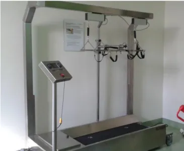
Figure 1 – Treadmill Runner 1™ (Theravet®) ... 1
Figure 2 – Examples of used devices. ... 2
Figure 3 – Cat during an UWT session ... 5
Figures 4 (left) and 5 (right) – Comparison of dogs with different average SG ... 9
Figure 6 – Canine patient with partial cranial cruciate ligament rupture performing UWT following stifle arthroscopy ...14
Figure 7 – Anterior view of a canine patient performing UWT. ...15
Figure 8 – View of the ramp and the lifting platform, used to facilitate patient access to the water treadmill ...16
Figure 9 – The Keiper™ water treadmill, from the Water Walker® brand ...16
Figure 10 – The custom-built water treadmill at the Vetmeduni ...17
Figure 11 – Foot ground contact diagram of the walk (left) and the trot (right) gait ...19
Figure 12 – Example of a normal pattern of vertical force distribution of a hindlimb over time, during the stance phase of a stride ...23
Figure 13 – Pressure plate setting in the motion analysis room ...26
Figure 14 – Example of two valid trials from one of the participant dogs ...27
LIST OF TABLES
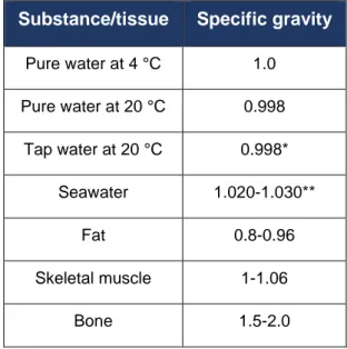
Table 1 – Specific gravity values of water and several main body tissues at atmospheric pressure ... 8Table 2 – Percentage of weight-bearing of dogs standing at different water depths ... 9
Table 3 – Breed, gender, age, body mass and BCS of all dogs taking part in this study ...25
Table 4 – Number of underwater treadmill therapy sessions performed by each dog, and their respective elapsed therapy time on the day of gait analysis ...28
Table 5 – Mean individual and group SIPFz and SIIFz for each contralateral limb pair, before and after UWT, with respective standard deviation ...30
Table 6 – Mean values and respective standard deviation of SISPD before and after UWT. 31 Table 7 – Mean values and respective standard deviations of SIPCA before and after UWT ...32
Table 8 – Mean individual and group values and respective standard deviation of step length before and after UWT, in meter (m) ...32
Table 9 – Mean values and respective standard deviations of mean velocity before and after UWT, in meter per second (m/s) ...33
vi
LIST OF GRAPHS
Graph 1 – Frequency of the main modality used for each patient, in percentage (n=143)... 4
Graph 2 – Distribution of sessions using one therapeutic modality or a combination of two to three modalities ... 4
Graph 3 – Distribution of sessions according to whether UWT therapy was performed alone or combined with other modalities ... 4
Graph 4 – Number of patients according to the different combinations of UWT with other modalities ... 5
Graph 5 – Exponential regression for the variation of step length according to height ...33
LIST OF ANNEXES
Annex I - General clinical and morphometric data of the candidates and its respective descriptive statistics. ...49Annex II – Template of Information and consent for pet owners ...50
Annex III – Water temperature measurements ...50
Annex IV – Collected kinetic data normalized to percentage of total force (%TF) ...50
Annex V – Normality tests. ...50
Annex VI – Paired t-test results ...50
Annex VII – Step length and withers height descriptive statistics, correlations and curve fit of several regression models ...50
vii
LIST OF ABBREVIATIONS AND SYMBOLS
°C – Degrees Celsius ρ – Density
%TF – Percentage of total force BCS – Body condition score cm – Centimeter
EMS – Electrical muscle stimulation
ESWT – Extracorporeal shockwave therapy GRF – Ground reaction force
IFz – Vertical impulse
LLLT – Low level laser therapy m/s – Meter per second
Kg – Kilogram
NSAID – Non-steroidal anti-inflammatory drug PCA – Pressure contact area
PFz – Peak vertical force Ppm – Parts per million SD – Standard deviation SG – Specific gravity SI – Symmetry index
SPD – Stance phase duration TF – Total force
TENS – Transcutaneous electrical nerve stimulation US – Therapeutic ultrasound
1
1. TRAINEESHIP REPORT
As part of the Integrated Masters Degree in Veterinary Medicine from the Faculty of Veterinary Medicine of the University of Lisbon I completed a 4-month training between the 26th of February of 2015 and the 30th of June of 2015, in an approximate total of 680 hours, in the section for Physical Therapy and Rehabilitation, headed by Dr Barbara Bockstahler (DVM, PD, DECVSMR (Small Animals), DACVSMR (Canine), FTA, CCRP, EBVS® European Specialist in Veterinary Sports Medicine and Rehabilitation) at the Clinic for Small Animal Surgery and Ophthalmology, University of Veterinary Medicine (Vetmeduni), Vienna, Austria. Throughout the training, I acquired skills concerning physical therapy case diagnosis and planning, resourcing from referral reports, anamnesis and complementary exams, which included MRI scan, CT scan, X-ray and motion analysis using a pressure plate and a camera. I had the opportunity to assist to and train physical examination, mainly within the neurologic and orthopaedic disciplines, comprising a significant number of geriatric and postoperative patients. Additionally, I partook in therapy planning, involving the devices and exercises, according to each case and its progression throughout sessions. I learned to work with the various tools in the department, which I operated daily.
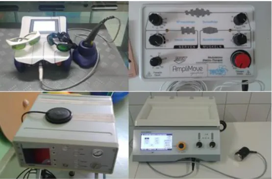
On-site rehabilitative equipment included two underwater treadmills (treadmill 1: Keiper™ model, Water Walker® brand; treadmill 2: custom-built, no brand), and a land treadmill with an adjustable sling suspension system (Runner 1™, Theravet®) (Figure 1). Other therapeutic on-site gears were a low level laser therapy (LLLT) device with Multiwave Locked System® (Mphi VET™ model, ASAlaser®), two different models of electrical stimulators which providedjjjj electrotherapy in the form ofjjjj
transcutaneous electrical nerve stimulation (TENS) and electrical muscle stimulation (EMS) (AmpliMove synchro™, Knop®;PT-2010-N™, S+B medVET®), a therapeutic ultrasound (US) apparatus (Vetri-combi™, Physiomed®) and an extracorporeal shockwave therapy (ESWT) device (Swiss DolorClast VET™, EMS®) (Figure 2). The department was also equipped with
Figure 1 – Treadmill Runner 1™ (Theravet®), equipped with a sling suspension system.
Figure 2 – Examples of used devices. Top left: LLLT device Mphi VET™ (ASAlaser®) with protective goggles for the operator and patient; top right: electrotherapy device AmpliMove synchro™ (Knop®); bottom left: ESWT device Swiss DolorClast VET™ (EMS®); bottom right: US device Vetri-combi™ (Physiomed®).Figure 3 – Treadmill Runner 1™ (Theravet®), equipped with a sling suspension system.
2
a nuclear magnetic resonance therapy machine (ProVet Station™, MBST®). However, it was not operated during the traineeship period, as it was mostly used for research purposes.
Additional tools were available to complement therapy, namely, therapy balls and rolls, balance boards, vertical weave poles, cavalletti rails, elastic bands, hot and cold packs, and vests and flotation equipment for aquatic therapy. Assistive devices for ambulation such as carts, slings, harnesses, boots, and joint protectors were available for pet owners to borrow and purchase. Contacts for reliable manufacturers of veterinary custom-made carts, orthoses, and prostheses were provided as well.
The Physical Therapy and Rehabilitation department provided the Surgery, Internal Medicine, and Intensive Care units with ambulatory physical therapy treatment for the inpatients, where I practised post-operatory and critical care handling. Also, I studied and trained massage techniques, and assisted in shockwave therapy, neural therapy, and acupuncture sessions. With the tutoring of Dr Marion Mucha (DVM, CCRP, CVA, CVPP) I learned basic principles of neural and acupuncture therapies, mostly regarding pain and stress management, as well as acupuncture needle handling. During the traineeship period, I attended a webinar lectured by Dr Mila Speciani (DVM, EBW, GP Cert WVA&CPM) from
Figure 2 – Examples of used devices. Top left: LLLT device Mphi VET™ (ASAlaser®) with protective goggles for the operator and patient; top right: electrotherapy device AmpliMove synchro™ (Knop®); bottom left: ESWT device Swiss DolorClast VET™ (EMS®); bottom right: US device Vetri-combi™ (Physiomed®).
Graph 1 – Frequency of the main modality used for each patient, in percentage (n=143).Figure 4 – Examples of used devices. Top left: LLLT device Mphi VET™ (ASAlaser®) with protective goggles for the operator and patient; top right: electrotherapy device AmpliMove synchro™ (Knop®); bottom left: ESWT device Swiss DolorClast VET™ (EMS®); bottom right: US device Vetri-combi™ (Physiomed®).
3
ASAVET®, concerning biological and therapeutic effects of general laser therapy, its modalities and applications, and the Multiwave Locked System®.
Although not numbered for this dissertation, the main objectives for patients to attend the Physical Therapy and Rehabilitation Department were for pain management, muscular reinforcement, weight loss, and neurologic rehabilitation. These usually followed surgery, trauma (car accidents, falling off windows, balconies and stairs), or were part of a degenerative joint disease management program. Surgical patients were accompanied together with a surgeon, and physical therapy was practised alongside conservative treatment, before and after surgery, as needed, adjusted to each case. Besides traditional acupuncture, a form of permanent acupuncture with gold bead implantation was used in surgery. It was mainly aimed at musculoskeletal conditions with chronic pain and/or inflammation, such as degenerative joint disease and osteochondritis.
In cases regarding severe pain management, Gabapentin was the elected drug to provide analgesia, and it was on occasion complemented with nonsteroidal anti-inflammatory drug (NSAID) therapy, as needed. Patients with neurologic disorders affecting micturition were administered Terazosin regularly, to prevent urostasis and subsequent urinary tract infection. A minor component of outpatients came from other hospitals and clinics as referrals. These were usually referred due to the specialised equipment the department provided, and to get a second opinion.
The overall number of patients observed during the traineeship period consisted of 139 dogs and 4 cats, comprising a total of 143 patients. From this group, one healthy dog attended the department for exercising purposes, as physical conditioning for rescue dog training. All the remaining animals presented a clinical condition background.
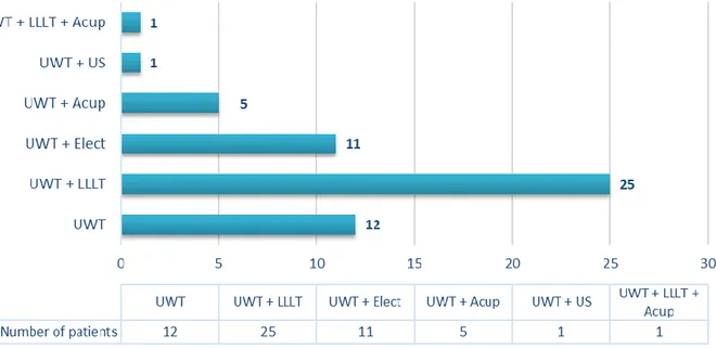
As shown in Graph 1, underwater treadmill therapy (UWT) was the most commonly practiced modality (n=55, dogs=54, cats=1). However, this graphic depicts only the main modality for each session, as most cases included a combination of two or more therapeutic modalities (Graph 2).
4
It is currently widely accepted that a rehabilitation program including a combination of different therapeutic modalities works synergistically in generating a better outcome when compared to using a single modality (Bockstahler 2004; Millis et al. 2004; Robertson 2013; Monk 2016). During the course of this study, UWT was more frequently used in combination with other modalities than it was as a single modality (Graph 3).
Graph 2 – Distribution of sessions using one therapeutic modality or a combination of two to three modalities.
Graph 4 – Distribution of sessions according to whether UWT therapy was performed alone or combined with other modalities.Graph 5 – Distribution of sessions using one therapeutic modality or a combination of two to three modalities.
Graph 3 – Distribution of sessions according to whether UWT therapy was performed alone or combined with other modalities. Graph 6 – Number of patients according to the different combinations of UWT with other modalities. UWT– underwater treadmill therapy; LLLT– low level laser therapy; Acup – acupuncture; US– therapeutic ultrasound; Elect – electrotherapy.Graph 7 – Distribution of sessions according to whether UWT therapy was performed alone or combined with other modalities.
Graph 1 – Frequency of the main modality used for each patient, in percentage (n=143). LLLT– low level laser therapy; US– therapeutic ultrasound therapy; ESWT– extracorporeal shockwave therapy; UWT– underwater treadmill therapy.
Graph 2 – Distribution of sessions using one therapeutic modality or a combination of two to three modalities.Graph 3 – Frequency of the main modality used for each patient, in percentage (n=143). LLLT– low level laser therapy; US– therapeutic ultrasound therapy; ESWT– extracorporeal shockwave therapy; UWT– underwater treadmill therapy.
5
As seen in Graph 4, UWT was routinely the first phase of the session and would either be followed by laser therapy or electrotherapy (usually TENS). It was common practice to massage the patients at the end of the session, for 10 to 20 minutes, depending on the intensity of the exercise, presence of pain or an increase in muscular tension. A warm-up massage prior to UWT was also performed on geriatric individuals and on patients with considerable muscular soreness or tension.
At the time of the traineeship, veterinary chiropractic was becoming increasingly popular in Vienna, particularly in dogs and horses. Many dogs that were on a physical therapy program also attended private chiropractors; some regularly once a week, others solely when the owners noticed muscular discomfort or postural abnormalities.
Considering the cat patients observed during the traineeship (n=4), laser therapy and acupuncture were the most frequent treatment choices (n=3), and one cat performed underwater therapy (Figure 3). Both massage and passive range of motion were fairly accepted therapy complements.
Patients attended the department typically twice a week, usually with a
Graph 4 – Number of patients according to the different combinations of UWT with other modalities. UWT– underwater treadmill therapy; LLLT– low level laser therapy; Acup – acupuncture; US– therapeutic ultrasound; Elect – electrotherapy.
6
minimum two-day interval between sessions. During the traineeship period, only two dogs did three sessions weekly. Depending on the patient’s condition, its improvement, and owner’s compliance, sessions would become progressively less frequent, up to one session monthly or total therapy cease. However, some cases, mainly associated with palsy and unsuccessful post-hemilaminectomy recovery, were kept on lifelong therapy to maximise the chances of better living quality.
Being the physical therapy team part of the research Movement Science Group, I also had the opportunity to assist to a project’s results briefing. It regarded the conception of an apparatus (Vienna Equine Surface Tester – “The BALL”) designed to measure the mechanical properties of surfaces and floors, and its potential on future research on the racing horse health and performance, which was meanwhile published (Schramel and Peham 2016) 1.
On March I wrote an Animal Use Protocol with the guidance of Dr Bockstahler, which was later presented to the University’s Ethics and Animal Welfare Committee, to obtain approval for the use of patients to represent the population sample for this dissertation.
1 Schramel J and Peham C. 2016. Vorrichtung zur Bestimmung der mechanischen Eigenschaften von Oberflächen und
7
2. LITERATURE REVIEW
2.1.
AQUATIC PHYSICAL THERAPY
According to Geytenbeek (2008), the concept of aquatic physical therapy applies to the exercise of physical therapy in a water medium, with the goal of rehabilitating or achieving a particular physical conditioning objective. Hydrotherapy is a broader concept, which comprises all types of water-based therapy performed by a variety of professional specialities, including balneotherapy, spa therapy, whirlpool, colonic irrigation, Kneipp therapy, and hydrokinesiotherapy.
Exercising in an aqueous environment is physically different from doing the same in land. In water, there are additional resistance forces involved in locomotion, and the body is subjected to buoyant and extra pressure forces. Therefore, land exercises cannot be identically mimicked in water, and to do so would imply not using the assets of exercising in water (Millis et al. 2004; Monk 2016).
To develop an efficient aquatic therapy program, it is essential first to acknowledge several intrinsic properties of water, as well as principles that apply to the immersion and movement in the water. (Lindley and Watson 2010; NARCH 2015).
2.1.1.
Basic properties of water
2.1.1.1. Fluid mechanics
a. Density and specific gravity
The density (ρ) of a substance is the quantity of mass existing per unit of volume. It is mathematically defined as the division of mass by volume. For a heterogeneous body (p.e., a mammal), the considered density is the average of all its body components (OpenStax 2017; Ling et al. 2018).
In aquatic physical therapy, the average density of a body is what fundamentally determines whether it floats or sinks in water. To determine such, a ratio is calculated that compares the density of a substance (an animal, in this case) with the density of a reference substance (water, in this case). This ratio is referred to as specific gravity (SG), also known as relative density (Monk 2016; OpenStax 2017; Ling et al. 2018).
Pure water has a SG of 1.0 at 4.00 °C, where water is at its maximum density, and it is considered the standard reference substance (Table 1). Besides temperature, density (and therefore SG) also varies with pressure and the presence of dissolved substances (OpenStax 2017; Ling et al. 2018).
8
Table 1 – Specific gravity values of water and several main body tissues at atmospheric pressure. A fat SG of 0.96 means it’s 0.04 less dense than water. Muscle and bone SG of 1.06 and 2.0 means it’s 1.06 and 2 times denser than water, respectively. *Varies from area to area, due to differences in its dissolved substances. **Varies with salinity content and temperature. (Adapted from Kerth 2013; Walker 2015; Monk and Goff 2016; Ling et al. 2018).
Substance/tissue Specific gravity
Pure water at 4 °C 1.0 Pure water at 20 °C 0.998 Tap water at 20 °C 0.998* Seawater 1.020-1.030** Fat 0.8-0.96 Skeletal muscle 1-1.06 Bone 1.5-2.0
The variations in water SG are negligible when applied to aquatic physical therapy practice. Hence, a SG value of 1 is standardly used to refer to pool2 and tap water.
If the SG of a body is lower than 1, it will float on water; if higher than 1, it will sink (Millis et al. 2004; Lindley and Watson, 2010). If SG is exactly 1, the body will remain suspended at its present depth (OpenStax 2017). This phenomenon is further discussed in the buoyancy topic below.
The SG also determines the degree of immersion of a body in water (Figures 4 and 5). If a body has a SG of 0.8, 80% of it will be submerged in water, while the remaining 20% will sit above the water surface (Monk 2016). An individual’s SG is influenced by its body condition. As such, the effects of SG on aquatic therapy are:
- Animals with higher body condition score (BCS) float more easily in the water;
- Lean or heavily muscled animals tend to sink, and thus need to make more effort to move in the water. They may require additional assistance or flotation equipment; - Animals with osteoporosis will have a lower bone SG and, consequently, tend to float
more easily. (Bockstahler et al. 2004; Mikail 2006; Monk 2016).
2Pool water has a standard chlorine content of 0.001% to 0.003% in veterinary aquatic therapy practices, so it’s considered to have a SG similar to tap water (NARCH 2015).
9 b. Buoyancy
Buoyancy is the upward force of the water on an immersed or floating body. It is experienced as a thrust of the body towards water surface (Bockstahler et al. 2004; Mikail 2006). According to Archimedes’ principle, the value of the buoyancy of a body equals the weight of the water it displaces. In its turn, the weight of water displaced depends on the SG of the body. The buoyant force is always present on any body in water, whether it floats or not. (OpenStax 2017; Ling et al. 2018).
In aquatic physical therapy, buoyancy substantially reduces the weight the animal must carry (Bockstahler et al. 2004). The interaction between weight-bearing and water depth in dogs was assessed by Levine et al. in 2002 (Table 2).
Table 2 – Percentage of weight-bearing of dogs standing at different water depths. Comparing to normal weight-bearing on land, the results obtained showed the higher the water level, the less weight-bearing occurs (Levine et al. 2002). Additional investigation in various breeds and sizes of dogs is needed, as well as the study of variations on weight-bearing during ambulation (Monk 2016).
Immersion depth Weight-bearing (%)
Tarsus 91
Stifle 85
Hip 38
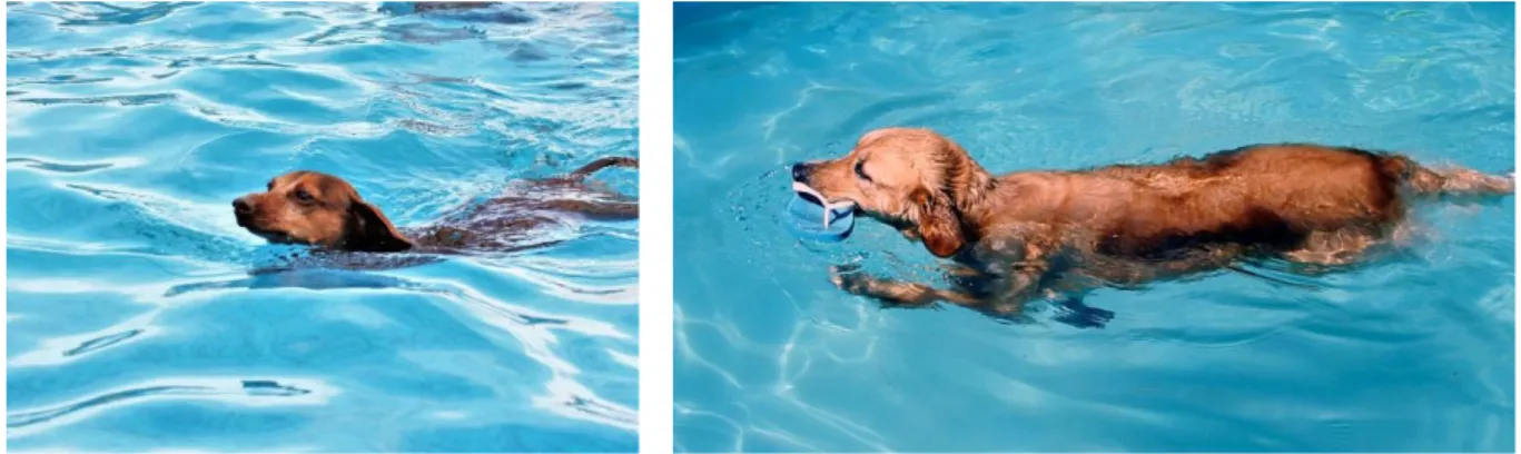
Figures 4 (left) and 5 (right) – Comparison of dogs with different average SG. The dog in figure 4 is deeper immersed in water, compared to the dog in figure 5.
https://www.needpix.com/photo/download/1522834/nature-animals-pets-dogs-browndog-dachshund-wienerdog-swimming-bluewater
10
Since ambulatory limitations are typically due to an inability to support weight normally during a full gait cycle, animals may show improved ambulation when load-bearing is reduced. (Shmalberg 2018).
The implications of buoyancy and reduced weight-bearing on animal patients are: - Unloading of painful joints by raising the water level above these (Bockstahler et al.
2004; Lindley and Watson 2010; Steiss 2010);
- Allowing ambulation when land-based exercise is contraindicated (Jackson et al. 2002). It includes starting ambulation earlier in recovery, for example in intervertebral disc protrusions and postoperative rehabilitation following cruciate ligament repair;
- Provide a milder transition to land-based exercise, post-surgery or injury (Shmalberg 2018);
- Rehabilitation of muscular weakness (Steiss 2010; NARCH 2015).
Buoyancy can additionally be used to increase resistance to movement, namely with flotation devices (NARCH 2015). If an anatomical part moves parallel to the water surface, buoyancy eases the movement acting as support. If it moves perpendicularly, buoyancy works as a resistance to movement. (Mikail 2006).
Besides buoyancy, a body immersed in water is also subjected to gravity, which acts as an opposing force. If the centre of buoyancy and the centre of gravity are not aligned in the same vertical plane, the animal will not be in equilibrium and will tend to tip forward or tilt sideways (Lindley and Watson 2010; Monk 2016).
In practice, animals with an amputated or a flexed limb will tend to rotate down to the opposite side to reach equilibrium. Patients with spinal injury or asymmetrical tone may struggle to control trunk rotation. Flotation devices are generally used to aid but need to be placed accordingly to compensate the imbalance (Monk 2016).
b. Hydrostatic pressure
Hydrostatic pressure is the pressure force effected on an immersed body due to the weight of a liquid (Ling et al. 2018). It is directly proportional to the immersion depth and fluid density: the greater the depth and fluid density, the greater the pressure. (Bockstahler et al. 2004).
Implications for animal patients:
- Water exerts resistance to thoracic expansion when inhaling. Therefore, caution is advised when submerging patients with cardiac or respiratory conditions (Mikail 2006; NARCH 2015);
- Hydrostatic pressure is beneficial for swollen joints and oedematous tissue located in the distal portion of limbs, which are deeper submerged, and aids venous return. It also
11
decreases nociception due to phasic stimuli exerted to the sensory receptors on the skin, thus allowing more movement with a lesser sensation of pain (NARCH 2015).
c. Viscosity and resistance
The viscosity of a fluid refers to the frictional resistance to flow, which is dependent on the cohesive forces at a molecular level. Resistance consists of the force exerted by a solid body moving through the fluid and is dictated by viscosity (Bockstahler et al. 2004).
Implications for animal patients:
- Water provides resistance that promotes muscular strengthening and cardiovascular fitness;
- There is a possible increase in sensory awareness; - Assistance in stabilising unstable joint/s;
- There is a greater prevention of falling by increasing the time for an animal to react, improving its willingness to move. This is particularly relevant in spinal patients (NARCH 2015);
- Both buoyancy and hydrostatic pressure provide a supporting feeling to the patient while submerged in water (Mikail 2006).
d. Turbulence
Water turbulence consists of an irregular water flux which increases water-resistance to the patient’s movement when compared to a continuous unidirectional water flux. Generating water turbulence through manual water agitation or jet streamers creates variations of pressure in different body parts. It additionally has a massaging effect, enhancing blood and lymph circulation, and gently removing wound exudates and debris (Mikail, 2006).
e. Surface tension
Water molecules tend to have greater adherence among themselves near the surface. This surface tension creates a higher resistance to movement on the surface (Bockstahler et al. 2004).
Implications for animal patients:
- Exercising near the surface is more difficult, and must be accounted for with debilitated animals (Bockstahler et al. 2004);
12
2.1.1.2. Other properties
a. Temperature
Millis et al. (2004) recommend that healthy dogs exercise with a water temperature range between 26-28 °C in pools, and approximately 25,5-32 °C in aquatic treadmills.
Heated water – The main physiological effects of exercising in water temperatures above normal body temperature are: increase of respiratory frequency, increase of cardiac frequency, increase in the heart returning blood flow, decrease of blood pressure, increase in muscular blood flow, increase in peripheral circulation, increase in metabolic rate, general muscular relaxation and increase in joint flexibility. The increase in circulatory flow enhances oxygen intake and carbon dyoxide and lactic acid removal, thus reducing muscular discomfort. Due to the increased strain in cardiac output, caution is advised when exercising cardiac and geriatric patients in heated water (Millis et al. 2004; Mikail 2006).
Cold water – Water temperatures below normal body temperature are apparently well tolerated by dogs, particularly individuals with thicker coats (Millis et al. 2004). The main physiological effects of exercising in cold water are cellular metabolism decrease, reduction in capillary permeability and pain relief. The cold water also helps patients exercising with active inflammation (Mikail 2006). Subjects that perform UWT in cold water show lower heart rates than individuals exercising on land treadmills at the same velocity and length of time (Millis et al. 2004).
b. Salinity
Besides increasing the density of water, the addition of salts to water also increase osmolarity and osmotic pressure. Water with a high content in salt helps “draining” swollen tissues and open exudative wounds (Mikail 2006). The salinity changes the specific gravity much more than the temperature does (Walker 2015).
c. Oxygenation
A higher concentration of oxygen in water enhances tissue healing, similarly to the increase that occurs in hyperbaric chambers. (Sen 2009; Ladizinsky and Roe 2010). The mean oxygen content in water at room temperature is around 2,4 parts per million (ppm) and increases to 8-11 ppm when at 2°C. Using jet streamers can also contribute to improving water oxygenation (Mikail 2006).
13
2.1.2.
Physiological effects of immersion
Body immersion in water at body temperature leads to well documented physiological changes. Some of those changes have already been discussed previously in this dissertation, and include a shift in blood volume from peripheric to central circulation caused by hydrostatic pressure, increased cardiac volume output, reduced heart rate and inspiratory volume changes (Millis et al. 2004; NARCH 2015). Although no clear mechanism has been described, it is also proposed that immersion reduces sympathetic nervous system activity. Sympathetic nervous system activation has been associated with some of the detrimental consequences of chronic stress and illness (Becker et al 2009).
2.1.3.
Physiological effects of exercising in water
Exercising in water differs greatly from exercising on land (Monk 2016). Studies in humans described that metabolic requirements, oxygen uptake and heart rate were greater while exercising in water compared to performing the same exercises on land (Whitley and Schoene 1987; Johnson et al. 1977). Both in dogs and humans, it has been described that peak heart rate, blood lactate and oxygen uptake are lower in individuals exercising in water, which means that exhaustion is achieved with a smaller work rate when compared to exercising on land (Becker 2004; Mikail 2006).
A study by Nganvongpanit et al. (2014) reported that swimming significantly improved the range of motion in dogs affected by hip osteoarthritis. Another study by Marsolais et al. (2003), suggested that swimming promoted significantly greater range of motion of the stifle and tarsal joints than walking in dogs following CrCL rupture correction surgery. Yet another one by Preston and Wills (2018) concluded that aquatic therapy increases range of motion and step length in labrador retrievers with elbow pathology and can be of benefit for canine elbow dysplasia management.
Energy expenditure when exercising in water can vary significantly compared to the same exercise on land. On one hand, buoyancy reduces bodyweight and therefore reduces the amount of energy required to counter gravity. On the other hand, water viscosity, friction and turbulence require increased work to overcome resistance during movement. Water temperature also impacts energy expenditure, as thermoregulation mechanisms will need to balance body temperature if the water is too cold. Shivering due to low temperatures also increases energy consumption (Monk 2016).
14
2.1.4.
Underwater treadmill therapy
Underwater treadmill therapy is a modality that, like swimming, reduces weight-bearing on joints and adds resistance to movement due to the water’s attributes, while also enabling the execution of a range of motion similar to normal gait in land (Schmalberg 2018). Therefore, this type of aquatic therapy is selected when there is a need to exercise the patient in a normal gait pattern, especially in those cases where walking on ground would result in repeated or serious injury due to muscle weakness or improper balance (Millis et al. 2004).
2.1.4.1. Indications
The major indications for UWT are rehabilitation following orthopaedic surgery or neurological injury and improving joint and muscle strength (Bockstahler et al. 2004) (Figure 6). Animals that have been recumbent for long periods of time benefit from aquatic therapy as a means to strengthen muscle mass while bearing reduced weight (Lindley and Watson 2010). It may also be useful in patients with peripheral oedema, muscle spasm, and to improve confidence in dogs reluctant to walk on ground. Due to having a greater muscle and cardiovascular demand compared to land treadmill, it is also indicated for athletic conditioning and weight management (Millis et al. 2004).
The recommended water temperature varies with the condition treated: neurologic patients are usually recommended to walk with warmer water (approximately 29,5-32 °C), while orthopaedic patients and dogs in conditioning regimens require less warm temperatures (approximately 25,5-28,5 °C) (Millis et al. 2004).
Figure6 – Canine patient with partial cranial cruciate ligament rupture performing UWT following stifle arthroscopy.
Figure 10 – Anterior view of a canine patient performing UWT.Figure11 – Canine patient with partial cranial cruciate ligament rupture performing UWT following stifle arthroscopy.
15
2.1.4.2. Contraindications and precautions
Aquatic therapy is not recommended in the case of open wounds, infections, cardiac or respiratory conditions, uncontrolled epilepsy, urinary incontinence and diarrhoea (Mikail 2006). Dogs with surgical sutures that have not healed completely and do not have a waterproof cover should not be subjected to underwater therapy. Caution should be exercised in patients with laryngeal paralysis, skin or ear problems, epilepsy, and mild systemic compromise. Geriatric patients and those with very high BCS should be monitored closely during UWT (Monk 2016). If a dog exhibits fear or aversion to water and needs aquatic therapy, it is advised to first introduce the animal slowly to the room and encourage playing and exercising before actually starting therapy. Same animals might not be able to perform aquatic therapy due to the possibility of injuring themselves or the operators (Lindley and Watson 2010).
2.1.4.3. Comparing underwater treadmill therapy and swimming
When swimming, almost the entire body is immersed, as only the head and part of the neck are outside the water. Additionally, there is no contact between the patient and the ground, requiring a constant motion of the limbs to maintain the head above the surface (Mikail 2006). As such, swimming is the modality of choice for improving range of motion and in patients with severe osteoarthritis, as they are able to exercise while bearing no weight on their limbs (Schmalberg 2018). With the ribcage immersed in water, increased respiratory work is required to overcome the pressure exerted by the water during inhaling. This results in a better cardiorespiratory capacity, and improved venous return and cardiac output (Mikail 2006).
While swimming is certainly beneficial in some situations, underwater treadmill therapy is the
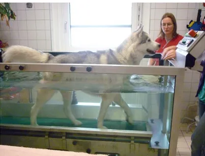
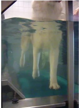
better choice when the goal is improving proprioception and dynamic balance, as direct contact with the ground and the need to maintain a walking stance forces the patient to work its muscles and joints, while supporting less weight (Schmalberg 2018) (Figure 7).
Figure 7 – Anterior view of a canine patient performing UWT.
16
2.1.4.4. Underwater treadmills used for the traineeship and study
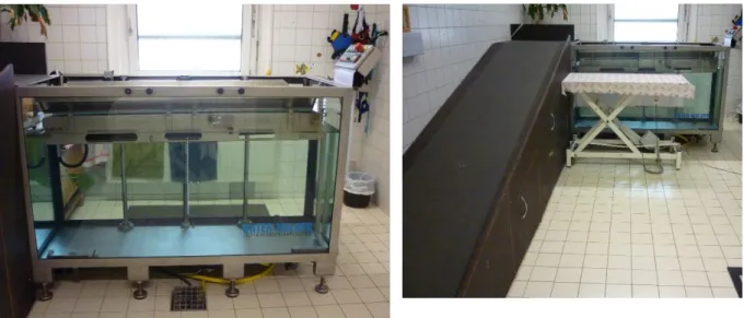
As previously stated in the traineeship report, there were two underwater treadmills at the Physical Therapy and Rehabilitation section at the Vetmeduni.
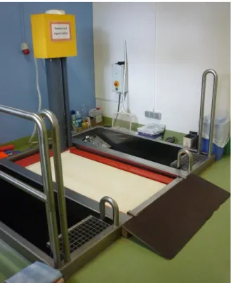
The Keiper™ treadmill (Figure 8) was set above floor level, with a ramp and a lifting platform (Figure 9), so the patients could access it. It was equipped with a spindle lift to change the incline, a water heating system, and a counter-current jet streamer that provided the option to increase water resistance during exercise. The physiotherapist stood by one of the sides of the treadmill, within reach of both the controls and the patient, and the owner/caretaker stood at the front facing the patient for motivation and positive reinforcement.
The custom-built treadmill (Figure 10) was purposely designed for the Vetmeduni. It worked through an exterior motor with adjustable speed to run the treadmill, and a hydraulic system to lift and lower the treadmill in and out of water. It was set below floor level, with sideways access on both sides. The treadmill was lifted to floor level for the patients to access through a small incline ramp. There was also a custom-made harness lift system to facilitate access. The physical therapist stood on the right-side pit to access both controls and patient, and the patient’s caretaker was positioned either on the left-side pit or outside, facing the patient.
Figure 8 – The Keiper™ water treadmill, from the Water Walker® brand.
Figure 9 – View of the ramp and the lifting platform, used to facilitate patient access to the water treadmill.
17 Comparing both treadmills, the Keiper™ treadmill offered a more accurate speed control and better assessment of the patient’s locomotion during exercise through its transparent panels. It was preferable for nervous dogs since some dogs stressed when they were lowered below floor level. The custom-built treadmill was more easily accessible for patients with ambulatory limitations and more practical for hoisting larger and heavier individuals. It also allowed better control over limb movement when assistance for walking underwater was needed.
Regarding maintenance, the Keiper™ was more environmentally and cost-friendly through multiple re-using of
the water with filter systems, enabling greater intervals between cleaning, but being more time-consuming to clean. The custom-built treadmill had no filtering system to use between therapy sessions. It was easier to clean but required much more regular maintenance, which implied being emptied of water every time. Both treadmills were used for this study since each one presented assets that best suited the different types and conditions of the dogs.
Figure 10 – The custom-built water treadmill at the Vetmeduni.
18
2.2.
CANINE GAIT ANALYSIS
Biomechanics is described as the study of the principles of mechanics applied to biological systems, particularly regarding structure and function. It concerns the effects of forces on body motion (Hatze 1974; Karduna 2007). Knowledge on the mechanical demands and constraints that occur during locomotion enables the assessment of compensations, secondary neuromuscular and skeletal problems and pathology that follow the failure of one or more elements. It is therefore essential in the diagnosis of numerous musculoskeletal and neurologic conditions and in the delivery of physical therapy. (Adrian 2016; Carr and Dycus 2016).
There are two primary types of locomotion: gait and nonrepetitive motions (Millis et al. 2004). Gait is a repetitive sequence of movements which drive an animal forward. It consists of a way to translocate a body from one point to another in space, developed to minimise unwanted displacements that translate into energy costs (Hildebrand 1977). Nonrepetitive motions involve single events such as sitting, jumping, and movement initiation (Millis et al. 2004).
2.2.1.
Normal gait
Currently, sound dogs are considered to use four main gaits: walk, trot, canter and gallop (Zink and Carr 2018). Some authors also include swim, particularly the paddle (Millis et al. 2004; Catavitello 2015).
The dynamic of a gait is influenced by the properties of the surface, its incline, ground movement (i.e., treadmill), submersion in water, and the curvature of the path. Body conformation and breed deeply influence an individual’s moving performance as well (Bertram et al. 2000; Millis et al. 2004; Mölsa et al. 2010).
As repetitive locomotion, gait is composed of a series of strides. A stride consists of a cycle of body motion that starts with the contact of one foot and ends with that same foot again contacting the ground. In a stride, each limb undergoes a step cycle. Each limb’s complete step cycle is divided in stance phase and swing phase. Stance phase initiates with ground contact, where braking forces take place, and is followed by propulsion. After stance phase, swing phase starts, in which the foot is suspended and not contacting the ground (Millis et al. 2004).
In quadrupedal mammals, normal gait can be divided into symmetric and asymmetric gaits, according to how the body moves during each stride. In symmetric gait, the movements of the limbs are mirrored in the sagittal plane by their contralateral parts. In asymmetric gait, the left and right side of the body do not mirror each other (Alexander 1984; Budsberg et al.
19
1993). Examples of normal symmetric gaits are the pace, walk and trot; of asymmetric, are the rack, gallop and canter.
In a healthy standing dog, approximately 30% of its weight is supported by each of the forelimbs, and 20% is supported by each of the hindlimbs (Zebas et al. 1991). Such weight distribution positions the centre of gravity at mid-chest level, behind the scapula. This proportion equilibrium results that when moving forward in a plane ground surface, the forelimbs are mainly responsible for braking, while the hindlimbs are in charge of the majority of propulsion (Millis et al. 2004).
Because the experimental part of this dissertation involves solely the analysis of the walking gait and uses the trot for comparison purposes, the literature review will focus on these gait types.
2.2.1.1. Walk
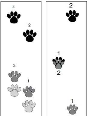
The walk is the slowest gait a sound dog performs. It is also the only gait where there is a phase with three feet simultaneously contacting the ground.
A full walking cycle (Figure 11, left) follows the pattern: right hindfoot, right forefoot, left hindfoot, left forefoot. Simply put, it starts with a hindfoot, followed by its ipsilateral forefoot, and then the same is performed by the contralateral limbs. Each hindfoot steps right ahead of where the forefoot was before (Zink and Carr 2018). The centre of gravity remains central throughout the whole cycle (Prydie and Hewitt 2015).
Due to its speed, the walk is considered the easiest gait to visually assess. Though, it has the disadvantage of difficulting the detection of subtle lameness and that dogs are more prone to being distracted at walking speed (Millis et al. 2004).
Figure 11 – Foot ground contact diagram of the walk (left) and the trot (right) gait. The numbers represent the sequence of footfall. The black prints depict the forefeet, the dark-grey depict the hindfeet, and the light-grey depict the forefoot from the precedent stride. (Adapted from Zink and Carr 2018).
Figure 18 – Example of a normal pattern of vertical force distribution of a hindlimb over time, during the stance phase of a stride. Each coloured line represents a different trial of the same hindlimb. The PFz of each trial is their
20
2.2.1.2. Trot
In the trotting gait, the two diagonal legs contact the ground simultaneously. It follows the pattern: left forefoot with right hindfoot, then right forefoot with left hindfoot. As observed in Figure 11 (right), two of the feet contact the ground in the same spot. It occurs every time one of the hindfeet moves forward, stepping into the place where the ipsilateral forefoot was moments before (Prydie and Hewitt, 2015; Zink and Carr 2018). In most breeds, when a dog is trotting there is a moment of suspension between the contact of each diagonal pair (Elliott 2009). The trot is considered the best gait to visually detect lameness. This is due to being the only type of gait in which the limb contacting the ground is never assisted by its contralateral limb (Millis et al. 2004).
2.2.2.
Lameness
Lameness consists of a disturbance of normal gait, that affects the weight-bearing of one or more limbs. It can be of anatomical or pathologic nature. Anatomical lameness can be genetic (e.g. improper body conformation) or acquired (e.g. vitamin D deficiency). It may or not be generated by pain. Pathological lameness can occur due to neural (e.g. cauda equina) or musculoskeletal causes (e.g. hip dysplasia). Typically, musculoskeletal lameness is triggered by pain (Gillette 2011).
The presence of lameness promotes complex adjustments in locomotion, which may generate secondary conditions (Wilson and Smith 2016). For instance, in the presence of unilateral lameness, a decrease in load in the lame limb is expected. This decrease is compensated by the transition of forces to other extremities (Bockstahler et al. 2009).
Analysing symmetrical gait types is recommended to assess lameness, as it is easier to detect (Colborne et al. 2011; Oosterlinck et al. 2011). Because of their symmetry and speed, the walk and trot are the two types most frequently used in lameness evaluation. Moreover, dogs tend to naturally perform them when incentivised to move (Weigel et al. 2005).
2.2.3.
Methods of gait analysis
Gait analysis is currently performed resourcing both to subjective and objective methods (Carr and Dycus 2016).
Observational gait analysis, also known as subjective gait analysis, involves the visual assessment of the patient’s locomotion at all angles, usually walking and trotting. It is recommended to be performed prior to other physical examinations, since the palpation of limbs and joints may influence subtle lameness. This method of gait analysis can include videotaping, and either a numerical rating score or a visual analogue scale. It is considered
21
quick and inexpensive and is currently the most common practice when assessing lameness in a clinical setting (Huntingford and Fossum 2019).
Instrumented gait analysis, commonly named objective gait analysis, is considered to encompass some of the most accurate methods to assess locomotion. It allows the quantification of changes in gait, and the validated methods possess a higher sensitivity power in detecting subtle lameness (Huntingford and Fossum 2019). Owing to these attributes, it has been increasingly used in the developing of treatment plans and in the monitoring of patient progress (Carr and Dycus 2016).
Numerous methods for gait analysis have been developed in the last decades. Among the most well-established techniques in veterinary practice are kinematic and kinetic gait analysis (Griffon 2008).
Kinematic analysis evaluates the characteristics of motion from a spatial perspective. It involves the positioning of reflective markers in joint landmarks, and the use of 3D cameras to subsequently measure position, velocity, and acceleration of the body, limbs and joints. 2D systems are also available but are less effective (Weigel et al. 2005). It is mostly used for measuring changes in stride length and in joint angles during gait (Millis et al. 2004).
Kinetic gait analysis quantifies the forces involved in locomotion, mainly in relation to the ground. It includes the measurement of braking, propulsive, horizontal and peak vertical forces; vertical, braking, and propulsive impulses; rates of loading and pressure distribution within the paw (Millis et al. 2004). It is the most common technique for describing normal and abnormal locomotion (Schnabl-Feichter et al. 2017). Kinetic data can be collected using one or multiple force plates, or a pressure-sensitive walkway (also referred to as “pressure plate”) (Gillette and Angle 2008). Both are considered well-validated reliable measuring tools (Bockstahler, Skalicky et al. 2007; LeQuang, Maitre, Colin et al. 2010; LeQuang, Maitre, Roger et al. 2010).
It is common for both the kinematic and kinetic methods to incorporate the time dimension in the analysis of their parameters (Gillette and Angle 2008).
Several studies comparing subjective and objective lameness assessment indicate that due to its subjective nature, observational gait analysis may not be able to consistently detect subtle lameness (Oosterlinck et al. 2011; Lane et al. 2015; Carr and Dycus 2016). Nevertheless, observational gait analysis is still a practical tool in clinical practice and should integrate a complete orthopaedic, neurological and rehabilitation examination procedure.
For the experimental part of this dissertation, a pressure plate with a video recorder was used to perform kinetic gait analysis, including temporospatial variables.
22
2.2.3.1. Kinetic gait analysis
In kinetic gait analysis, the measured forces occur along three axes: vertical (z), horizontal (y) and transverse (x). Because these are all reaction forces occurring at the point of contact with the ground, they are referred to as ground reaction forces (GRFs) (Zink and Carr 2018).
The assessment of GRFs is based on Newton’s third law of motion, which states that for every action force occurs an equal, collinear, and opposite reaction force (Weigel et al. 2005). The reaction force along the vertical (z) is the force that occurs perpendicular to a plane ground surface. It represents the weight-bearing dimension of the resultant force. The reaction force along the horizontal (y) axis is composed by the propulsion and the braking components. Propulsion occurs in the positive direction of the resultant force, whereas braking occurs in the negative direction. Lastly, the reaction force along the transverse (x) axis concerns mediolateral forces. It is considered minor and is usually not quantified in most kinetic studies in dogs (Millis et al. 2004).
According to the parameters measured in this dissertation’s clinical study, the following text will be dedicated to describing peak vertical force, vertical impulse, paw pressure contact area, and symmetry indexes. The parameter of stance phase duration was previously mentioned in the normal gait subchapter.
a. Ground reaction forces
Peak vertical force (PFz) represents the maximum force of the vertical GRF of a limb during stance. It is a punctual moment in time in the force time curve of a stride.
Vertical impulse (IFz) is composed of the sum of the vertical force of a limb over time. Mathematically, it is represented by the area under the force-time curve (Zink and Carr 2018)
Both PFz and IFz variables are depicted in Figure 12.
b. Paw pressure contact area
This variable represents the surface area of a paw that is contacting the ground (Millis et al. 2004).
c. Limb symmetry
As in previous human (Soudan 1982) and equine (Merkens et al. 1985) locomotion studies, in 1993 Budsberg et al. documented that healthy dogs did not present perfect right-left symmetry at a trot. Instead, they presented small but consistent variations in weight-shifting.
23
The percentage of asymmetry accepted in nonlame dogs has been the focus of numerous canine gait analysis studies. Symmetry or asymmetry indexes are currently considered the gold standard in the assessment of lameness in several species (Budsberg et al. 1993; Schnabl-Feichter et al. 2017). They can be calculated for each of the discussed kinetic parameters. Namely symmetry indexes of PFz and IFz are used in canine gait analysis (Oosterlinck et al. 2011), as they are considered the parameters with the higher reliability, and have been investigated by several researchers for a SI cut-off value to distinguish lame from non-lame individuals (Budsberg et al. 1993; Fanchon et al. 2007; Volstad et al. 2017). Typically lower PFz and IFz show lower values in the affected limb of a lame dog (Gillette and Angle 2008), which can generate an imbalance in a limb pair and therefore be categorized as an asymmetry.
Figure 12 – Example of a normal pattern of vertical force distribution of a hindlimb over time, during the stance phase of a stride. Each coloured line represents a different trial of the same hindlimb. The PFz of each trial is their respective highest point marked in the graph. The IFz of each trial is the area under their respective curve. The stance phase of a stride typically has an “M” shape, due to the peak of the first ground contact, and then the peak of the propulsion before the foot leaves the ground (Millis et al. 2004). This graph was automatically translated using the gait analysis dedicated software available at the Vetmeduni (Pressure Analyzer 1.3.0.2; Michael Schwanda®).
24
3. MATERIALS AND METHODS
3.1.
Introduction
Computer-assisted gait analysis is currently considered a cornerstone in the veterinary biomechanical field (Gillette and Angle 2008; Carr and Dycus 2016). Kinetic analysis is one method of gait analysis which evaluates weight-bearing alterations by measuring reaction forces. It is a reliable tool in the diagnosis of locomotion disorders, and evaluation of different treatment effects, supporting adjustments to produce a better outcome (McLaughlin 2001; LeQuang, Maitre, Roger et al. 2010). It is expected that as adherence to veterinary physical therapy and rehabilitation grows, the use of gait analysis in varied settings will increase as well (Weigel et al. 2005; Feeney et al. 2007; Griffon 2008).
The benefits of underwater treadmill therapy in dogs have been extensively recognised and discussed in veterinary literature (Levine et al. 2002; Jackson et al. 2002; Bockstahler et al. 2004; Dunning et al. 2004; Millis et al. 2004; Chauvet et al. 2011; Monk 2016; Bertocci et al. 2018). However, much data is still required to more accurately understand its influence on canine locomotion, being one of the cases individuals recovering from orthopaedic lameness.
3.2.
Objective
The aim of this study was to use a pressure plate to compare ground reaction forces (GRFs) in a heterogeneous population of dogs with lameness due to appendicular orthopaedic condition(s), before and after a physical therapy session on a water treadmill. It was proposed to measure changes in symmetry variation of vertical ground reaction forces, stance phase duration, and paw pressure contact area between contralateral limb pairs, as well as variation in step length. It was also proposed to investigate whether a correlation existed between the withers height and the step length of each dog.
3.3.
Candidates
Fourteen client-owned dogs enrolled in the clinical study, from April 2015 to July 2015, for voluntary participation. Eligible candidates were selected by medical record investigation. Inclusion criteria comprised a clinical history of lameness due to an orthopaedic condition originated in one or both contralateral limb pairs, and ongoing underwater treadmill treatment. Exclusion criteria included simultaneous orthopaedic conditions in both fore and hindlimbs, abnormal findings on a routine physical examination, previous neurologic diagnosis or abnormal findings on neurologic examination, or a diagnosis of a non-orthopaedic condition.
One dog was excluded due to limb suspension during locomotion, which affected the consistency of measurements; 4 dogs were excluded after measurements, as they were no
25
longer considered lame. The 9 candidates that met the inclusion criteria were divided into two groups, based on whether the orthopaedic condition occurred in the forelimbs (Group A) or in the hindlimbs (Group B), as shown in Table 3. Each dog was assigned a number, to identify them throughout the study.
In Group A, mean age ± standard deviation (SD) was 5.13 ± 3.29 years, ranging from 0.5 to 8.08 years. Mean body mass ± SD was 23.55 ± 14.02 kilogram (kg); mean BCS ± SD was 6.25 ± 1.71, and 3 dogs were appraised as overweight (BCS ≥ 6/9) using the Purina® Body Condition Tool for Dogs (Laflamme 1997). There were 2 individuals affected in one forelimb, and 2 in both forelimbs. Regarding medication, participant number 4 was receiving Cimicoxib (2.5 mg/kg daily) to alleviate the symptoms of bilateral elbow dysplasia aggravated by omarthrosis and cubarthrosis.
Group B participants mean age ± SD was 6.42 ± 4.17 years, ranging from 2.33 to 11.50 years. Mean body mass ± SD was 27.36 ± 12.56 kg; mean BCS ± SD was 5.60 ± 0.89, and 2 dogs were considered overweight. There were 3 dogs affected in one hindlimb and 2 in both hindlimbs. Participant number 2 was on oral Carprofen as needed (1.6 mg/kg). It was diagnosed with cranial cruciate ligament rupture and treated with unilateral tibial tuberosity advancement surgery 98 days before measurements day.
Table 3 – Breed, gender, age, body mass and BCS of all dogs taking part in this study. F – sexually intact female; FS – spayed female; M – sexually intact male; MN – neutered male.
Figure 20 –Pressure plate setting in the motion analysis room. The measurement area is marked with white tape and covered under a rubber mat. In the corner, is the video camera used to film
the trials.Table 3 – Breed, gender, age, body mass and BCS of all dogs taking part in this study. F – sexually intact female; FS – spayed female; M – sexually intact male; MN – neutered male.
Group A
Dog Breed Gender Age (years) Body mass (kg) BCS (x/9)
1 Mixed breed FS 5,25 42,0 8
2 Mixed breed FS 8,08 26,3 7
3 Irish Terrier F 0,50 10,0 4
4 Mixed breed M 6,67 15,9 6
Group B
Dog Breed Gender Age (years) Body mass (kg) BCS (x/9)
1 Icelandic Sheepdog MN 3,42 18,8 5
2 Mixed breed FS 11,5 32 7
3 Dogo Argentino X Labrador FS 4,58 40,0 5
4 King Charles Spaniel M 2,33 10 6