INTRODUCTION
Orofacial clefts are among the most common congeni-tal malformations of the craniofacial region,1–4 which in-clude cleft palate only (CP) and cleft lip with or without palate (CL/P). Their estimated incidence worldwide is about 1 in 1,500–2,000 births for CP and 1 in 700–1,000 births for CL/P, showing a considerable sex, ethnic, and geographic variation.1–3,5,6 For instance, the highest
incidence rates for CL/P were reported in Native Ameri-cans and Asians (China, Japan), and the lowest in AfriAmeri-cans and Southern Europeans. On the other hand, incidence rates for CP seem similar in Europeans, Africans, Native Americans, and Asians.3,5–7
Although orofacial clefts most commonly appear as isolated conditions, with a generally favorable outcome for the patients, it has long been known that they may Background: Orofacial clefts are among the most common congenital craniofa-cial malformations and may be associated with other birth defects. However, the proportion and type of additional anomalies vary greatly between studies. This study assessed the prevalence and type of associated congenital malformations in children with orofacial clefts, who attended the largest cleft lip and palate tertiary referral center in Portugal.
Methods: Consecutive children with orofacial clefts who attended at least 1 con-sultation in our Clefts Unit between 1981 and 2012 were studied. Demographic and clinical data regarding the number and type of associated malformations were retrospectively collected and analyzed.
Results: Of the 701 patients studied, 219 (31.2%) had associated congenital mal-formations. These malformations were more frequent in children with cleft pal-ate (43.4%) than in children with cleft lip and palpal-ate (27.5%) or with cleft lip only (19.4%). Within the group with associated anomalies, 73 cases (33.3%) had condi-tions related with known chromosomal defects, monogenic syndromes or sequences, and 146 cases (66.7%) had multiple congenital anomalies of unknown origin. From those, head and neck malformations were the most common (60.3%), followed by malformations in the cardiovascular (28.3%) and musculoskeletal systems (26%). Conclusions: The overall prevalence of associated malformations of nearly 1 in 3 children with orofacial clefts stressed the need for a comprehensive evaluation of these patients by a multidisciplinary cleft team. Moreover, one-third of the children had multiple congenital anomalies of known origins. Thus, early routine screen-ing for other malformations and genetic counselscreen-ing might be valuable for orofa-cial clefts management. (Plast Reconstr Surg Glob Open 2018;6:e1635; doi: 10.1097/ GOX.0000000000001635; Published online 9 February 2018.)
Alice V. Pereira, MD* Nuno Fradinho, MD* Sara Carmo, MD, MSc† Juliana M. de Sousa, MD, MSc* David Rasteiro, MD* Regina Duarte, MD† Maria J. Leal, MD†
Associated Malformations in Children with
Orofacial Clefts in Portugal: A 31-Year Study
Pediatric/Craniofacial
Disclosure: The authors have no financial interest to declare in relation to the content of this article. No funding was received for this article and the Article Processing Charge was paid for by the authors.
Copyright © 2018 The Authors. Published by Wolters Kluwer Health, Inc. on behalf of The American Society of Plastic Surgeons. This is an open-access article distributed under the terms of the Creative Commons Attribution-Non Commercial-No Derivatives License 4.0 (CCBY-NC-ND), where it is permissible to download and share the work provided it is properly cited. The work cannot be changed in any way or used commercially without permission from the journal. From the *Serviço de Cirurgia Plástica Reconstrutiva, Hospital de
São José, Centro Hospitalar de Lisboa Central, Lisboa, Portugal; and †Serviço de Cirurgia Pediátrica, Hospital de Dona Estefânia, Centro Hospitalar de Lisboa Central, Lisboa, Portugal.
Received for publication August 30, 2017; accepted November 17, 2017.
Presented at the 49th National Congress of the Spanish Society of Plastic, Reconstructive and Aesthetic Surgery (SECPRE) and 44th National Congress of the Portuguese Society of Plastic, Reconstructive and Aesthetic Surgery (SPCPRE) 2014 in Granada, Spain. Ethical standards: This study has been approved by the appropriate ethics committee and has therefore been performed in accordance with the ethical standards laid down in the 1964 Declaration of Helsinki and its later amendments.
PRS Global Open
•
2018
be frequently associated with other congenital malforma-tions.8–11 In these cases, the outcome depends primarily on the presence and type of associated malformations.12 However, the proportions of patients with orofacial clefts with additional abnormalities varies greatly between stud-ies, from 1.5% to 64.2%.8–11,13–22 Also, there is no consensus on the type of malformations that are most commonly as-sociated with orofacial clefts.8,10,14,21,22
The interplay of different environmental and genetic risk factors has been proposed as an underlying mecha-nism for orofacial clefts. However, a single major risk factor for these congenital malformations has not been identified yet, suggesting a more complex etiology than the oligogenic model originally proposed.23–27 Moreover, consanguinity and a positive family history for orofacial clefts also play a role. Those whose parents have a close de-gree consanguinity and those with a positive family history for clefts are subject to higher risks for congenital mal-formations.25,28,29 Hence, the identification of specific co-occurring congenital malformations with orofacial clefts is important for improving the definition of the etiology of this pathology.1,27,30
A combination of epidemiological and clinical ap-proaches may enhance our understanding of the causes and pathogenesis of congenital malformations with im-plications for the prevention, diagnosis, prognosis, treat-ment, and counseling as well in the development of public health policies. Portugal has several advantages for epidemiological studies on orofacial clefts and associated congenital malformations. Indeed, it has a relatively ho-mogenous population, and the treatment is centralized in few centers. According to the European Network for the Epidemiological Surveillance of Congenital Anoma-lies (EUROCAT) report, the prevalence of cleft lip with or without cleft palate was 7.8 per 10,000 births between 1980 and 2015 in Southern Portugal.31 Approximately half of the patients in this country is or has been at some point referred to our cleft lip and palate tertiary care center. Therefore, the aim of this study was to assess the preva-lence and type of associated congenital malformations in patients with orofacial clefts who attended our tertiary re-ferral center in Portugal.
PATIENTS AND METHODS
Study Design
This was a retrospective study carried out at the Clefts Unit of the Paediatric Surgery Department at Dona Estefânia Hospital - Central Lisbon Hospital Cen-tre in Lisbon, Portugal. This unit comprises a tertiary referral center for the multidisciplinary care of orofacial clefts patients in Southern Portugal and the Portuguese Islands being the largest in the country. It also receives some patients referred from the Portuguese-speaking African countries.
Data were collected retrospectively from the medical records on all consecutive pediatric patients with orofacial clefts, who had at least 1 appointment at the Clefts Unit between January 1, 1981, and December 31, 2012.
Patients
Eligible study participants were children (< 18 years old) with typical orofacial clefts, ie, CP, cleft lip only (CL), and cleft lip and palate (CLP). Orofacial clefts were de-fined as failures in developing embryonic facial and pala-tal processes to either completely merge or fuse, which results in a predictable series of postnatal deformities. Patients were excluded if they had atypical clefts, includ-ing median, transverse, oblique, and other Tessier types of orofacial clefts32 or those whose clinical files did not explicitly refer to the presence or absence of associated malformations.
Data Collection and Variables
Data were collected from the Cleft Patient Data Sheet, usually completed by the physician in the first appoint-ment by direct interview of the patient or parents and by physical examination. Data were also collected from all available patients’ medical records (electronic and pa-per), including prenatal consultation, maternity, neonatal unit, outpatient clinic, pediatrics, and pediatric surgery files.
Variables under study included the following: date of birth, sex, follow-up period, occurrence and laterality of the orofacial cleft, associated malformations and respec-tive molecular diagnosis, family history of orofacial clefts, consanguinity between the parents, and prenatal ultra-sound diagnosis.
Orofacial clefts were described according to Tessier’s anatomical classification.32 Their occurrence was catego-rized as unilateral or bilateral, and complete, incomplete, or microform (eg, submucous cleft palate). Cases of oro-facial clefts were categorized as: without associated mal-formations, whenever no other congenital abnormalities were identified; or with associated malformations, whether 1 or more congenital abnormalities, unrelated to orofacial clefts, were also present. Dental anomalies were excluded from this study as associated malformations because most of these anomalies are closely related to orofacial clefts.
Cases of orofacial clefts with associated malformations were further divided into 4 categories according to their etiology: recognized causes, such as chromosomal syn-dromes (ie, involving clinically significant structural and/ or numerical chromosomal abnormalities), monogenic syndromes (ie, related to a single gene), or sequence (ie, occurrence of associated anomalies due to a single known structural defect), or multiple congenital anoma-lies (MCAs) of unknown origin. For this study, MCA cases were defined as cases with 2 or more structural malforma-tions (other than the cleft) that could not be explained by an underlying syndrome or sequence. The MCA were grouped according to the organ system or the anatomic region primarily affected.
Each case of orofacial cleft was referred to a consulta-tion with a geneticist, and the following diagnostic genetic tests were performed as appropriate: until 2009, karyotype and fluorescence in situ hybridization for the 22q11.2 region; from 2009, karyotype and multiplex ligation-dependent probe amplification for the main microdele-tion/microduplication syndromes, including the 22q11.2
region; from 2011, comparative genomic hybridization array; and gene-targeted sequencing, as the molecular causes for specific monogenic syndromes have been iden-tified.
Statistical Methods
The collected data were analyzed using the SPSS software (version 20.0). Continuous variables were sum-marized by mean and minimum-maximum. Categorical variables were expressed as number and percentage of cases in each group (ie, with and without associated mal-formations) and compared using the Chi-square test or Fisher’s exact test, as appropriate. Due to the study design, no sample calculation was performed. The statistical sig-nificance was concluded at the 5% level.
RESULTS
A total of 1,059 patients with orofacial clefts has had at least 1 appointment at the tertiary referral center during the study period of 31 years. After applying the eligibility criteria, 8 subjects were excluded because they had atypi-cal clefts, and 350 subjects were excluded because they had incomplete medical records (not referring explicitly to the presence or absence of associated malformations). Only the data of the remaining 701 patients were included in our analysis. Of those patients, 393 (56.1%) were males and 308 (43.9%) were females. Patients were followed up until a mean age of 15 years old (minimum 1 year and 2 months; maximum 33 years).
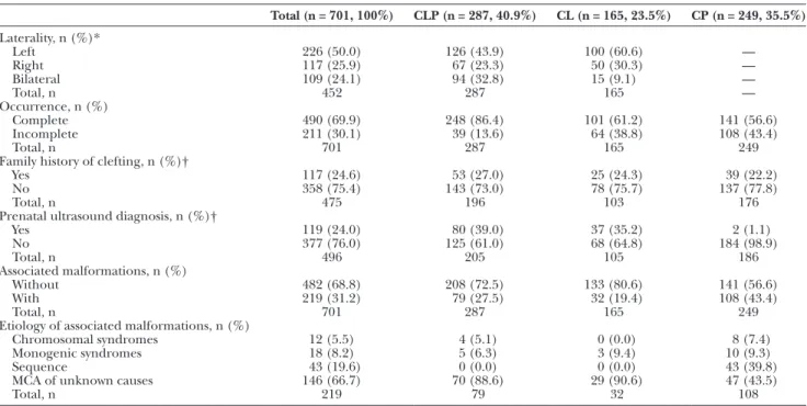
The prevalence and characteristics of the orofacial clefts and associated malformations are shown in Tables 1 and 2. Overall, the most frequent orofacial cleft was CLP, recorded in 287 (40.9%) patients, followed by CP and
CL, recorded in 249 (35.5%) and 165 (23.5%) patients, respectively.
In the 452 children with CL/P, 50.0% (n = 226) had left-sided cleft, 25.9% (n = 117) had right-sided cleft, and 24.1% (n = 109) had bilateral cleft. About 17.3% (n = 78) of the children with CL/P had a family history of clefting, compared with 15.7% (n = 39) of the 249 children with CP. In addition, a prenatal ultrasound diagnosis of cleft was obtained in 24.0% of our study population, compris-ing mostly cases with CL/P (Table 1).
As shown in Table 1, the prevalence of orofacial clefts without associated malformations was 482 (68.8%), whereas 219 (31.2%) cases were found to have an associ-ated malformation that required follow-up or treatment. Moreover, associated malformations were more frequent in children who had CP (in 43.4% of those) than in dren with CLP (27.5%) or CL (19.4%). Of the 219 chil-dren with associated malformations, 108 (49.3%) had CP, 79 (36.1%) had CLP, and 32 (14.6%) had CL (Table 2).
Regarding gender, the group without associated malfor-mations had 280 males (58.1%) and 202 females (41.9%), whereas the group with associated malformations had 105 males (47.9%) and 114 females (52.1%) (Table 2).
As shown in Table 2, family history of clefting was present in 13.9% of the children without associated mal-formations and in 22.8% of the children with associated malformations. Cases with associated malformations had a statistically significant lower proportion of prenatal ultra-sound diagnosis when comparing with the group without associated malformations (13.2% versus 18.7%; P < 0.001). Finally, parental consanguinity was present in 1.2% of the children without associated malformations and in 2.3% of children with associated malformations (Table 2).
Table 1. Prevalence and Characteristics of Orofacial Clefts in the Study Population
Total (n = 701, 100%) CLP (n = 287, 40.9%) CL (n = 165, 23.5%) CP (n = 249, 35.5%) Laterality, n (%)* Left 226 (50.0) 126 (43.9) 100 (60.6) — Right 117 (25.9) 67 (23.3) 50 (30.3) — Bilateral 109 (24.1) 94 (32.8) 15 (9.1) — Total, n 452 287 165 — Occurrence, n (%) Complete 490 (69.9) 248 (86.4) 101 (61.2) 141 (56.6) Incomplete 211 (30.1) 39 (13.6) 64 (38.8) 108 (43.4) Total, n 701 287 165 249 Family history of clefting, n (%)†
Yes 117 (24.6) 53 (27.0) 25 (24.3) 39 (22.2) No 358 (75.4) 143 (73.0) 78 (75.7) 137 (77.8) Total, n 475 196 103 176 Prenatal ultrasound diagnosis, n (%)†
Yes 119 (24.0) 80 (39.0) 37 (35.2) 2 (1.1) No 377 (76.0) 125 (61.0) 68 (64.8) 184 (98.9) Total, n 496 205 105 186 Associated malformations, n (%) Without 482 (68.8) 208 (72.5) 133 (80.6) 141 (56.6) With 219 (31.2) 79 (27.5) 32 (19.4) 108 (43.4) Total, n 701 287 165 249 Etiology of associated malformations, n (%)
Chromosomal syndromes 12 (5.5) 4 (5.1) 0 (0.0) 8 (7.4) Monogenic syndromes 18 (8.2) 5 (6.3) 3 (9.4) 10 (9.3) Sequence 43 (19.6) 0 (0.0) 0 (0.0) 43 (39.8) MCA of unknown causes 146 (66.7) 70 (88.6) 29 (90.6) 47 (43.5) Total, n 219 79 32 108
*Considering that cleft palate does not exhibit laterality.
PRS Global Open
•
2018
In both groups of patients, isolated cleft or with associ-ated malformations, the left side was the most affected. However, the group with associated malformations had a significantly lower proportion of left side involvement (19.6% versus 23.9%) and a higher proportion of right side (13.2% versus 11.0%) and bilateral involvement (15.5% versus 8.7%), compared with the group without associated malformations (P < 0.001).
Regarding the etiology of the associated malforma-tions, 146 (66.7%) patients had MCA of unknown ori-gin and 73 (33.3%) patients had recognized conditions. Among these, 12 patients (16.4%) had identified chro-mosomal syndromes, 18 patients (24.7%) had monogenic
syndromes, and 43 patients (58.9%) had sequences. The most frequent chromosomal anomaly was the 22q11.2 de-letion syndrome, also known as the velocardiofacial or Di-George syndrome, occurring in 8 CP patients, followed by trisomy 13, trisomy 21, 21q deletion, and Klinefelter syn-drome, in 1 patient each. The most frequently identified monogenic syndrome was the Van der Woude syndrome (n = 6), followed by Treacher-Collins syndrome (n = 5), Goldenhar syndrome (n = 2), orofacial digital syndrome type 1 (n = 2), Apert syndrome (n = 1), Gorlin syndrome (n = 1), and Kabuki syndrome (n = 1). Finally, the Pierre Robin sequence was identified in 43 patients.
Among the 219 patients with associated malforma-tions, 90 cases (41.1%) had 1 unrelated associated malfor-mation, whereas 2 associated malformations were found in 62 cases (28.3%), and 3 or more associated malforma-tions were recorded in 67 cases (30.6%).
The number of individuals with a certain organ sys-tem affected among those with associated malformations is shown in Figure 1. Head and neck anomalies were the most frequent associated malformations, in 60.3% (n = 132) of the patients with associated malformations, and among them, eye and ear were the most affected or-gans. Cardiovascular malformations were the second most common anomalies, accounting for recorded malforma-tions in 28.3% (n = 62) of the patients with associated malformations, of which atrial and ventricular septal de-fects, followed by patent ductus arteriosus, were the most prevalent. Musculoskeletal anomalies were the third most common malformations, occurring in 26.0% (n = 57) of patients, and among them, most were cases of polydactyly and limb reductions. In 11.4% (n = 25) of patients, uro-logic anomalies were also found, being cryptorchidism the most common. In 9.6% (n = 21) of the associated malfor-mations cases, malformalfor-mations of the digestive system and abdominal wall occurred, mostly inguinal and umbilical
Table 2. Prevalence and Characteristics of Associated Malformations in Children with Orofacial Clefts
Associated Malformations P Without (n = 482) (n = 219)With Gender, n (%) Male 280 (58.1) 105 (47.9) Female 202 (41.9) 114 (52.1) 0.182 Orofacial cleft, n (%) CP 141 (29.3) 108 (49.3) CL 133 (27.6) 32 (14.6) CLP 208 (43.2) 79 (36.1) < 0.001 Laterality, n (%) Left 115 (23.9) 43 (19.6) Right 53 (11.0) 29 (13.2) Bilateral 42 (8.7) 34 (15.5) < 0.001 Family history of clefting 67 (13.9) 50 (22.8) 0.384 Prenatal ultrasound diagnosis 90 (18.7) 29 (13.2) < 0.001 Parental consanguinity 6 (1.2) 5 (2.3) 0.765 Etiology
Chromosomal syndromes 12 (5.5) Monogenic syndromes 18 (8.2) Sequence 43 (19.6) MCA of unknown origin 146 (66.7)
hernias. Finally, malformations of the central nervous sys-tem appeared in 6.4% (n = 14) of the associated malfor-mations, of which the majority were reduction deformities of the brain.
DISCUSSION
We investigated the prevalence and type of associ-ated congenital malformations in 701 children with CP and CL/P who attended a tertiary referral center during a 31-year period. These patients represented most of the cases of orofacial clefts born in Southern Portugal and the Portuguese Islands between 1981 and 2012. During this period, the overall incidence of clefts was around 5.5 per 10,000 total births in Southern Portugal, according to the literature.8 In our study, we found a higher prevalence of orofacial clefts in males, which is in agreement with pub-lished data,1–3,15,33 with a 1.3:1 ratio of affected boys to girls. A prenatal diagnosis of orofacial cleft was obtained in only 24.6% of the study sample, and most of these cases correspond to CL/P. In fact, not all the subjects had an ultrasound performed, particularly the older ones, as rou-tine obstetric ultrasound examinations were implemented in Portugal in the early 90s. In addition, although the di-agnostic accuracy of ultrasound examinations has been improving over the past years, routine screening for the palate is technically more difficult than for the lip and is not included in most centers’ protocols.34 Therefore, prospective parents should be advised that palatal involve-ment might be underdiagnosed prenatally.
The frequency of associated congenital malformations in children with orofacial clefts was 31.2%, which was slightly above the 25.5% and 27.5% previously reported by 2 Portuguese studies,14,35 but in agreement with the in-ternational literature that reports a range from 1.5% to 64.2%.8,10,11,13,16–22,36–42 This wide variation might be in part attributed to the fact that most studies do not report all infants born within a certain geographical area, but only those referred to a specific unit (frequently tertiary). An-other possible explanation for this variation is the lack of agreement on what should be regarded as a congenital defect. In our study, we have included abnormalities that could lead to function impairment and, in this sense, re-quire either continual medical follow-up or treatment.
Similar to the results of our study, previous studies in-dicated that the orofacial cleft type most frequently asso-ciated with other malformations was CP.1,8,10,11,13,14,16–22,37–42 Moreover, we found a significantly higher proportion of bilateral involvement in the cases with associated malfor-mations than in the cases without associated malforma-tions, in agreement with several studies suggesting that more extensive clefts are associated with a higher risk of occurrence of other congenital malformations.8–10
In our study, most associated MCA were recorded in the head and neck region, accounting for 60.3% of the patients with malformations. The second most common MCA associated with orofacial clefts were those affecting the cardiovascular system, followed by the musculoskel-etal, the urologic, the digestive, and the central nervous systems. From embryological studies, we know that the
development of facial structures is intimately related and interdependent with the development of other structures, and we also know that several components may be affected by the same etiopathogenic factors in pathological con-ditions. Also, failure in the adequate development of 1 anatomic structure may compromise the normal develop-ment of several dependent ones, as well illustrated by the Pierre Robin sequence.43 Thus, associated malformations in children with orofacial clefts may involve several ana-tomic systems, even in areas far from the cleft. Indeed, in agreement with our study, the head and neck region, the musculoskeletal, cardiovascular, and central nervous sys-tems are the most cited in the literature. However, there are divergent reports regarding which system and which congenital malformation is exactly the most common in orofacial cleft infants.8–11,13,14,16,18,19,21,39
Once again, different results between studies may be due to different sampling methods, how long after birth the orofacial clefts cases were examined, differences in case definition and inclusion/exclusion criteria, or dif-ferences between the populations analyzed, which could themselves have different incidences of clefts and other congenital malformations. For instance, our long follow-up period (until patients were 15 years old on average) may have increased the proportion of minor or non–life-threatening conditions over serious life-non–life-threatening ones, which lead to death early in life. We believe that this may be responsible for the relatively low proportion of the re-corded central nervous system malformations in our study. In addition, more recent studies may be influenced by the fact that several formerly regarded MCA of unknown ori-gin are now recognized as part of a specific syndrome, se-quence or chromosomic abnormality. On the other hand, we have been increasing our ability to diagnose morpho-logic anomalies with the development of more accurate imaging, genetic and molecular tests.
Potential limitations of this study arise from its retro-spective observational nature, as our analysis was based on the available medical records over a long period of 31 years, which might have led to variations in case inves-tigation, genetic diagnostic procedures, and complete-ness of reporting. Orofacial cleft patients were excluded from this study due to missing data on the presence or absence of associated malformations. Nevertheless, we found a significant homogeneity among registries and most patients had a complete Cleft Patient Data Sheet with all the variables under study. Another limitation might be the study setting, which was hospital-based. However, clefting is a condition that requires hospital treatment, and therefore, we considered that our study population was representative of the Portuguese orofa-cial cleft patients.
The main strengths of this study include a well-defined geographical area (South of Portugal and the Islands), a large sample size (n = 701), a long follow-up period, the classification into without associated malformations, chro-mosomic syndromes, monogenic syndromes, sequences or MCA of unknown origin, and the examinations by a clinical geneticist of the orofacial clefts cases with associ-ated malformations.
PRS Global Open
•
2018
CONCLUSIONS
This study provides a basis for research of the etiol-ogy of orofacial clefts. The presence and nature of differ-ent synchronous malformations might indicate differdiffer-ent mechanisms of abnormal prenatal development. Identifi-cation of smaller subgroups or clusters may be important in etiological studies to elucidate the environmental and genetic risk factors and the interaction between them.
The overall prevalence of associated malformations (nearly 1 in 3 infants) emphasizes the need for a more comprehensive evaluation of children with orofacial clefts. An early screening routine for other congenital malfor-mations, particularly those of the head and neck, cardio-vascular, skeletal, and central nervous systems, should be considered in all orofacial clefts patients, especially when considering lip surgery within the first days of life, as many severe defects may not be diagnosed during the neonatal period by clinical examination alone. Genetic counseling might be also valuable, particularly in the orofacial cleft cases with associated malformations. Strict cooperation be-tween cleft team members is essential to comprehensively cover all aspects of the management of the patient with orofacial clefts.
Alice Varanda Pereira, MD
Serviço de Cirurgia Plástica Reconstrutiva Hospital de São José Centro Hospitalar de Lisboa Central Rua José António Serrano 1150–199 Lisboa, Portugal E-mail: [email protected]
REFERENCES
1. Calzolari E, Bianchi F, Rubini M, et al; EUROCAT Working Group. Epidemiology of cleft palate in Europe: implications for genetic research. Cleft Palate Craniofac J. 2004;41:244–249. 2. Derijcke A, Eerens A, Carels C. The incidence of oral clefts: a
review. Br J Oral Maxillofac Surg. 1996;34:488–494.
3. Mossey P, Castilla E, eds. Global Registry and Database on Craniofacial
Anomalies: Report of a WHO Registry Meeting on Craniofacial Anomalies, Baurú, Brazil, 4–6 December 2001. Geneva, Switzerland:
World Health Organization; 2001.
4. Schutte BC, Murray JC. The many faces and factors of orofacial clefts. Hum Mol Genet. 1999;8:1853–1859.
5. Butali A, Mossey PA. Epidemiology of orofacial clefts in Africa: meth-odological challenges in ascertainment. Pan Afr Med J. 2009;2:5. 6. Natsume N. Incidence of cleft lip and palate among Japanese
newborns, 1982 to 1984. Plast Reconstr Surg. 1987;79:499–501. 7. Yi NN, Yeow VK, Lee ST. Epidemiology of cleft lip and palate
in Singapore—a 10-year hospital-based study. Ann Acad Med
Singapore. 1999;28:655–659.
8. Calzolari E, Pierini A, Astolfi G, et al. Associated anomalies in multi-malformed infants with cleft lip and palate: an epidemio-logic study of nearly 6 million births in 23 EUROCAT registries.
Am J Med Genet A. 2007;143A:528–537.
9. Hagberg C, Larson O, Milerad J. Incidence of cleft lip and pal-ate and risks of additional malformations. Cleft Palpal-ate Craniofac J. 1998;35:40–45.
10. Milerad J, Larson O, Haqberg C, et al. Associated malformations in infants with cleft lip and palate: a prospective, population-based study. Pediatrics. 1997;100:180–186.
11. Stoll C, Alembik Y, Dott B, et al. Associated malformations in cases with oral clefts. Cleft Palate Craniofac J. 2000;37:41–47. 12. Jones MC. Facial clefting. Etiology and developmental
13. Beriaghi S, Myers SL, Jensen SA, et al. Cleft lip and palate: asso-ciation with other congenital malformations. J Clin Pediatr Dent. 2009;33:207–210.
14. Duarte R, Leal MJ. [The range of congenital malformations asso-ciated with cleft lip and palate]. Acta Med Port. 1999;12:147–154. 15. Ipdtoc Working Group. Prevalence at birth of cleft lip with or with-out cleft palate: data from the International Perinatal Database of Typical Oral Clefts (IPDTOC). Cleft Palate Craniofac J. 2011;48:66–81. 16. Jhawar D, Prasad B, Sharma S, et al. Congenital anomalies as-sociated with cleft lip and/or palate (CL/P): an epidemiological study. Int J Oral Maxillofac Surg. 2007;36:988–989.
17. Jones MC. The risk that an apparently isolated cleft lip with or without cleft palate will be associated with anomalies that impact outcome: follow up of 32 cases ascertained through prenatal di-agnosis. Proc Greenwood Genetic Center. 2000;19:122–123.
18. Lilius GP. Clefts with associated anomalies and syndromes in Finland. Scand J Plast Reconstr Surg Hand Surg. 1992;26:185–196. 19. Perrotin F, de Poncheville LM, Marret H, et al. Chromosomal
de-fects and associated malformations in fetal cleft lip with or with-out cleft palate. Eur J Obstet Gynecol Reprod Biol. 2001;99:19–24. 20. Shaw GM, Carmichael SL, Yang W, et al. Congenital
malforma-tions in births with orofacial clefts among 3.6 million California births, 1983-1997. Am J Med Genet A. 2004;125A:250–256. 21. Shprintzen RJ, Siegel-Sadewitz VL, Amato J, et al. Anomalies
associated with cleft lip, cleft palate, or both. Am J Med Genet. 1985;20:585–595.
22. Wyszynski DF, Sárközi A, Czeizel AE. Oral clefts with associ-ated anomalies: methodological issues. Cleft Palate Craniofac J. 2006;43:1–6.
23. Aylsworth AS. Genetic considerations in clefts of the lip and pal-ate. Clin Plast Surg. 1985;12:533–542.
24. Murray JC. Gene/environment causes of cleft lip and/or palate.
Clin Genet. 2002;61:248–256.
25. Murthy J, Bhaskar L. Current concepts in genetics of nonsyn-dromic clefts. Indian J Plast Surg. 2009;42:68–81.
26. Shi M, Wehby GL, Murray JC. Review on genetic variants and maternal smoking in the etiology of oral clefts and other birth defects. Birth Defects Res C Embryo Today. 2008;84:16–29.
27. Stanier P, Moore GE. Genetics of cleft lip and palate: syndromic genes contribute to the incidence of non-syndromic clefts. Hum
Mol Genet. 2004;13 Spec No 1:R73–R81.
28. Aquino SN, Paranaíba LM, Martelli DR, et al. [Study of patients with cleft lip and palate with consanguineous parents]. Braz J
Otorhinolaryngol. 2011;77:19–23.
29. Rittler M, Liascovich R, López-Camelo J, et al. Parental consan-guinity in specific types of congenital anomalies. Am J Med Genet. 2001;102:36–43.
30. Lidral AC, Murray JC. Genetic approaches to identify disease genes for birth defects with cleft lip/palate as a model. Birth
Defects Res A Clin Mol Teratol. 2004;70:893–901.
31. European Surveillance of Congenital Anomalies. EUROCAT prevalence data tables. A1 - cases and prevalence (per 10,000 births) for the following registries: S Portugal, from 1980 - 2015. Available at http://www.eurocat-network.eu/prevdata/resultsP-df.aspx?title=A1&datefrom=1980&dateto=2015&allanom=&allr egf=false&allrega=false&anomalies=®istriesf=28®istriesa= &winx=1256&winy=894. Accessed May 8, 2017.
32. Tessier P. Anatomical classification facial, cranio-facial and latero-facial clefts. J Maxillofac Surg. 1976;4:69–92.
33. Lisi A, Botto LD, Rittler M, et al. Sex and congenital mal-formations: an international perspective. Am J Med Genet A. 2005;134A:49–57.
34. Knox G, Braithwaite F. Cleft lips and palates in Northumberland and Durham. Arch Dis Child. 1963;38:66–70.
35. Fino D, Almeida JMR. [Congenital malformations: retrospective study in 184282 newborns]. Arquivo Clínico da Maternidade Alfredo
36. Croen LA, Shaw GM, Wasserman CR, et al. Racial and ethnic variations in the prevalence of orofacial clefts in California, 1983-1992. Am J Med Genet. 1998;79:42–47.
37. Magdalenić-Mestrović M, Bagatin M. An epidemiological study of orofacial clefts in Croatia 1988-1998. J Craniomaxillofac Surg. 2005;33:85–90.
38. Matthews MS, Cohen M, Viglione M, et al. Prenatal counseling for cleft lip and palate. Plast Reconstr Surg. 1998;101:1–5. 39. Sárközi A, Wyszynski DF, Czeizel AE. Oral clefts with associated
anomalies: findings in the Hungarian Congenital Abnormality Registry. BMC Oral Health. 2005;5:4.
40. Shafi T, Khan MR, Atiq M. Congenital heart disease and asso-ciated malformations in children with cleft lip and palate in Pakistan. Br J Plast Surg. 2003;56:106–109.
41. Vallino-Napoli LD, Riley MM, Halliday J. An epidemiologic study of isolated cleft lip, palate, or both in Victoria, Australia from 1983 to 2000. Cleft Palate Craniofac J. 2004;41:185–194.
42. Wyse RK, Mars M, al-Mahdawi S, et al. Congenital heart anoma-lies in patients with clefts of the lip and/or palate. Cleft Palate J. 1990;27:258–264; discussion 264.
43. Sperber GH. First year of life: prenatal craniofacial development.