Universidade de Aveiro 2014
Secção Autónoma de Ciências da Saúde
Sílvia Isabel
Ferreira da Silva
A PPA é uma proteína fundamental na
espermatogénese
Universidade de Aveiro 2014
Secção Autónoma de Ciências da Saúde
Sílvia Isabel
Ferreira da Silva
A PPA é uma proteína fundamental na
espermatogénese
APP is a critical protein in spermatogenesis
Dissertação apresentada à Universidade de Aveiro para cumprimento dos requisitos necessários à obtenção do grau de Mestre em Biomedicina Molecular, realizada sob a orientação científica da Professora Doutora Odete Abreu Beirão da Cruz e Silva, Professora Auxiliar com Agregação da Secção Autónoma de Ciências da Saúde da Universidade de Aveiro, e co-orientação da Doutora Dânia Sofia da Silva Teixeira, Investigadora do Centro de Biologia Celular (Secção Autónoma de Ciências da Saúde) da Universidade de Aveiro.
Este trabalho contou com o apoio do Centro de Biologia Celular (CBC) da Universidade de Aveiro, e é financiado por fundos FEDER através do Programa Operacional Factores de Competitividade – COMPETE e por Fundos nacionais da FCT – Fundação para a Ciência e a Tecnologia no âmbito do projeto PTDC/BEX-BCM/0493/2012.
o júri
presidente Professora Doutora Ana Gabriela da Silva Cavaleiro Henriques
Professora Auxiliar Convidada da Secção Autónoma de Ciências da Saúde da Universidade de Aveiro
Professora Doutora Odete Abreu Beirão da Cruz e Silva
Professora Auxiliar com Agregação da Secção Autónoma de Ciências da Saúde da Universidade de Aveiro
Professora Doutora Maria de Lourdes Gomes Pereira
agradecimentos Um agradecimento muito especial à Professora Doutora Odete Abreu Beirão da Cruz e Silva pela oportunidade de realização deste trabalho e pela orientação dada, que permitiu o meu enriquecimento pessoal, científico e profissional.
À Doutora Dânia Sofia da Silva Teixeira pela ajuda e apoio dado ao longo de todo o percurso deste trabalho.
Ao Professor João Carlos Sousa, do ICVS da Universidade do Minho, por ter fornecido os tecidos sem os quais este trabalho não seria possível. Muito obrigado pela disponibilidade e por toda a ajuda dada.
Ao Centro de Biologia Celular da Universidade de Aveiro e à FCT pelo financiamento deste estudo.
Muito obrigado aos colegas de laboratório de Neurociências, em especial à Filipa Martins, Joana Oliveira, Joana Rocha, Regina Cerqueira e Ana Marote, pelo suporte, amizade e companhia.
Aos colegas do Mestrado de Biomedicina Molecular, em especial à Rita Cardoso, Margarida Oliveira e Cláudia Nunes. Obrigado por todos os momentos partilhados.
Obrigada às minhas companheiras de casa, Inês Santana e Liliana Carvalho, pela amizade.
Por último, muito obrigada à minha família, em especial à minha mãe, que me apoia incondicionalmente.
palavras-chave proteína precursora de amilóide, proteína tau, fosforilação, proteína
serina/treonina fosfatase, espermatogénese.
resumo A proteína precursora de amilóide (PPA) e a proteína Tau estão relacionadas
com os marcos histológicos da doença de Alzheimer, uma doença neurodegenerativa progressiva e complexa. A PPA é uma glicoproteína integral transmembranar que tem duas predominantes vias de processamento proteolítico, a via não-amiloidogénica e a via amiloidogénica. A proteína Tau encontra-se associada aos microtúbulos, sendo a afinidade dessa ligação regulada pela fosforilação de resíduos de serina e treonina. A expressão da PPA e da proteína Tau também já foi reportada no testículo. No entanto, ainda não foram estabelecidas as suas funções e modificações pós-traducionais neste tecido. Assim, analisamos a expressão destas duas proteínas e o seu padrão de fosforilação durante a espermatogénese usando como modelos murganhos e ratos wild-type. Através de imuno-histoquímica revelamos que a PPA, a proteína Tau, a proteína serina/treonina fosfatase (PP) 1α e a PP1γ são expressas em toda a espermatogénese. Fosforilação da PPA e da proteína Tau foi detetada durante a meiose. Em contraste com a localização da PPA-total, a PPA fosforilada na treonina 668 está especialmente localizada no núcleo dos espermatócitos. Estes resultados sugerem que a fosforilação da PPA e da proteína Tau contribui para a espermatogénese, especialmente na meiose. No entanto, são necessários estudos futuros para validar os nossos resultados e desvendar a função específica da PPA e da proteína Tau durante a espermatogénese e na meiose.
keywords amyloid precursor protein, tau protein, phosphorylation, serine/threonine
protein phosphatase, spermatogenesis.
abstract The Amyloid precursor protein (APP) and Tau protein are related to
histopathological hallmarks of Alzheimer’s disease, a progressive and complex neurodegenerative disease. APP is a type 1 integral transmembrane glycoprotein. There are two predominant proteolytic processing pathways of APP, the nonamyloidogenic pathway, and the amyloidogenic pathway. Tau is one of the microtubule-associated proteins, and its binding affinity for microtubules is regulated by phosphorylation of serine and threonine residues. APP and Tau protein expression has also been reported in the testis. However, their function and posttranslational modifications in the testis have not been established. Thus, we analyzed the expression of these two proteins and their phosphorylation patterns during spermatogenesis using wild-type mice and rats as models. Through immunohistochemistry we revealed that APP, Tau protein, Serine/Threonine Protein Phosphatase (PP) 1α and PP1γ are expressed throughout spermatogenesis. APP and Tau are phosphorylated during meiosis. In contrast to total-APP localization, phosphorylated APP at Thr668 was specially localized in spermatocyte nuclei. These results suggest that phosphorylation of APP and Tau protein contribute to spermatogenesis, especially in meiosis. However, further research is required to validate our results and unravel the specific function of APP and Tau protein during spermatogenesis and meiosis.
Index
List of Figures ... III List of Tables ... V Acronyms, Abbreviations, and Initials ... VI
1. Introduction ... 1 Alzheimer’s Disease ... 3 1.1.1. AD Hallmarks... 4 1.1.2. AD Forms... 5 1.1.3. AD Risk Factors ... 6 APP ... 7
1.2.1. APP Proteolytic Processing ... 9
1.2.2. APP Functions ... 13
1.2.3. APP Post-translational Modifications ... 14
Tau ... 15
1.3.1. Tau Phosphorylation ... 18
1.3.2. Tauopathies ... 21
Spermatogenesis ... 22
Humans vs. Rodents ... 25
AD-related Proteins and Fertility ... 27
1.5.1. APP and Fertility ... 27
1.5.2. Tau and Fertility ... 29
1.5.3. Other AD-related proteins and Fertility ... 29
2. Objectives ... 31
3. Materials and Methods... 35
Animals ... 37
Antibodies ... 37
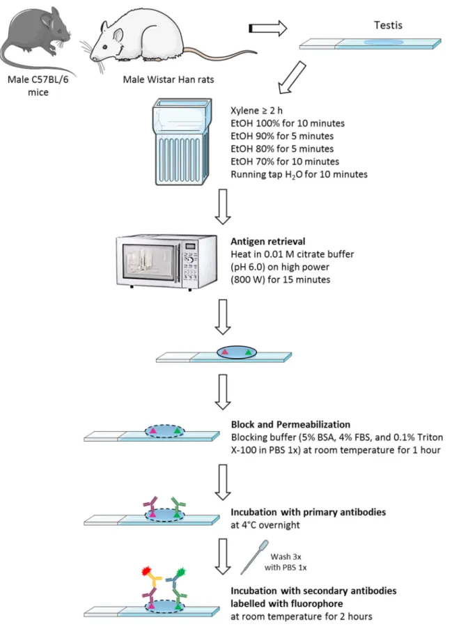
Immunohistochemistry ... 39
Fluorescent IHC (Paraffin-Embedded Tissue) Protocol ... 40
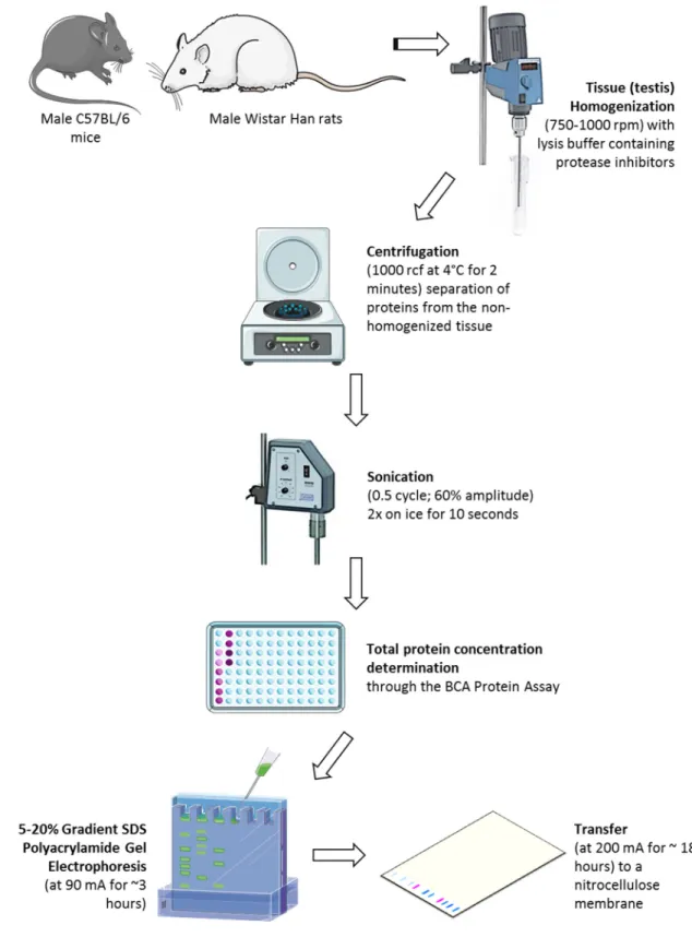
Western Blot Analysis ... 43
3.4.2. Total Protein Concentration Determination ... 44
3.4.3. Gradient SDS Polyacrylamide Gel Electrophoresis ... 45
3.4.4. Immunoblotting ... 46
4. Results – Setting up fluorescent immunohistochemistry ... 51
Abstract ... 53
Results – Detecting Autofluorescence ... 53
Results – Reducing the Autofluorescence ... 55
5. Results – Tau protein, PP1α and PP1γ ... 59
Abstract ... 61
Results – Tau ... 61
Results – Tau Phosphorylation ... 62
Results – PP1α ... 64
Results – PP1γ ... 65
Discussion ... 66
6. Results – APP and its phosphorylation ... 69
6.1.Abstract ... 71
6.2.Results – APP ... 71
6.3.Results – APP Phosphorylation ... 74
6.4.Results – γ-Tubulin ... 76
6.5.Results – APP Western Blot Analysis ... 77
6.6.Discussion ... 79 7. Conclusion ... 83 Future perspectives: ... 86 8. References ... 87 9. Appendix ... 103 Immunohistochemistry Solutions... 105
Western Blot Analysis Solutions ... 106
List of Figures
Figure 1: Alzheimer disease histopathological features in the brain. ... 4
Figure 2: Schematic diagram of APP and its homologues domain organization. ... 8
Figure 3: Schemes of the amyloid precursor protein (APP) and its two proteolytic processing pathways and principal proteolytic derivatives. ... 10
Figure 4: Structure of α-secretase. ... 11
Figure 5: Structure of β-secretase. ... 11
Figure 6: Scheme of the γ-secretase complex... 13
Figure 7: Schematic representation of the human Tau gene, the human Tau primary transcript and the six human central nervous system (CNS) Tau isoforms. ... 17
Figure 8: Schematic representation of the functional domains of the longest Tau isoform (2+3+10+). ... 18
Figure 9: Schematic representation of part of a seminiferous tubule with its surrounding cells. ... 23
Figure 10: Spermiogenesis diagram. ... 25
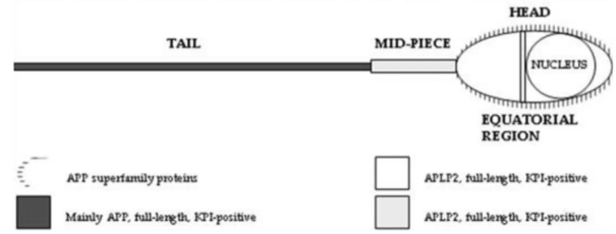
Figure 11: Schematic representation of the amyloid precursor protein (APP) family members expression in the human sperm. ... 28
Figure 12: Schematic representation of the fluorescent immunohistochemistry protocol performed in this study, using paraffin-embedded tissue. ... 42
Figure 13: Diagram of the western blot protocol performed in this study, using mouse and rat testes. ... 48
Figure 14: Schematic representation of the immunoblotting protocol performed in this study. ... 49
Figure 15: Autofluorescence of testis from 2-month-old mouse. ... 54
Figure 16: Autofluorescence of testis from 29-month-old rat. ... 54
Figure 17: Autofluorescence of testis from 2-month-old mouse, after antigen retrieval pre-treatment. ... 56
Figure 18: Autofluorescence of testis from 26-month-old rat, after antigen retrieval pre-treatment. ... 57
Figure 19: Tau protein expression in testis from 6-month-old mouse. ... 62
Figure 20: Phosphorylated Tau (Ser202 and Thr205) expression in testis from 3-month-old mouse. ... 63
Figure 21: Phosphorylated Tau (Ser202 and Thr205) expression in testis from 15-month-old
mouse. ... 63
Figure 22: PP1α expression in testis from 2-month-old mouse. ... 64
Figure 23: PP1γ expression in testis from 2-month-old mouse. ... 65
Figure 24: APP expression in testis from 2-month-old mouse. ... 72
Figure 25: APP expression in testis from 2-month-old mouse. ... 72
Figure 26: APP expression in testis from 6-month-old mouse. ... 73
Figure 27: Phosphorylated APP (Thr668) expression in testis from 3-month-old mouse. 74 Figure 28: Phosphorylated APP (Thr668) expression in testis from 15-month-old mouse. ... 75
Figure 29: γ-Tubulin expression in testis from 26-month-old rat. ... 76
Figure 30: APP expression through western blot analysis of testes from 2-month-old (n = 2) and 6-month-old (n = 1) mice, and of testes from 5-month-old (n = 2), 26-month-old (n = 3) and 29-month-old (n = 3) rats. ... 77
Figure 31: Relative protein expression of APP in testes from 2-month-old (n = 2) and 6-month-old (n = 1) mice (on the left), and in testes from 5-6-month-old (n = 2), 26-6-month-old (n = 3) and 29-month-old (n = 3) rats (on the right), based on the quantification of immunoblot in Figure 30. ... 78
List of Tables
Table 1: Some of phosphorylation sites of APP and its homologues, and the different
enzymes that mediate the phosphorylation. ... 15
Table 2: Most studied protein kinases involved in Tau phosphorylation (reviewed in 72,80). ... 19
Table 3: Protein kinases involved in Tau phosphorylation and the respective
phosphorylation sites (reviewed in 88). ... 20
Table 4: Protein phosphatases involved in Tau dephosphorylation and the respective
dephosphorylation sites (reviewed in 88). ... 21
Table 5: Characteristics that differentiate spermatogenesis and testes of humans and
rodents, which are constant and specific in each species. ... 26
Table 6: Summary of the primary antibodies used for detection of the target proteins in the
different assays. ... 38
Table 7: Summary of the secondary antibodies used for detection of the various primary
antibodies used in immunohistochemistry. ... 41
Table 8: Protein standards used in BCA Protein Assay method... 45 Table 9: Relative amounts of APP species in rat testis and their expected size. ... 81 Table 10: Summary of relative expression of addressed proteins throughout
Acronyms, Abbreviations, and Initials
a.a. amino acids
AD Alzheimer’s Disease
ADAM A Disintegrin And Metalloprotease
AICD Amyloid Precursor Protein Intracellular Domain
Ala Alanine
Aph Anterior Pharynx Defective
APL β-Amyloid-Like Protein (Caenorhabditis elegans) APLP Amyloid Precursor-Like Protein
APOE Apolipoprotein E gene APP Amyloid Precursor Protein
APPL β-Amyloid-Like Protein (Drosophila melanogaster)
APS Ammonium Persulphate
Asp Aspartic acid
Aβ β-Amyloid Peptide
BACE β-Site Amyloid Precursor Protein-Cleaving Enzyme
BCA Bicinchoninic Acid
BSA Bovine Serum Albumin
CDK Cyclin-Dependent Kinase
CHAPS 3[(3-Cholamidopropyl)dimethylammonio]-propanesulfonic acid
CNS Central Nervous System
CS GAG Chondroitin Sulphate Glycosaminoglycan C-terminal Carboxy-terminal
CTF Carboxy-Terminal Fragment
CuBD Copper-Binding Domain
DE Acidic Region
DNA Deoxyribonucleic acid
ECL Enhanced Chemiluminescence
FPKM Fragments Per Kilobase of transcript per Million fragments mapped
FSH Follicle Stimulating Hormone
GSK Glycogen Synthase Kinase
HFBD/GFLD Heparin-Binding/Growth-Factor-Like Domain
HRP Horseradish Peroxidase
Ig Immunoglobulin
IHC Immunohistochemistry
Ile Isoleucine
JNK c-Jun N-terminal Kinase
KPI Kunitz Protease Inhibitor Domain
Leu Leucine
LH Luteinizing Hormone
MAP Microtubule-Associated Protein MAPT Microtubule-Associated Protein Tau MCI Mild Cognitive Impairment
Met Methionine
MPM Metaphase Protein Marker
mRNA messenger Ribonucleic Acid
MT Microtubule
NFTs Neurofibrillary Tangles N-terminal Amino-Terminal
PBS Phosphate Buffered Saline
PDPK Proline-Directed Protein Kinases
Pen Presenilin Enhancer
PGCs Primordial Germ Cells PHF Paired Helical Filament
PMSF Phenylmethylsulfonyl Fluoride
PP Serine/Threonine Protein Phosphatase
PS Presenilin
PSEN Presenilin gene
RC Random Coil
sAPP soluble Amyloid Precursor Protein
SDS Sodium Dodecyl Sulphate
Ser Serine
TACE Tumour Necrosis Factor-α Converting Enzyme
TBS Tris Buffered Saline
TBS-T Tris Buffered Saline-Tween
TEMED N,N,N′,N′-Tetramethylethylenediamine Thr Threonine TM Transmembrane Tyr Tyrosine Val Valine ZnBD Zinc-Binding Domain
Alzheimer’s Disease
Over the last decades societies have been growing older, with the most intensive growth in the number of individuals above 85 years old. This scenario represents one of the biggest challenges for health care and social systems. Gradual improvement of health care, arising from advances in science research, and the social support given to senior citizens has significantly contributed to this beneficial tendency. Still, ageing populations contribute to an increase in the ratio of age-related diseases, and consequently, an increase in the burden for the health care system, thus changing the main concerns of science research 1.
Alzheimer’s disease (AD) was first described in 1907 by Dr. Alois Alzheimer, a German psychiatrist and neuropathologist, as “an unusual illness of the cerebral cortex” 2. It’s a progressive and complex neurodegenerative disease, and is characterized by an irreversible memory loss and intellectual function decline, severe enough to interfere with daily functions (reviewed in Thies and Bleiler, 2013 3).
AD is the most common type of dementia, accounted for an estimated 60% to 80% of cases. According to Alzheimer’s Association (http://www.alz.org/) the term dementia describes “a variety of diseases and conditions that develop when neurons die or no longer function normally”, consequently causing “changes in one’s memory, behaviour, and ability to think clearly”. Besides AD, other types of dementia include vascular dementia, dementia with Lewy bodies, frontotemporal lobar degeneration, mixed dementia, Parkinson’s disease, Creutzfeldt-Jakob disease, and normal pressure hydrocephalus. Each type of dementia is associated with distinct symptom patterns and brain abnormalities. AD affects people in different ways, but the common symptoms are: memory loss that disrupts daily life; challenges in planning or solving problems; difficulty completing familiar tasks; confusion with time or place; trouble understanding visual images and spatial relationships; new problems with words in speaking or writing; misplacing things and losing the ability to retrace steps; decreased or poor judgment; withdrawal from work or social activities; changes in mood and personality. Later symptoms include impaired judgment, disorientation, confusion, behaviour changes, and difficulty speaking, swallowing, and walking (reviewed in Thies and Bleiler, 2013 3).
World Health Organization estimated that 44.4 million people worldwide were living with dementia in 2013, and there are 7.7 million new cases of dementia each year 4. In 2010, Western Europe was the global burden of disease region with the highest number of people with dementia (7.0 million), followed by East Asia with 5.5 million, South Asia with 4.5 million and North America with 4.4 million 5. According to the Alzheimer Association, in the United States of America an estimated 5.2 million of people of all ages have AD in 2013 (reviewed in Thies and Bleiler, 2013 3). Of those with AD, about 4% are less than 65 years old, 13% are 65 to 74 years old, 44% are 75 to 84 years old, and 38% are 85 years or older
6 (reviewed in Thies and Bleiler, 2013 3). By 2050, the total estimated prevalence expected is 13.8 million of people in United States of America 6 (reviewed in Thies and Bleiler, 2013 3) and 115.4 million of people worldwide 7.
1.1.1. AD Hallmarks
AD has as microscopic hallmarks brain abnormalities the extracellular senile plaques (also known as neuritic plaques) and intracellular neurofibrillary tangles (NFTs), as represented in Figure 1 8. These histological features go along with decreases in synaptic density, increases in neuronal loss, neuroinflammatory glial activity and neurodegeneration 1. For a neuropathological diagnosis of AD, both senile plaques and NFTs are mandatory 9, and can only be confirmed post-mortem at the autopsy 10.
Senile plaques are neurotoxic and consist of a dense amyloid core of β-amyloid peptide (Aβ) of 40 and 42 amino acids (a.a.) in length 11. These plaques are surrounded by dystrophic neurites and, precipitate and deposit outside and around neurons, predominantly those in the limbic system and cortex. This mechanism will finally lead to neuronal death, which is proposed to be responsible for phenotypical dementia in affected patients 9.
Tau is a microtubule-associated protein (MAP), and the major constituent of NFTs. Tau’s function is regulated by phosphorylation. In AD, Tau protein is abnormally hyperphosphorylated, which leads to Tau polymerization, disruption of microtubule (MT) dynamics, and impaired axonal transport. That consequently results in the formation of intraneuronal NFTs and finally neuronal death 9.
Figure 1: Alzheimer disease histopathological features in the brain.
A) Plaques with dystrophic neurites surrounding amyloid cores (arrows). B) Neurofibrillary tangle is present within one neuron, and several extracellular tangles are also present (arrows) (taken from 12).
Nowadays, the “amyloid hypothesis” is generally supported. According to this hypothesis the abnormal production and aggregation of Aβ, principally the more fibrillogenic Aβ42 isoform, are the primary pathogenic events in AD, and NFTs are further downstream in the neuropathogenesis. This hypothesis is controversial, because senile
plaques and NFTs have been found in the brain of non-demented persons as well, although in lower abundance. Additionally, the precise sequence and repercussion of the different biological processes involved in AD are not yet entirely clear. Though neurodegeneration, senile plaques, and NFTs are broadly accepted as part of the AD, it remains difficult to determine to what magnitude these factors contribute to dementia and to what magnitude they are interconnected with each other. However, some studies have shown that soluble oligomers of Aβ can disrupt synaptic function, facilitate neuronal dysfunction in AD, and are both essential and sufficient to disrupt learning behaviour in a way that is both potent, rapid, and transient 9.
1.1.2. AD Forms
There are two forms of AD: the familial form, which has an earlier onset (symptoms tend to develop before age 65), faster progression, and affects around 1% of the patients; and the sporadic form, which has a later onset (symptoms develop at age 65 or older), and is the most common condition. Nonetheless, the clinical and histopathological features of both forms are undistinguishable 1,3.
The familial AD is caused by mutations in three genes: the amyloid precursor protein (APP) gene, and the presenilin genes, PSEN1 and PSEN2, which are involved in the cleavage of APP and production of Aβ 1,3. Mutations in the substrate and in the proteases can contribute to amyloid deposition, which forms one of the corner stones of the “amyloid hypothesis” 13. Mutations in the APP gene, on chromosome 21q21.3, are estimated to represent about 5% of familial AD. To date, a total of 51 mutations have been identified in the APP gene, but not all are pathogenic. Two well-studied mutations found in familial AD families have been expressed in transgenic mice. In the first case, the “London mutation”, a missense mutation, a valine is replaced by an isoleucine at codon 717 (V717I). In the second case, the “Swedish mutation”, where, immediately upstream of the Aβ domain, mutations at codons 670 and 671 resulted in a double base pair substitution, whereby a lysine and a methionine are replaced by aspartic acid and leucine (K670D/M671L, APPsw) 14.
Mutations in the PSEN1 gene, on chromosome 14q24.3, are the most common cause of autosomal dominant early-onset AD, accounting for up to 70% of cases 15, with about 207 mutations identified in this gene. While in the PSEN2 gene, on chromosome 1q31-q42, only 25 mutations have been described so far, and not all have been confirmed to be pathogenic, accounting for less than 5% of early-onset AD cases 16,17. Mutations in PSEN1 and PSEN2 genes result in the production of abnormal presenilin (PS) proteins, which interfere with the γ-secretase complex function, mainly due to a defective PS1. Consequently this alters APP processing, because PS/γ-secretase can cleave APP at different positions. That leads to variable effects on the spectrum of Aβ peptides released,
which are the overproduction of the longer and toxic version of Aβ peptides (≥Aβ42) and the underproduction of the shorter version of Aβ peptides (≤Aβ40). Copies of the toxic peptide fragment stick together and build up in the brain, forming the senile plaques 13.
The MAPT gene is located on chromosome 17q21.1, and codes for human microtubule-associated protein Tau 18. No mutations have been identified in familial AD. However, exonic and intronic mutations were identified in the Tau gene that were associated to frontotemporal dementia with parkinsonism-17, a familial dementia related to AD. These findings prove that dysfunction of Tau in itself can cause neurodegeneration and consequently contribute to dementia. Besides frontotemporal dementia, Tau mutations can cause diseases as varied as subcortical gliosis, cortico-basal degeneration and pallido-ponto-nigral degeneration 14.
1.1.3. AD Risk Factors
AD is a complex multifactorial disease attributable to various interrelated and interacting environmental and genetic factors. Many factors contribute to the development of AD. Advanced age is the greatest risk factor for AD, but this disease is not a typical part of ageing. Another risk factor is the family history, because individuals who have a first-degree relative with AD are more likely to develop the disease than those who do not. People with mild cognitive impairment (MCI), especially MCI involving memory problems, are more likely to develop dementia than people without it. Cardiovascular diseases and factors that increase the risk of cardiovascular diseases, like smoking, alcohol intake, high body mass index, diabetes mellitus, high cholesterol, and hypertension, are also associated with a higher risk of developing AD and other dementias. A healthy heart and healthy blood vessels help ensure that the brain is supplied with the oxygen- and nutrient-rich blood, required for normal brain function. Apolipoprotein E gene (APOE) is one of the genetic risk factors for AD, and is associated with sporadic AD. The APOE gene, on chromosome 19q13.2, has three allelic variants, ε2, ε3 and ε4, and studies suggest that the ε3 allele neither increases nor decreases one’s risk of AD, but having the ε2 allele may decrease one’s risk. However, having the ε4 allele increases the risk of developing AD and of developing it at a younger age. People who inherit two copies of the ε4 allele have an even higher risk of developing AD, although inheriting the ε4 allele does not guarantee that a person will develop this illness 3,19.
On the preventive side, physical activity and consumption of a diet low in saturated fats and rich in fruits, vegetables and vegetable-based oils are two factors that protect the heart as well the brain and reduce the risk of developing dementia. Other protective factors are education, and social and cognitive engagement. This can be explained by the fact that having more years of education, and remaining socially and cognitively active builds a “cognitive reserve”. According to this hypothesis, having more years of education, and
remaining socially and cognitively active increases the connections between neurons in the brain and empowers the brain to compensate more fully for the early brain changes of AD or other dementias by using alternative routes of neuron-to-neuron communication to complete a cognitive task 3,19.
APP
APP is evolutionary highly conserved and it´s family includes the mammalian amyloid precursor-like protein (APLP) 1 and 2, the Drosophila melanogaster β-amyloid-like protein APPL, and the Caenorhabditis elegans β-amyloid-like protein APL-1 (reviewed in Jacobsen and Iverfeldt, 2009 20).
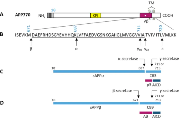
All APP family members are type 1 integral transmembrane (TM) glycoproteins, and are composed by a large ectoplasmic amino-terminal (N-terminal) region, a single membrane-spanning domain, and a shorter cytoplasmic carboxy-terminal (C-terminal) region 21,22 (reviewed in Jacobsen and Iverfeldt, 2009 20). The APP sequence can be divided into several distinct domains, as demonstrated in Figure 2. The ectoplasmic region of APP, which represents the larger part of the protein, can be divided into the E1 and E2 domains. The E1 domain can be further divided into a number of subdomains, including a heparin-binding/growth-factor-like domain (HFBD/GFLD), a copper-binding domain (CuBD) and a zinc-binding domain (ZnBD). The E1 domain is followed by an acidic region rich in aspartic acid and glutamic acid (DE) and a Kunitz protease inhibitor domain (KPI), which is not present in APP695 (an APP alternatively spliced isoform). The E2 region consists of another HFBD/GFLD and a random coil (RC) region. The cytoplasmic region of APP contains a protein interaction motif, namely the YENPTY sequence (including the NPXY internalization signal), which is conserved in all APP homologues. The sequences of all APP homologues can be divided into similar domain structures as APP (Figure 2) (reviewed in Jacobsen and Iverfeldt, 2009 20).
Figure 2: Schematic diagram of APP and its homologues domain organization.
All APP family members are type I integral membrane proteins that have relatively large extracellular domains and short intracellular domains. They contain heparin-binding/growth-factor-like domains (HBD/GFLD), copper- and zinc-binding domains (CuBD and ZnBD), an acidic domain (DE), and a protein interaction motif (YENPTY) in the carboxy-terminal. Other abbreviations: APL-1, Caenorhabditis elegans β-Amyloid-Like Protein; APLP, Amyloid Precursor-Like Protein; APP770, an Amyloid Precursor Protein alternatively spliced isoform; APPL, Drosophila melanogaster β-Amyloid-Like Protein; Aβ, β-Amyloid Peptide; KPI, Kunitz Protease Inhibitor Domain; RC, Random Coil (adapted from 20).
APP is ubiquitously expressed and is encoded by a single gene on chromosome 21q21.3. However, APP exists in 11 isoforms produced by alternative splicing of three exons in a 19-exon gene, of which exons 7, 8 and 15 can be alternatively spliced 23, and the different isoforms can be preferentially expressed in different cell types 20. There are three major isoforms of APP in mammals: APP695 (exons 1–6, 9–18), APP751 (exons 1–7, 9–18), and APP770 20,23. The main difference between APP isoforms is the presence or absence of the KPI domain and a chondroitin sulphate glycosaminoglycan (CS GAG) attachment site. The APP695 isoform is mainly found in cells of neuronal origin 20, while APP751 and APP770 are the predominant isoforms expressed in non-neuronal cells 24. In mouse the presence of three APP splicing forms (APP695, APP751, and APP770) was described, the tissue distributions of these three forms are similar to that seen in humans 25,26.
1.2.1. APP Proteolytic Processing
There are two proteolytic processing pathways of APP, the nonamyloidogenic pathway, and the amyloidogenic pathway, represented in Figure 3. Both pathways are mediated by at least three cleavage events. The nonamyloidogenic processing of APP is initiated through cleavage by α-secretase, which leads to the release and secretion of the soluble N-terminal ectodomain sAPPα from the cell surface leaving an 83 a.a. long C-terminal membrane-bound fragment (C83). This fragment can be further processed by γ-secretase, leading to the release and secretion of a small non-pathogenic peptide p3 and a free APP intracellular domain (AICD). In the amyloidogenic pathway, APP is initially cleaved by β-secretase and releases the large soluble N-terminal ectodomain sAPPβ. The remaining C-terminal stub of 99 a.a. (C99) can then be further processed by γ-secretase, in a similar way to the nonamyloidogenic pathway, generating Aβ and AICD. The AICD can be translocated into the nucleus, where it may function as a transcription factor. Aβ is a normal soluble cellular metabolite encompassing two main forms with different C-terminal regions, Aβ40 and Aβ42 20,23.
γ-Secretase cleavage between Val711 and Ile712 (in the APP770 isoform) generates Aβ40, and cleavage between Ala713 and Thr714 results in production of Aβ42. After γ-secretase cleavage, the remaining C-terminal fragments are 57 or 59 a.a. long (C57 and C59), and have been identified as AICD. Consequently, the final processing step has been suggested to be a result of three cleavage events. The C99 fragment is first cleaved at the ε-site between Leu720 and Val721, followed by cleavage at the ζ-site at the Val717-Ile718 bond, and finally the peptide is cut at the γ-site 8,20.
Figure 3: Schemes of the amyloid precursor protein (APP) and its two proteolytic processing pathways and principal proteolytic derivatives.
A) The largest of the known APP alternative splice forms, APP770, comprising 770 amino acids (a.a.). Regions of interest: a) a 17-residue signal peptide occurs at the N-terminal; b) two alternatively spliced exons of 56 and 19 a.a. are inserted at residue 289; the first contains a serine protease inhibitor domain of the Kunitz type (KPI); c) a single transmembrane domain (TM) at a.a. 700–723. The β-amyloid peptide (Aβ) includes 28 residues just outside the membrane plus the first 12–14 residues of the TM. B) The a.a. sequence within APP that contains the Aβ and TM regions. The underlined residues represent the Aβ42 peptide. Three-digit
numbers represent codon numbers (APP770 isoform). C) The nonamyloidogenic pathway. The first arrow indicates a site (after residue 687; same as white dot in Aβ box in A) of cleavage by α-secretase that enables secretion of the large, soluble ectodomain sAPPα into the medium and retention of the 83-residue C-terminal fragment (C83) in the membrane. C83 can undergo cleavage by γ-secretase principally at residue 711 or residue 713 to release the p3 peptides. D) The amyloidogenic pathway. The alternative proteolytic cleavage after residue 671 by β-secretase that results in the secretion of the slightly truncated sAPPβ molecule and the retention of a 99-residue C-terminal fragment in the membrane. C99 can also undergo cleavage by γ-secretase to release the Aβ peptides. Cleavage of both C83 and C99 by γ-γ-secretase at the ε-site (in B) releases the APP intracellular domain (AICD) into the cytoplasm. The order and interdependency of the γ- and ε-cleavages have not been established (adapted from 8).
Although APP processing has been widely studied, all the processing events and the enzymes involved have not yet been fully elucidated.
a) α-Secretase
α-Secretase is a zinc metalloproteinase that cleaves APP at the Lys687-Leu688 bond (in the APP770 isoform) 8,20, and metabolizes about 90% of APP 9. Several enzymes can act as α-secretase, and they are all members of the ADAM (A Disintegrin And Metalloprotease)
family. The enzymes are ADAM9, ADAM10 and ADAM17, also known as tumour necrosis factor-α converting enzyme (TACE) 9,20. ADAMs are type 1 integral membrane proteins, ~750 a.a. long, with a multidomain structure, including signal peptide, pro-domain, catalytic metalloprotease domain, disintegrin/cystein-rich domain, TM domain, and a short cytoplasmic domain 20, represented in Figure 4. All members of this family have four potential functions: cell fusion, cell adhesion, intracellular signalling and proteolysis 23. Apparently, they have redundant α-secretase cleavage activities but differential expression patterns 9.
Figure 4: Structure of α-secretase.
Processing of APP by α-secretase is mediated by a series of proteases, mostly members of the ADAM (A Disintegrin And Metalloprotease) family, which are all membrane bound and consist of several extracellular domains, including a signal peptide (SP), a pro-domain (Pro), a catalytic metalloprotease domain, a disintegrin domain (which in some instances provides interaction with integrin receptors), and a cysteine-rich domain. Other abbreviations: TM, Transmembrane (adapted from 27).
b) β-Secretase
The APP cleavage at the Met671-Asp672 bond (in the APP770 isoform) is carried out by β-secretase (Figure 5), an aspartyl protease, which constitutes the first step towards Aβ generation. BACE (β-Site Amyloid Precursor Protein-Cleaving Enzyme) 1 and BACE2 are the two β-secretases that have been identified. BACE is a single domain integral protein with the active site located on the ectoplasmic side of the membrane. The optimal pH of BACE activity is approximately 4.5, indicative that the β-site cleavage of APP takes place in the more acidic cellular compartments, like the endosomes. BACE cleavage has also been proposed to occur in lipid rafts (reviewed in Jacobsen and Iverfeldt, 2009 20). BACE1 is widely expressed in the brain 20, particularly in the neurons 9.
Figure 5: Structure of β-secretase.
β-Secretase activity is specifically mediated by the β-Site Amyloid Precursor Protein-Cleaving Enzyme 1 (BACE1). BACE1 is a membrane-bound enzyme that is synthesized with a signal peptide (SP), a pro-domain (Pro), and a catalytic domain. Other abbreviations: TM, Transmembrane (adapted from 27).
c) γ-Secretase
γ-Secretase is an aspartyl protease, with a low sequence specificity that cleaves the substrate within its TM domain. The protein is a complex holoenzyme that consists of
several individual enzymes, including PS1 or PS2, anterior pharynx defective 1 (Aph-1), nicastrin, and presenilin enhancer 2 (Pen-2), as demonstrated in Figure 6. The stoichiometry (proportions in which elements are combined) of the γ-secretase components is likely 1:1:1:1, but was also suggested the possibility that also larger multimers are formed or that additional proteins are incorporated into the complex. PS is a polytopic protein with nine TM domains, and is considered to possess the active site of the enzyme. The core of the catalytic site of PS consists of two highly conserved aspartate residues (Asp257 and Asp385 in human PS1) within TM6 and TM7. Nicastrin is an evolutionarily conserved type I integral membrane protein that is highly glycosylated. This protein has one TM and a large ectodomain, proposed to function as a gate keeper to the PS1 active site, restricting access of substrates to the complex. Nicastrin also functions as a substrate receptor and is important for the assembly process of the γ-secretase complex. Aph-1 has a seven TM structure with the C-terminal located in the cytoplasm. Pen-2 displays a hairpin-like structure with two TM domains and both termini located at the luminal side. The functions of Aph-1 and Pen-2 are not yet completely understood. Still, Aph-1 has been implicated in stabilization of PS1 and plays a significant role during assembly of the complex. Additionally, Pen-2 stabilizes the final complex and is involved in endoproteolysis of PS1 13,20.
PS/γ-secretase cleaves with remarkable relaxed sequence specificity TM domains of several proteins, like APP, APLP-1, APLP-2, Notch 1, 2, 3 and 4, among others (reviewed in Wakabayashi and De Strooper, 2008 13). It is essential that the substrates have a type I conformation of the TM domain and a very short (<50 a.a.) extracellular ectodomain 28,29. Bulky extracellular ectodomains greater than 200-300 a.a. residues prevent γ-secretase cleavage and are usually removed by membrane bound (metallo)-proteases or “sheddases” at the cell surface. This first cleavage can be triggered by ligand binding, as is the case for Notch, whereas for other substrates, e.g., APP, shedding is largely constitutive. γ-Secretase cleavage occurs at different positions in the membrane domain of its substrates, generating a series of small peptides, like Aβ or p3, and AICD (APP), Nβ and NICD (Notch), etc., which are secreted at the extracellular side of the membrane (reviewed in Wakabayashi and De Strooper, 2008 13).
Both PS proteins (PS1 and PS2) share approximately 63% homology with the highest similarity in the TM domains, where most of the familial AD-linked mutations are found (reviewed in Parent and Thinakaran, 2010 30). Both PS have been targets of intense study. Knockouts, knockins, and transgenic models have been used to study these proteins 9.
Figure 6: Scheme of the γ-secretase complex.
γ-Secretase is a highly hydrophobic complex composed of four integral membrane proteins: presenilin enhancer 2 (Pen-2), presenilin (presenilin 1 or presenilin 2), nicastrin, and anterior pharynx defective 1 (Aph-1). Presenilin 1 and presenilin 2, and Aph-1a and Aph-1b are encoded by different genes, and all subunits can combine to generate at least four different complexes. Alternative splicing of these genes adds further complexity. Presenilin provides the active site with two catalytic aspartates located in transmembrane (TM) domains 6 and 7 (indicated as stars). Nicastrin and Aph-1 first form a stable subcomplex, followed by incorporation of presenilin and Pen-2 in the early secretory compartment. Once all four components assemble, presenilin undergoes endoproteolysis by unknown “presenilinase” activity within the large intracellular loop between TM6 and TM7 (indicated by an arrow), and the amino-terminal fragment (NTF) and the carboxy-terminal fragment (CTF) remain noncovalently bound. Only fully assembled complexes reach the cell surface and endocytic compartments (adapted from 13).
1.2.2. APP Functions
Despite all the findings with respect to APP and its homologues, its biological functions are still not fully understood, and will require further research in the future. It is clear that proteins from the APP family have redundant and partially overlapping functions, and their biological functions are not restricted to the nervous system. Several in vitro and
in vivo studies suggest that APP, APLP1, and APLP2 are involved in the developing and adult
nervous system, cell adhesion and migration, neurite outgrowth, neuronal survival and apoptosis, synaptogenesis, vesicular transport, neuronal migration, modulation of synaptic plasticity, axonal transport, and can act as modulators of insulin and glucose homeostasis. In particular, APP functions as a cell surface receptor and is involved in transcription regulation through protein-protein interactions (reviewed in Jacobsen and Iverfeldt, 2009 20).
1.2.3. APP Post-translational Modifications
The APP family proteins are post-translationally modified in several different ways. All family members undergo N-glycosylation within its extracellular domain. Both APP and APLP2 have been shown to be subject to O-glycosylation within its extracellular domain, sialylation, and CS GAG modification 20,24. Some post-translational modification sites are conserved in all homologues, like the APP N467 N-glycosylation site. Whereas others sites are only conserved within the mammalian homologues, like the APP K695 sumoylation site 20. N-glycosylation affects the processing of APLP1, once when it’s blocked with tunicamycin additional APLP1 fragments are generated. However, the processing of APP and APLP2 was unaffected by the blocking of N-glycosylation 31. In contrast, sialylation affects APP processing, since overexpression of sialyltransferase enhanced secretion of Aβ 32.
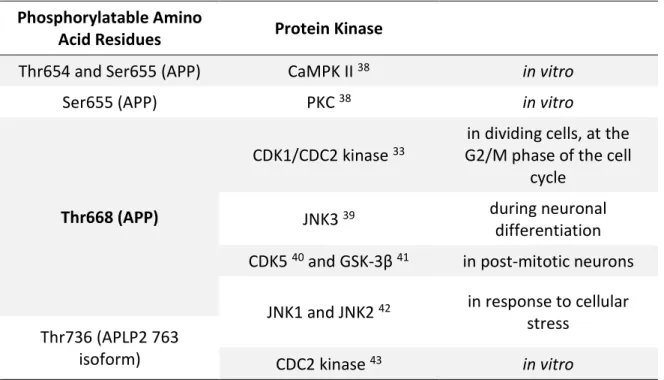
All APP family members have been shown to be phosphorylated, and some of the most studied phosphorylation sites are shown in Table 1. APP is a phosphoprotein that has several phosphorylatable a.a. residues in its intracellular and extracellular domains 20,24. Among the intracellular domain sites that can be phosphorylated, Thr654, Ser655, and Thr668 (numbering for the APP695 isoform) have been shown to be phosphorylated in adult rat brain 33,34. The Tyr653, Ser655, Thr668, Ser675, Tyr682, Thr686 and Tyr687 were found to be phosphorylated in post-mortem brain tissue of AD patients 35. Phosphorylation at these sites has been described to impact APP cellular localization. In addition, Thr668 phosphorylated APP has been found to be enriched in endocytic compartments and to co-localize with BACE in AD brains 35.
Since these residues are located in specific protein interacting sites its phosphorylation may interfere with protein binding and thus interfere with APP and AICD functions. Additionally, some phosphorylation sites may affect the processing of APP 20. Lee
et al. (2003) reported that Aβ production was reduced in rat embryonic cortical neurons
when treated with a cyclin-dependent kinase (CDK) 5 inhibitor, which reduced Thr668 phosphorylation, or when transfected with Thr668Ala mutant APP 35. In another study, Thr668 phosphorylation of APP was reported to induce increased β-cleavage, but decreased γ-cleavage, resulting in decreased Aβ production 36. Phosphorylation of APP at Tyr687 was suggested to regulate α-cleavage of APP since overexpression of the mutant Tyr687Ala decreased the level of the C83 fragment 37.
Alignment of the APP family members shows that the APP Thr668 phosphorylation site is conserved in all APP homologues: APLP1 (Thr624), APLP2 (Thr736), APPL (Thr855), and APL-1 (Thr660). Three other in vivo phosphorylated sites in APP, Tyr682, Thr686, and Tyr687 (which are part of the YENPTY motif), are also conserved in all APP homologues. Tyr653, another in vivo phosphorylated site in APP, is conserved in both mammalian homologues, but not in APPL or APL-1 (reviewed in Jacobsen and Iverfeldt, 2009 20).
Table 1: Some of phosphorylation sites of APP and its homologues, and the different enzymes that mediate the phosphorylation.
Phosphorylatable Amino
Acid Residues Protein Kinase
Thr654 and Ser655 (APP) CaMPK II 38 in vitro
Ser655 (APP) PKC 38 in vitro
Thr668 (APP)
CDK1/CDC2 kinase 33
in dividing cells, at the G2/M phase of the cell
cycle
JNK3 39 during neuronal
differentiation CDK5 40 and GSK-3β 41 in post-mitotic neurons
JNK1 and JNK2 42 in response to cellular stress
Thr736 (APLP2 763
isoform) CDC2 kinase 43 in vitro
Abbreviations: APLP, Amyloid Precursor-Like Protein; APP, Amyloid Precursor Protein; CaMPK, Calcium/Calmodulin-dependent Protein Kinase; CDC, Cell Division Cycle; CDK, Cyclin-Dependent Kinase; GSK, Glycogen Synthase Kinase; JNK, c-Jun N-terminal Kinase; PKC, Protein Kinase C; Ser, Serine; Thr, Threonine. Residue in bold type was the one addressed in this study.
APP phosphorylation has been shown to regulate intracellular signaling via adaptor proteins. Thr668 phosphorylation was shown to induce large conformational changes of the intracellular domain 44. It was later shown that the peptidyl-, prolyl cis/trans isomerase Pin1 binds to Thr668-phosphorylated APP both in vitro and in vivo and thereby accelerates the production of the trans conformation of Pro669 45. This proline residue is conserved in APLP2 (Pro737) and APL-1 (Pro662), but not in APLP1 or APPL. It was further demonstrated that the Pin1-catalysed isomerization of APP regulated its processing, since overexpression or knockout of Pin1 reduced or increased the secretion of Aβ, respectively. It has further been shown that Pin1 knockout in mice resulted in age-dependent neuropathy 46.
Tau
Tau protein regulates MT assembly, dynamic behaviour, and spatial organization under physiological conditions 47,48. Tau protein has been shown to regulate axonal transport of organelles, including mitochondria 49. Proteins from the MAP family are present in several species, such as C. elegans 50,51, Drosophila 52,53, goldfish 54, bullfrog 55,
rodents 56,57, bovines 58,59, goat 60, monkeys 60, and human 61,62. In human, MAPs are mainly present in neurons 63, although non-neuronal cells usually present trace amounts. For example, glial cells express Tau protein mainly in pathological conditions 64, and it is possible to detect Tau mRNA and proteins in several peripheral tissues like heart, lung, kidney, pancreas, testis, muscle, and fibroblasts 65–67.
The gene encoding human Tau protein MAPT is unique and located on chromosome 17q21.1 68, spans approximately 150 kb, and contains 16 exons 69 (Figure 7). The exons 4A, 6 and 8 are specific to peripheral Tau protein, therefore are never present in any mRNA of human brain 58,60. Tau is encoded by a single gene from which nine isoforms are produced through alternative splicing 70, six of which (range from 352 to 441 a.a. and an apparent molecular weight of 48–67 kDa) are the isoforms expressed in human central nervous system (CNS) 61,62,71.
Figure 7: Schematic representation of the human Tau gene, the human Tau primary transcript and the six human central nervous system (CNS) Tau isoforms.
The human Tau gene is located over 100kb on chromosome 17q21.1. It contains 16 exons, with exon -1 being part of the promoter (upper panel). Tau primary transcript contains 13 exons, since exons 4A, 6 and 8 are not transcribed in human brain (middle panel). Exons -1 and 14 are transcribed but not translated. Exons 1, 4, 5, 7, 9, 11, 12, 13 are constitutive, and exons 2, 3, and 10 are alternatively spliced, giving rise to six different mRNAs, translated in six different CNS Tau isoforms (lower panel). These isoforms differ by the absence or presence of one or two 29 amino acids (a.a.) inserts encoded by exon 2 (yellow box) and 3 (green box) in the amino-terminal part, in combination with either three (R1, R3 and R4) or four (R1–R4) repeat-regions (black boxes) in the carboxy-terminal part. The fourth microtubule-binding domain is encoded by exon 10 (light blue box) (lower panel). The adult Tau isoforms include the longest 441-a.a. component (2+3+10+), the 410-a.a. component (2+3+10-), the 412-a.a. component (2+3-10+), the 381-a.a. component (2+3-10-) and the 383-a.a. component (2-3-10+). The shortest 352-a.a. isoform (2-3-10-) is found only in the foetal brain, and thus is referred as foetal Tau isoform (adapted from 72).
Tau can be subdivided into four regions (Figure 8): i) an acidic region in the N-terminal part; ii) a proline-rich region; iii) a region responsible for binding with MTs (MT-binding domains); and iv) a C-terminal region 73. There are two aspects that separate Tau isoforms, these are: the absence or presence of one or two inserts (29 or 58 a.a.) in the N-terminal part of the molecule; and the presence of either three or four repeat-regions in the C-terminal part (3R and 4R Tau), an outcome of alternative splicing of exon 10 58,61,62,74. There
is evidence that each of these isoforms may have particular biological roles, once they are differentially expressed during development. For example, the only Tau isoform present during foetal stages is the one characterized by the absence of N-terminal inserts and the presence of three C-terminal repeats, 3R Tau (exons 2, 3 and 10 are spliced out). Adult human CNS expresses equal amounts of 3R and 4R isoforms (with one or two N-terminal inserts and three or four C-terminal repeats). These findings demonstrate developmental regulation of exon 10 splicing 75,76. Therefore, it is highly probable that Tau isoforms have specific functions related to the absence or presence of regions encoded by the cassette exons 2, 3 and 10. Moreover, the relative levels of six Tau isoforms expressed in adult human CNS (3R and 4R isoforms) differ between neurons in different brain regions. Granular cells of the dentate gyrus (hippocampal formation) don’t express Tau mRNAs containing exon 10 (4R Tau), only 3R Tau 61,62. Tau isoforms may be differentially distributed in neuronal subpopulations 72.
Figure 8: Schematic representation of the functional domains of the longest Tau isoform (2+3+10+).
The projection domain, including an acidic and a proline-rich region, interacts with neural plasma membrane and cytoskeletal elements to determine spacings between microtubules in axons. The amino-terminal part is also involved in signal transduction pathways by interacting with proteins as phospholipase C-γ (PLC-γ) and Src-kinases. The carboxy-terminal part, referred to as microtubules binding domain, regulates the rate of microtubules polymerization and stabilization. It is also involved in the binding with functional proteins as protein phosphatase 2A (PP2A) or presenilin 1 (PS1) (adapted from 72).
1.3.1. Tau Phosphorylation
The exons 9-12 of Tau gene encode four imperfect repeat domains (encompassing 31–32 residues) that mediate the interaction between Tau and tubulin 77. Within neurons, Tau is predominantly found in axons 78 as a highly soluble phosphoprotein 79. Tau contains 85 putative phosphorylation sites: 45 are serines, 35 are threonines and only 5 are tyrosines 72,80,81. Most of these phosphorylation sites are on Ser-Pro and Thr-Pro motives 82. Table 2 summarizes the four groups of protein kinases that can phosphorylate Tau: i) the Proline-Directed Protein Kinases (PDPKs) which phosphorylate Tau on serine or threonine residues that are followed by a proline residue; ii) the non-PDPK group; iii) protein kinases that phosphorylate Tau on serine or threonine residues followed or not by a proline; iv) and the tyrosine protein kinases (reviewed in Buée et al., 2010 83).
Table 2: Most studied protein kinases involved in Tau phosphorylation (reviewed in 72,80).
Abbreviations: CaMPK, Calcium/Calmodulin-Dependent Protein Kinase; CDC, Cell Division Cycle; CDK, Cyclin-Dependent Kinase; CK, Casein Kinase; DYRK, Dual-Specificity Tyrosine Phosphorylation-Regulated Kinase; GSK, Glycogen Synthase Kinase; MAPK, Mitogen Activated Protein Kinase; MARK, Microtubule-Affinity Regulating Kinase; MSK, Mitogen- and Stress-Activated Protein Kinase; PDPK, Proline-Directed Protein Kinases; PK, Protein Kinase; SAP, Stress-Activated Protein; TTBK, Tau-Tubulin Kinase.
Tau binding affinity for MTs is regulated by the phosphorylation of serine and threonine residues 84,85. It is believed that through this binding, Tau plays a major role in stabilizing MTs. When phosphorylated, Tau protein is not able to polymerize tubulin into MTs and do not stabilize the latter 86.
Tau’s phosphorylation process is also developmentally regulated. During embryogenesis and early development, when only the shortest of the isoforms is expressed, Tau phosphorylation reaches high levels. By contrast, adult brain expresses six isoforms with relatively reduced phosphorylation levels compared with the foetal situation 87.
PDPK
•Cyclin-Dependent Kinases including CDC2, CDK1 and CDK5; • MAPK; • SAP Kinases. Non-PDPK • CaMPK II; • CK1, CK2 and CK1δ; • DYRK1A; • MARK; • Phosphorylase Kinase; • PKA, PKB, PKC and PKN; • Rho kinase; • TTBK1 and TTBK2.
Kinases that phosphorylate on Serine or Threonine residues followed or not by a Proline
•AGC kinases, such as MSK1 (recognition motifs RXRXXS/T); • GSK-3α and GSK-3β (recognition motifs SXXXS or SXXXD/E).
Tyrosine Protein Kinases
•Src kinases; • c-Abl; • c-Met.
The different levels of Tau phosphorylation are a dynamic process that results from the activity of several specific kinases (Table 3) and phosphatases (Table 4) towards specific sites.
Table 3: Protein kinases involved in Tau phosphorylation and the respective phosphorylation sites (reviewed in 88).
Protein Kinase Phosphorylation Sites
PKA
Ser195, Ser198, Ser199, Ser202, Thr205, Thr212, Ser214, Thr217, Thr231, Ser324, Ser235, Ser258, Ser262, Ser356, Ser409, Ser412, Ser413, Ser416, Ser422, Ser435
PKB Thr212, Ser214
PKC Ser258, Ser293, Ser324, Ser352
PKN Ser214, Ser258, Ser320, Ser352
RSK Thr212, Ser214
MSK1 Ser214, Ser262, Ser305
SGK1 Ser214
p70S6K Thr212, Ser214, Ser262
ROCK Thr245, Ser262, Thr377, Ser409
p110 kinase Ser262
CaMPK II Ser214, Thr217, Thr231, Ser235, Ser262, Ser396, Ser404
AMPK Thr231, Ser262, Ser396, Ser404
BRSK Ser262
MARK Thr17, Thr95, Ser113, Ser131, Thr149, Thr169, Ser184, Ser198, Ser208, Ser214,
Ser237, Ser238, Ser262, Ser320, Ser324, Ser356
CK1 Ser241, Ser258, Ser262, Thr263, Ser285, Ser289, Ser305, Ser341, Ser352,
Ser356, Thr361, Thr373, Thr386, Ser412, Ser416, Ser433, Ser435
TTBK1/2 Ser198, Ser199, Ser202, Ser208, Ser210, Ser422
CDK5 Thr181, Ser199, Ser202, Thr205, Thr212, Ser214, Thr217, Thr231, Ser235,
Ser396, Ser404
MAPK Ser46, Thr50, Thr153, Thr181, Ser199, Ser202, Thr205, Thr212, Thr217, Ser235,
Ser396, Ser404, Ser422
JNK Thr181, Ser199, Ser202, Thr205, Thr212, Thr217, Ser396, Ser404, Ser422
CDC2 Thr153, Ser202, Thr205, Thr212, Thr231, Ser235, Ser404
GSK-3β Ser46, Thr50, Thr181, Ser184, Ser199, Ser202, Thr205, Thr212, Ser214, Thr217,
Thr231
DYRK1A Thr181, Ser199, Ser202, Thr205, Thr212, Thr217, Thr231, Ser396, Ser400,
SAPK Thr50, Thr69, Thr153, Thr181, Ser199, Ser202, Thr205, Thr212, Thr217, Ser235,
Ser396, Ser404, Ser422
CK2 Thr39, Ser199, Ser396, Ser400
Abbreviations: AMPK, Adenosine Monophosphate-Activated Protein Kinase; BRSK, Brain-Selective Kinase; CaMPK, Calcium/Calmodulin-Dependent Protein Kinase; CDC, Cell Division Cycle; CDK, Cyclin-Dependent Kinase; CK, Casein Kinase; DYRK, Dual-Specificity Tyrosine Phosphorylation-Regulated Kinase; GSK, Glycogen Synthase Kinase; JNK, c-Jun N-terminal Kinase; MAPK, Mitogen Activated Protein Kinase; MARK, Microtubule-Affinity Regulating Kinase; MSK, Mitogen- and Stress-Activated Protein Kinase; p110 kinase, A 110 kDa Microtubule Affinity-Regulating Kinase; p70S6K, S6 Ribosomal Protein Kinase; PK, Protein Kinase; ROCK, Rho-Associated, Coiled-Coil-Containing Kinase; RSK, Ribosomal Protein S6 Kinase; SAPK, Stress-Activated Protein Kinase; Ser, Serine; SGK, Serum- and Glucocorticoid-Regulated Kinase; Thr, Threonine; TTBK, Tau-Tubulin Kinase. Residues in bold type were the ones addressed in this study.
Table 4: Protein phosphatases involved in Tau dephosphorylation and the respective dephosphorylation sites (reviewed in 88).
Protein
Phosphatase Dephosphorylation Sites
PP1 Ser198, Ser199, Ser202, Thr212, Thr217, Ser262, Ser396, Ser404,
Ser422
PP2A Ser46, Ser198, Ser199, Ser202, Ser205, Thr217, Thr231, Ser262,
Ser396, Ser404, Ser422
PP2B (calcineurin)
Ser46, Thr181, Ser199, Ser202, Thr217, Ser235, Ser262, Ser356, Ser396, Ser404, Ser422
PP5 Ser198, Ser199, Ser202, Thr231, Ser235, Ser262, Ser356, Ser396,
Ser404, Ser422
Abbreviations: PP, Serine/Threonine Protein Phosphatase; Ser, Serine; Thr, Threonine. Residues in bold type were the ones addressed in this study.
In addition to being phosphorylated, Tau can be O-GlcNAcylated, nitrated, and ubiquitinated 89.
1.3.2. Tauopathies
Tauopathies are a class of neurodegenerative disorders, that include AD, and are characterized by the presence of aggregates of abnormally phosphorylated Tau in the CNS 89–93. In tauopathies, the natively unfolded Tau protein forms intracellular fibrillar structures of aggregated, hyperphosphorylated, and ubiquitinated Tau, which are associated with synaptic and neuronal loss. Upon abnormal phosphorylation, Tau reduces its affinity for and dissociates from MTs 94–96. In AD brains Tau accumulates as paired helical filaments (PHF) in the neuronal perikarya and processes 97,98. It has been proposed that at
the single-cell level the defects start with a modification of Tau by phosphorylation, causing destabilization of MTs and consequently resulting in a “pre-tangle” stage 99. After this stage, the destabilization of MTs leads to loss of dendritic MTs and synapses, plasma membrane degeneration, and finally cell death 100.
Occurrence of fibrillar Tau inclusions in tauopathies strongly supports a key role in the observed clinical symptoms and pathology. Interestingly, Tau is required for Aβ’s toxicity both in vitro and in vivo. This was suggested by the resistance shown by primary cultured neurons from Tau knockout mice when exposed to Aβ 101 as well as by the rescuing premature mortality and preventing memory deficits observed in APP transgenic mice when crossed with Tau knockout mice 102. This supports a central role of Tau in mediating Aβ toxicity in the early pathogenesis of AD 103.
Spermatogenesis
Spermatogenesis is a dynamic and multistep process of male germ cell proliferation and differentiation by which spermatozoa are produced from primordial germ cells (PGCs) 104. This process in mammals is controlled by a complex system of paracrine and endocrine activity within a structurally well-organized tissue 105.
The formation of sperm begins with the embryonic PGCs migrating into the undifferentiated gonad as a response to soluble stem-cell factor. PGCs then differentiate into prospermatogonia and stay in a quiescent state within the testicular seminiferous tubules. At the onset of puberty, gonadotropic stimulation induces spermatogenesis (the meiotic divisions by which spermatogonia develop into sperm; Figure 9) followed by spermiogenesis (the differentiation of haploid round spermatids into flagellated sperm cells; Figure 10) 106. The mammalian process of spermatogenesis comprises three phases of cellular events, which are: i) the proliferative phase (spermatogonia), in which diploid spermatogonial germ cells divide by mitosis producing new germ cells, to maintain the germ cell pool, and committed cells (primary spermatocytes); ii) the meiotic phase (spermatocyte), in which primary spermatocytes undergo meiosis, reducing both the chromosome number and amount of deoxyribonucleic acid (DNA), and producing haploid daughter spermatids; iii) spermatid phase (spermiogenesis), in which spermatids undergo huge morphological and biochemical changes differentiating into mature sperm cells (spermatozoa) 105,107,108. Methylation of imprinted genes occurs between the spermatogonial and the spermatocyte phases. During the last stage of spermiogenesis, the nucleus flattens and condenses, as nonhistone basic proteins such as protamines displace the typical histones that associate with nuclear DNA, transcriptional activity in the spermatid is silenced, and nucleosomal structure is lost. At the same time, the remaining
cytoplasm is jettisoned as a “cytoplasmic droplet” 106. The resulting sperm cells ultimately separate from the adherent Sertoli cells and, once released into the lumen of the seminiferous tubule, passively migrate to the epididymis for further maturation 105. These maturation processes are required for fully functional spermatozoa (capable of motility and fertilization). Sperm cells are stored until they are ejaculated from the male reproductive tract. By the last stages of spermiogenesis, when sperm are released into the lumen of the seminiferous tubule, their ribosomes are nearly absent, and their endoplasmic reticulum has been lost from the cytoplasm. Given that they have no machinery to produce proteins, all of the factors that sperm will require for ascending the female reproductive tract must either be synthesized in advance and stored or must be provided from the outside – for example, by the cells of the cauda epididymis 106.
Figure 9: Schematic representation of part of a seminiferous tubule with its surrounding cells.
The seminiferous epithelium is formed by two cell populations: the cells of the spermatogenic lineage and the Sertoli cells (supporting cells). Surrounding the seminiferous tubule there is a layer of myoid cells, as well as connective tissue, blood vessels and interstitial cells. 1) Nearest the basement membrane are spermatogonia (diploid cells, 2n), which divide by mitosis to produce more germ cells and cells committed to meiosis (primary spermatocyte). 2) Primary spermatocytes replicate their deoxyribonucleic acid (DNA) shortly after they form and before meiosis begins, resulting in 2n 4d primary spermatocytes. Haploid secondary spermatocytes (1n 2d) are the result of the first meiotic division. 3) The secondary spermatocytes divide quickly again in the second meiotic division (no DNA replication precedes meiosis II) to become haploid spermatids (1n 1d). 4) These latter cells differentiate as sperm cells (spermiogenesis) that will be motile. These last events take place near the apical ends of the Sertoli cells at the lumen of the seminiferous tubules. Usually all stages of this process (spermatogenesis) do not take place within one small area of tubule as shown schematically here (adapted from 109,110).
Central to this system are the Sertoli cells (also called supporting or sustentacular cells), which in response to endocrine and paracrine stimulation by factors such as follicle stimulating hormone (FSH) and luteinizing hormone (LH)-stimulated testosterone provide
both paracrine regulation and structural support to the differentiating germ cells. Sertoli cells adhere to germ cells to form a highly complex epithelium, in which various tight and adherent junctions form the blood-testis-barrier and regulate germ cell location and movement toward the lumen during differentiation. As secretory cells, Sertoli cells produce growth and anti-apoptotic factors such as Steel (kit-ligand), as well as seminiferous tubule fluid with its proteins and other constituents. Sertoli cells also have the function of phagocytosis of the degenerating germ cells and the residual body (excess cytoplasm) that remains from the released sperm. These supporting cells are essential to control the diverse environmental niches in which male germ cells develop 105,111. Leydig cells (interstitial cells), other specialized cell type that also support spermatogenesis, are in the interstitial tissue of the testis, and are uniquely positioned to provide testosterone to the seminiferous tubules to drive spermatogenesis 108.
Sperm morphogenesis is accomplished inside the testis, but testicular sperm remain physiologically “immature”. During transit, mammalian sperm undergo epididymal maturation, but they remain unable to fertilize oocytes. Capacitation, the final preparatory step, occurs only when sperm have resided in the female genital tract for some time. This ill-defined process appears to be necessary for sperm to undergo a further morphological and physiological transition, the acrosome reaction, once they encounter the zona pellucida of the egg. The acrosome, a caplike structure covering the anterior portion of the sperm nucleus, contains multiple hydrolytic enzymes that are released by exocytosis prior to fertilization. Simultaneously, extensive changes occur in all sperm compartments (head and flagellum, membrane, cytosol, and cytoskeleton). Factors originating from epididymal fluid and seminal plasma are lost or redistributed, membrane lipids and proteins are reorganized, and complex signal-transduction mechanisms are initiated 106.
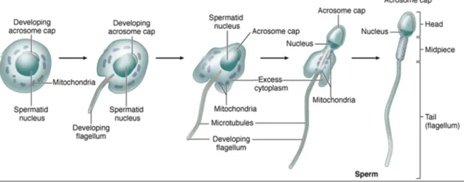
Figure 10: Spermiogenesis diagram.
Major morphological changes occur within spermatids as they undergo the differentiation process and become highly specialized sperm cells. These changes involve flattening of the nucleus, formation of an acrosome which resembles a large lysosome, growth of a flagellum (tail) from the basal body, reorganization of the mitochondria in the midpiece region, and shedding of unneeded cytoplasm as a residual body (taken from 110).
Humans vs. Rodents
Like in AD research, rodent (rat, mouse and hamster) models are also used in studies of gametogenesis. Despite both being mammals, there are several differences between humans and rodents spermatogenesis and testes. In Table 5 some characteristics that differentiate between human and the species addressed in this study (rat and mouse) are indicated. Spermatogonia, which constitute the first phase, are the most immature spermatogenic cells and reside on the basement membrane of the seminiferous epithelium. These cells are classified into three types in humans and four types in rodent, based on the appearance of the nuclei (presence and distribution of heterochromatin) 107,111. The human types are: type A dark spermatogonia; type A pale spermatogonia; and type B spermatogonia 107. The rodent types are: undifferentiated type A spermatogonia (A single, A paired, A aligned); differentiated type A spermatogonia (A1, A2, A3, A4); intermediate spermatogonia (In); and type B spermatogonia (B) (Table 5). However, presently, it is not possible to identify spermatogonial germ cells by routine microscopy, only by electron microscopy 111.