As leveduras do género Candida constituem um grupo muito diverso de fungos que inclui espécies comensais da microflora humana normal, podendo tornar-se patogénicas em determinadas condições de imunodepressão do hospedeiro. Apesar de várias espécies do género Candida estarem associadas a infecções em humanos, o principal agente é C. albicans, responsável por diversas candidíases superficiais das mucosas mas também por infecções sistémicas denominadas candidémias. Uma vez que a patogenicidade e susceptibilidade a antifúngicos varia entre estirpes da mesma espécie, é crucial uma identificação rápida e fiável da estirpe causadora da infecção para que possa ser aplicado o tratamento mais adequado.
O principal objectivo do presente trabalho consiste na descrição e caracterização de um novo microssatélite de C. albicans localizado no gene
IFF8 e que codifica uma proteína semelhante a Hyr1, um componente da
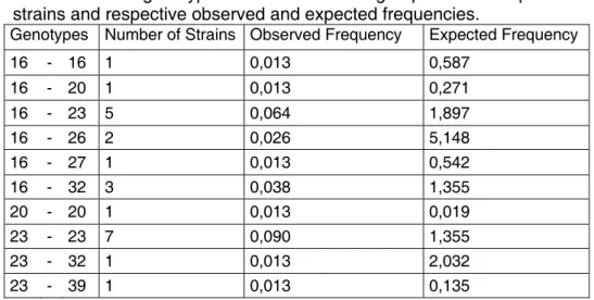
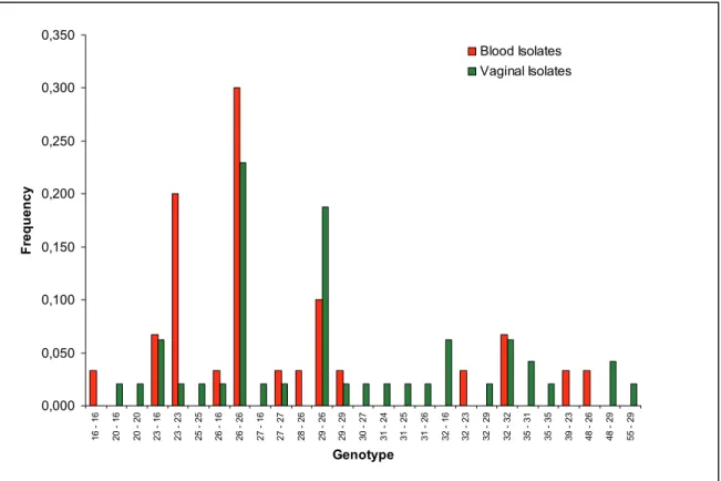
parede celular da forma de hifa de C. albicans. Para tal, utilizaram-se 78 isolados clínicos independentes provenientes de dois produtos biológicos, hemocultura e exsudados vaginais, e oriundos de pacientes internados em diversas instituições hospitalares. Foram identificados 16 alelos, com comprimentos entre 200 e 316 pares de bases, e 27 genótipos, o que resulta num poder de discriminação de cerca de 0.90. Foi ainda possível identificar genótipos específicos de isolados de hemoculturas, assim como de exsudados vaginais. Para determinar a origem da variabilidade alélica deste microssatélite a análise da estrutura dos diferentes alelos foi efectuada por sequenciação revelando a sua estrutura composta. Dada a localização deste microssatélite no gene IFF8 de C. albicans e uma vez que as proteínas GPI podem estar envolvidas na biossíntese e remodelação da parede celular, na adesão às células do hospedeiro e na virulência, a sua variabilidade poderá estar relacionada com o grau de virulência das estirpes em estudo.
Yeasts of the genus Candida include a variety of human commensal species that may become pathogenic in immunocompromised hosts. The predominant causal agent of candidiasis is C. albicans, responsible for superficial infections and systemic life threatening conditions. Since pathogenicity and antifungal susceptibility often vary between strains, it is crucial to rapidly and accurately identify the strain causing infection so that tha adequate treatment could be selected.
The aim of this work was to characterize a new microsatellite locus located inside IFF8 gene of C. albicans and coding for a GPI anchored protein similar to
Hyr1 which is a cell wall component of the C. albicans hypha. Seventy eight
independent C. albicans isolates from two biological products, blood cultures and vaginal exudates, and different health institutions were analysed. A total of 16 alleles, with lenghts varying between 200 and 316 bp were found and twenty seven genotypes were identified resulting in a discriminating power of 0.90. It was also possible to identify specific genotypes of blood culture isolates as well as specific of vaginal exudates isolates. Allele length variability was studied by sequence analysis showing that this is a compound microsatellite with a complex internal structure. Given the location of this microsatellite in C. albicans
IFF8 gene and since GPI proteins may be involved in cell wall biosynthesis and
remodelling, adhesion and virulence, its variability may be related to strain virulence.
AFLP Amplified fragment length polymorphism bp base pair
CGD Candida Genome Database CSC Centro de Saúde Carandá Braga DP Discriminatory power DNA Deoxyribonucleic acid
ECMM European Confederation of Medical Mycology EDTA Ethylenediamine tetraacetic acid
GPI Glycosylphosphatidylinositol GPIp GPI anchored protein
HIV Human Immunodeficiency Virus HYR1 HYphally Regulated gene 1 IFF Individual Protein File Family F
INSA Instituto Nacional Dr. Ricardo Jorge Lisboa HSJ Hospital São João Porto
ICU Intensive care unit
IPOL Instituto Português de Oncologia Lisboa IPOP Instituto Português de Oncologia Porto LOH Loss of heterozygocity
MLEE Multilocus Enzyme Electrophoresis MLST Multilocus sequence typing
PL Phospholipases PCR Polymerase chain reaction PGFE Pulsed field gel electrophoresis RAPD Randomly amplified polymorphic DNA REA Restriction endonuclease analysis RFLP Restriction fragment length polymorphism rpm Rotations per minute
SDA Sabouraud Dextrose Agar SDS Sodiumdodecyl sulphate SAP Secreted aspartyl proteases
SSLP Simple Sequence Length Polymorphism SSR Simple Sequence Repeats
STR Simple Tandem Repeats TE Tris EDTA
VNTR Variable Number of Tandem Repeats YPDA Yeast Peptone Dextrose Agar ZNF1 Zinc finger transcription factor
2. Virulence factors of Candida albicans 8 2.1. Adherence 9
2.2. Hydrolytic enzymes 9
2.2.1. Secreted Aspartyl Proteinases 10
2.2.2. Lipases 10 2.2.3. Phospholipases 11
2.3. Cell morphogenesis 11
2.4. Phenotypic switching 12
2.5. Other attributes 13
3. Genotyping of Candida albicans 13
4. Candida albicans microsatellite markers 21
5. Microsatellite DNA 23
6. IFF gene family 26
7. Importance and aims of this study 28
Materials & Methods 29
1. Origin and culture of the clinical isolates 31
2. Microsatellite selection and Primer design 31
3. Characterization of the selected microsatellite locus 32
3.1. Genomic DNA isolation 32
3.2. PCR amplification 33
3.3. Fragment size determination 34
3.3.1. Polyacrylamide gel electrophoresis 34
3.3.2. Genescan fragment determination 34
3.4. DNA sequencing 35
3.4.1. Allele sequence 35
3.4.2. Gene sequence 36
4. Differentiation of Candida albicans isolates using two microsatellite markers 36
2. Analysis of locus CAIV 44
3. Multilocus analysis 51
Conclusion & Final Remarks 57
References 63
1. Characteristics and medical relevance of the yeast Candida albicans
Over the past several decades, the incidence of fungal infections has increased, especially those acquired in health care associated settings, generally called nosocomial infections. Factors responsible for this rise include aging populations in countries with advanced medical technologies. As these trends continue, it can be predicted that the incidence of invasive nosocomial fungal infections will continue to increase.
The predominant nosocomial fungal pathogens include Aspergillus spp.,
Mucorales, Fusarium spp. and Candida spp. (Perlroth et al., 2007). Yeasts
belonging to the genus Candida have emerged as major fungal opportunistic pathogens in humans whose immune system is debilitated. As commensals
Candida species are harmless, they belong to the normal microbial flora of skin
and mucosal surfaces. However, if the balance of the normal flora is disrupted or the immune defences are compromised, these organisms can outgrow the mucosal flora and cause disease: frequently superficial infections of mucosa. Furthermore, in hospital settings, Candida species may cause life-threatening invasive infections in a growing population of vulnerable patients (Mavor et al., 2005).
Following reduction in the host immune status, two main types of infections can be observed: superficial and invasive candidiasis. Superficial infections of mucosal epithelial tissues are frequent in immunocompromised patients. In more severe cases, Candida species may enter the bloodstream (candidaemia) and can reach almost all organs of the body. Theoretically, any organ can be infected, however, the organs most commonly colonised are the kidney, the brain and the heart (Fridkin & Jarvis,1996).
Approximately two hundred species of Candida have been described but only a few are of medical importance (Ng et al., 1999). Candida albicans is the most frequently isolated species, but other species, such as C. tropicalis, C. krusei, C.
parapsilosis, and C. glabrata, have increasingly been recognized as pathogens
with wide distribution (Correia et al., 2004; Bassetti et al., 2006; Colombo et al., 2006; Snydman, 2006).
The risk of infections caused by yeasts of the genus Candida, generally denominated candidiasis, is influenced by host resistance as well as by external factors. Concerning host resistance, there is an association between diseases causing reduction in the host immune status and candidiasis. Patients are particularly predisposed to these infections when suffering from neutropenia, heart disease, cancer, and diabetes. Patients with late stage Human Immunodeficiency Virus (HIV) infection have also an extremely high incidence of candidiasis. However, HIV infection is not an independent risk factor because the increased incidence of disseminated candidiasis in patients infected with HIV is attributable to the increased incidence of the other usual risk factors. On the other hand, treatments themselves also have immunosuppressive effects that predispose patients to candidiasis, namely chemotherapy, radiotherapy, steroid drugs, and broad-spectrum antibiotics.
Additionally, disruption of normal skin barriers (burn injury, percutaneous catheter placement, abdominal surgery or parenteral nutrition) is an important risk factor for invasive Candida infections because it provides a direct route of entry for pathogens. Exogenous acquisition of candidaemia has been reported, with a significantly increased risk of Candida bloodstream infections associated with intravascular devices and parenteral nutrition (Tortorano et al., 2006).
Prior colonisation with Candida species is considered a prerequisite for subsequent deep-seated infection and has been demonstrated to be the leading risk factor for candidaemia (Tortorano et al., 2006). It has been shown that patients with higher colonization burdens have a proportionately higher risk of developing disseminated disease (Perlroth et al., 2007).
It should be highlighted that not all these predisposing factors equally favour superficial and invasive infections. For example, HIV infected individuals suffer extremely frequently from oral Candida infections, but rarely develop disseminated infections (Vargas & Joly, 2002).
It is not clear whether the strains that colonize healthy hosts are responsible for causing subsequent invasive disease when those hosts acquire the appropriate risk factors, or whether infections are caused by acquisition of more virulent strains from environmental sources in the nosocomial setting. Overall data suggest that in
most cases, the source of an infecting strain of C. albicans is endogenous flora, but that in certain circumstances transmission of more virulent strains may occur in the nosocomial setting. However, several reports of outbreaks of infection with
Candida species support the hypothesis that exogenous acquisition (transmission
of a strain from one patient to another) of the infecting yeast strain can occur (Ruiz-Diez et al., 1997; Shin et al., 2005).The increase of vulnerable patients has made Candida infections increasingly important, particularly in hospital settings. The frequency of candidaemia among hospitalised patients has doubled during the 1980s and 1990s (Blot & Vandewoude, 2004; Almirante et al., 2005). Through the late 1980s, the predominant species causing invasive Candida infections was C.
albicans. In Europe, more than half of all cases of candidemia were caused by C. albicans as monitored in three independent investigations (Mavor et al., 2005).
However, since the 1990s, there has been a steady increase in the relative frequencies of non-albicans species of Candida causing disseminated candidiasis (Perlroth et al., 2007). The relative contribution of different Candida species varies between countries (Tortorano et al., 2006). These circumstances have made
Candida infections an increasingly serious threat in European hospitals. Studies
reported a leveling off of the frequency of invasive Candida infections during the late 1990s due to the use of prophylaxis and more effective diagnostic methods and antifungal treatment (Blot & Vandewoude, 2004). Furthermore, most recently, several studies have been published contradicting the notion that the incidence of disseminated candidiasis is leveling off, and demonstrating a continued rise in its incidence since the turn of the 21st century (Perlroth et al., 2007).
The severity of candidaemia is confirmed by the high crude mortality rate found in
the European Confederation of Medical Mycology (ECMM) survey (38%)
(Tortorano et al., 2006).
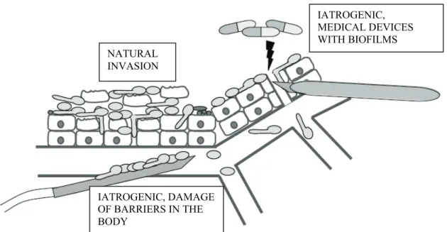
The three main routes of entry of Candida cells into the circulatory system are shown in Figure 1.
Figure 1. Candida albicans may enter the bloodstream by direct penetration from epithelial tissues (natural invasion), due to damage of barriers in the body or may spread from biofilms produced on medical devices (iatrogenic invasion). Adapted from Mavor et al., (2005).
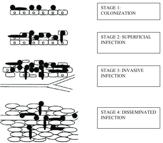
The first is the “natural” way through penetration of epithelial cells from mucosal surfaces into deeper tissues and blood capillaries or vessels. The second and third are “artificial” (iatrogenic) routes, which are available due to the use of medical devices or caused by damage of barriers (Mavor et al., 2005). Natural invasion is processed in four possible stages as shown in Figure 2.
NATURAL INVASION IATROGENIC, DAMAGE OF BARRIERS IN THE BODY IATROGENIC, MEDICAL DEVICES WITH BIOFILMS
Figure 2 – Stages of infection by natural invasion. Adapted from Naglik et al., (2003).
Not only are invasive Candida infections extremely common, they are also difficult to diagnose and treat (Blot & Vandewoude, 2004). Because of the difficulties in confirming the diagnosis with laboratory studies, empiric administration of therapy is often based on a clinical diagnosis of disseminated candidiasis typically made in a patient with symptoms and laboratory features consistent with infection, and who has risk factors for disseminated candidiasis.
Until the 1980s therapy for invasive candidiasis was limited to amphotericin B, but with the advent of new antifungal agents, such as azoles and echinocandins, less toxic therapeutic options are possible. In general, due to its favorable toxicity profile, high oral bioavailability, low cost, and impressive efficacy in randomized clinical trials, fluconazole therapy is preferred (Perlroth et al., 2007).
The antifungal agents available to treat candidiasis are limited in number and effectiveness, due basically to the limited number of identified targets that are clearly distinguished between eukaryotic fungal cells and mammalian cells. In addition to their efficacy, the clinical usefulness of these drugs is hampered by
STAGE 1: COLONIZATION STAGE 2: SUPERFICIAL INFECTION STAGE 3: INVASIVE INFECTION STAGE 4: DISSEMINATED INFECTION
their safety, as undesirable side effects on patients are often associated with antifungal drugs, and by the emergence of resistances that parallels their clinical use (Gozalbo et al., 2004).
2. Virulence factors of Candida albicans
In order to establish infection, opportunistic pathogens have to evade the host immune system, survive and divide in the host environment, and spread to new tissues. The characteristics that allow colonization and persistence in the host, conferring the ability to adapt to a variety of microhabitats and cause infections are generally termed virulence factors (Calderone & Fonzi, 2001; Yang, 2003).
During transition from commensal growth to superficial infections, and during penetration into deeper tissue and the bloodstream, C. albicans has to adapt to radically changing environments.
Most of C. albicans virulence factors are related to the cell wall which is a fungal specific, complex and dynamic structure essential to almost every aspect of its biology including pathogenicity (Gozalbo et al., 2004). Since it is the outermost cellular structure, it plays a major role in the interactions between the microorganism and the environment, including the host-fungal interactions. Several studies showed that yeast cell wall proteins are associated with a number of functions in cell physiology including cell surface integrity, flexibility and remodelling, cell morphology, growth, and budding (Calderone & Fonzi, 2001; Bates et al., 2006). Thus, this structure is inevitably related with virulence.
Candida albicans virulence factors include (i) attributes which allow cells to adhere
to the host, namely adhesins, (ii) hydrolytic enzymes that enable colonisation, invasion and tissue damage (such as secreted aspartyl proteinases (SAP), lipases (LIP), and phospholipases (PL)), (iii) morphogenesis (the reversible transition between unicellular yeast cells and, filamentous, growth forms) that permits shape and gene expression variation according to the pressures of the varying environments during infections, and (iv) phenotypic switching, which is accompanied by changes in antigen expression, colony morphology and tissue affinities (Calderone & Fonzi, 2001; Mavor et al., 2005).
2.1. Adherence
Adherence to host cells is seen as an essential early step in the establishment of disease. In addition, C. albicans can also adhere on the surfaces of medical devices and form biofilms increasing the possibility of contamination and systemic infection. It was observed that strains with the greater ability to form biofilms and to adhere to the host cells are the most virulent ones (Yang, 2003).
C. albicans displays several adhesins which are biomolecules that promote the
adherence to host cells or host-cell ligands. Most of them are glycoproteins, including proteins belonging to Als (agglutinin-like sequence) family which is one of the best characterized groups of cell surface proteins in C. albicans (Hoyer, 2001). These proteins are members of the glycosylphosphatidylinositol (GPI) cell wall proteins and share a central domain containing tandem repeats. They are homologous to -agglutinin, required for cell to cell recognition during mating in
Saccharomyces cereviseae, and have been shown to present adhesin functions to
mammalian cells (Hoyer, 2001; Yang, 2003). All family members have similar functions but seem to be specialized for different conditions encountered in various host environments. Als1, for example, is required in the early stages of morphogenesis, for full adherence and for full virulence in a hematogenously disseminated murine model, and Als5 (Ala1) was found to confer on S. cerevisiae cells the ability to adhere to extracellular matrix proteins (Alberti-Segui et al., 2004).
2.2. Hydrolytic enzymes
Another factor that contributes to the process of virulence is hydrolytic enzyme production, which is known to play a central role in the pathogenicity of protozoa, bacteria, and pathogenic yeasts. The secretion of hydrolases during infections may be required as a virulence attribute for the degradation of host surfaces (to aid adhesion, invasion, and destruction of host immune factors) in addition to nutrient acquisition. C. albicans possesses a large spectrum of hydrolytic enzymes with relatively broad substrate specificities, including secreted aspartyl proteases,
lipases, and phospholipases, which might be the reason for the outstanding position of this human pathogen.
2.2.1. Secreted Aspartyl Proteases
Although many microorganisms possess a variety of hydrolytic enzymes, proteinases particularly secreted aspartyl proteinases (SAP) are by far the most commonly associated with virulence. All proteinases catalyze the hydrolysis of peptide bonds (CO—NH) in proteins but can differ markedly in specificity. SAP are secreted primarily to provide nutrients for the cells; however, pathogenic fungi appear to have adapted this biochemical property to fulfil a number of specialized functions during the infective process. These more direct virulence functions may include digesting host cell membranes or damaging molecules of the host immune system to resist antimicrobial attack. They were also shown to contribute to several other virulence attributes of C. albicans during the infection process including hyphal formation, adhesion, and phenotypic switching (Naglik et al, 2003).
The SAP gene family has ten members and, as with the Als proteins, family members have similar functions but seem to be specialised for different conditions encountered in various host environments (Koelsch et al., 2000). For example,
Sap1–3 are required for mucosal infections and Sap4–6 for systemic infections
(Albrecht et al., 2006). Albrecht et al. (2006) showed that Sap9 and Sap10, in contrast to the other family members, are GPI-anchored aspartic proteinases that target proteins of fungal origin necessary for cell surface integrity, cell separation, and adhesion.
2.2.2. Lipases
Lipases are enzymes that are able to catalyse both the hydrolysis and synthesis of ester bonds of lipids. The most prominent role of extracellular lipases for a microorganism is also the digestion of lipids for nutrient acquisition. These enzymes might help both bacteria and fungi to grow in a carbohydrate-restricted
area or environments where lipids are the sole carbon source. Another putative role of these proteins is the enhancement of adhesion, cell growth and hyphal formation to support colonization.
In C. albicans, the lipases are also encoded by a gene family with ten members:
LIP1 to LIP10. In a similar manner to the SAP gene family, the LIP genes have
been shown to be differentially expressed at different stages and sites of infection (Stehr et al., 2003).
2.2.3. Phospholipases
Phospholipases (PL) hydrolyze one or more ester linkages of glycerophospholipids and are classified according to the specific ester bond cleaved. They belong to a group of enzymes capable to derange or destroy cell surface membranes after the adherence is realized ensuring their penetration into the host cells (Ivanovska, 2003). Results from several studies reveal that cells producing less PL are less virulent than cells producing more PL indicating that these enzymes can be classified as virulence factors (Calderone & Fonzi, 2001; Ghannoum, 2000). In addition, C. albicans strains isolated from blood produce higher levels of PL than commensal ones (Ibrahim et al., 1995).
In C. albicans, all four types (A, B, C and D) of PL have been detected but only proteins encoded by the PL B family seem to be extracellular. PLB1 appears to be responsible for most of the extracellular phospholipase activity and it has been shown to be required for virulence in an animal model of disseminated candidiasis (Calderone & Fonzi, 2001).
2.3. Cell morphogenesis
Cell morphogenesis is thought to be an important virulence factor of C. albicans. This species exhibits considerable morphogenetic plasticity, growing in either a yeast or hyphal form, or as pseudohyphae, previously thought to represent an intermediate stage between yeast cells and true hyphae but proved to be a distinct developmental form (Whiteway & Oberholzer, 2004). This morphogenetic
variability is linked to pathogenicity. It was shown that cells that are trapped in either the yeast or filamentous forms are significantly reduced in virulence in several infection models (Saville et al., 2006).
Both types of morphology may be present in infected tissue, and it is therefore possible that both may play important roles in the pathogenesis. Nevertheless, hyphal growth may be more critical since hyphae adhere more strongly to mammalian cells, promote tissue penetration, and provide a mechanism to escape the attack by macrophages (Ernst, 2000).
Although morphogenesis in C. albicans may be an important regulator of the specific response in the host, a variety of extracellular conditions can in turn induce yeast cells to form hyphae and pseudohyphae. The external signals that promote filamentous growth also include pH in neutral-basic range, elevated temperature (37ºC), nutrient starvation (e.g. iron), and exposure to an inductor (serum, N-acetylglucosamine, or proline) (Romani et al., 2003).
It is generally accepted that dimorphism is a virulence trait per se, but since changes in the cell surface accompany the morphological transition, it is intimately linked with other virulence factors.
2.4. Phenotypic switching
Many pathogenic protozoa, bacteria, and fungi have evolved strategies for alternative expression of surface-related phenotypes enabling their adaptation to changing environments and evasion of the immune response (Lan et al., 2002). In addition to the yeast-hypha transition, C. albicans is capable of undergoing a different type of morphological change, termed phenotypic switching, which involves the spontaneous and reversible generation of different morphological and physiological states (Soll, 1997). Besides changes in the morphology of the colonies, phenotypic switching also affects levels of resistance to antifungals, levels of proteinase secretion, adhesion, hyphal formation and sensitivity to neutrophils. Due to this influence over other virulence traits, phenotypic switching is referred to as “higher order” virulence attribute (Mavor et al., 2005).
Both processes, dimorphic transition and switching, confer on C. albicans the ability to generate variants which allow a better selective adaptation to changing environmental conditions, and particularly to evade the host immune system (Gozalbo et al., 2004).
2.5. Other attributes
Other traits have been implicated as important for the pathogenicity of C. albicans (Mavor et al., 2005). Increased antimicrobial drug resistance, for example, helps a pathogen evade clearance in the presence of antifungals. In C. albicans, resistance mechanisms include the mutation of the antifungal targets themselves and the upregulation of some multidrug transporters. Besides, several Candida species, including C. albicans, have an antioxidative stress response consisting of the production of antioxidants that defend them against reactive oxygen species during phagocytosis. In addition, the ability to assimilate iron is necessary for survival of all pathogens in the host and could hence be called a persistence factor, being frequently considered a virulence determinant.
The virulence factors required by C. albicans to cause infections may well vary and show distinct complex interactions depending on the type of infection, the site and stage of infection, and the nature of the host response.
3. Genotyping of Candida albicans
The typing of C. albicans at the strain level has become crucial for medical mycology. For this species, as for all microbial pathogens, a reproducible and discriminatory strain typing system is of benefit for clinical and epidemiological studies to provide information on sources, carriage, and transmission of infection and on relations between strain types and properties, such as virulence and antimicrobial resistance (Odds et al., 2006, Tay et al., 2005). Thus, studying the relatedness of clinical strains is relevant in clinical management (Chong et al., 2007, Chen et al., 2005).
A number of different approaches for typing C. albicans isolates have been developed since the early 1980s. Molecular typing methods should be reproducible, discriminatory, high throughput, easy-to-use, digitally portable and amenable to standardization and library typing (Soll, 2000). Most molecular typing methods are based on techniques such as pulsed field gel electrophoresis (PGFE), restriction fragment length polymorphism (RFLP), polymerase chain reaction (PCR), and sequencing.
Each of the developed methods described next has its own set of assets as well as limitations, ensuring the continuous search in this field aiming to overcome them.
Multilocus Enzyme Electrophoresis
Multilocus Enzyme Electrophoresis (MLEE) is a technique based on electrophoresis of total proteins under native conditions and subsequent visualization of the enzymes present in the gel by specific enzyme staining procedures. It has been extensively used to fingerprint C. albicans, among other
Candida species, and was shown to be useful to assess the mode of reproduction
of microorganisms (Pujol et al., 1997; Soll, 2000). The main drawback to this method is that, being an indirect analysis of DNA performed through analysis of proteins, only coding regions are monitored and it is not possible to differentiate DNA sequences that code for the same protein. Also, minor aminoacid variation may not affect electrophoretic mobility of the protein. On the other hand, MLEE is relatively laborious and time consuming, because it is necessary to combine data from at least ten enzymes that provide variability among isolates (Soll, 2000). For instance, in a study of clinical C. albicans strain discrimination using MLEE by Pujol et al. (1997), 21 enzymes were tested but only 13 exhibited variability and were therefore used in the analysis.
Pulsed field gel electrophoresis
For more than two decades, several PFGE-based typing methods have been widely used, including karyotyping (Chen et al., 2005). This analysis is based in the differential migration of entire chromosomes with different sizes in an agarose matrix under the influence of an electric field. After DNA fragments are separated according to size they are visualized by ethidium bromide staining. The high intraspecific variability of karyotypes of fungi makes this technique discriminatory and useful for population studies in pathogenic yeasts, particularly C. albicans. Because of the high chromosome instability of this species there are some questions regarding reproducibility of the method. Additionally, this technique is time-consuming so it is no longer frequently used for C. albicans strain typing (Soll, 2000).
Another related approach is PFGE of restriction fragments, also called restriction endonuclease analysis (REA) of genomic DNA. It consists in electrophoresis of fragments generated by rare-cutting restriction endonucleases, such as BssHII and SfiI useful for investigating microevolution of C. albicans clinical strains (Shin
et al., 2005). REA may be a more sensitive method than karyotyping in the
investigation of C. albicans infections but the use of more than one restriction enzyme may be necessary for optimal strain discrimination (Voss et al., 1995).
Restriction fragment length polymorphism
Restriction fragment length polymorphism (RFLP) techniques assay variations of the genome using restriction endonucleases that recognize and cut specific DNA sequences. There are basically two variants of this method; the first is called direct RFLP because it goes without probe hybridization, and the second is indirect RFLP because it is followed by specific probe hybridization.
One of the first DNA fingerprinting methods used to assess strain relatedness in a variety of infectious fungi, including C. albicans, was RFLP without any probe hybridization (Vazquez et al., 1991, Voss et al., 1995). In this technique, DNA extracted from spheroplasts is digested with one or more endonucleases, and
separated by electrophoresis in an agarose gel. The banding pattern of digested DNA is then visualized, usually by staining with ethidium bromide. This pattern is based on different fragment lengths determined by the restriction sites identified by the particular endonucleases employed. Low-intensity bands in most RFLP patterns are poorly resolved and, therefore, comparisons usually rely only on differences between intense bands, representing mainly ribossomal and mitochondrial DNA sequences. These fragments do not provide enough information to assess the relatedness of moderately close isolates. Even so, there is abundant evidence from the many RFLP studies of C. albicans that the method has been successful in identifying the same strain in independent isolates and in distinguishing among unrelated isolates.
The RFLP pattern of eukaryotic cell DNA as described above is poorly resolved primarily because all restriction fragments are stained. To allow visualization of particular fragments in the pattern, one can probe a Southern blot of the RFLP gel with a labelled DNA sequence that recognizes one or more fragments as a result of sequence homology. Any region of DNA can be used, including repetitive regions, if variations can be visualized with probes. Ribossomal and mitochondrial DNA probes have not been generally used in broad epidemiological studies of the infectious fungi mainly because of the presence of homologous genes and low discriminating power (DP). To date, the most successful and popular hybridization probes for the fungi have been cloned fragments containing repetitive genomic sequences. In a Southern blot of endonuclease digested genomic DNA, such a probe will hybridize to repetitive sequences dispersed throughout the genome, thus identifying variability among isolates at a variety of dispersed loci. It will also hybridize to additional sequences that are less variable, including sequences that vary as a result of allelic polymorphisms. Finally, it will hybridize to some hypervariable sequences, revealing microevolutionary changes within a strain. The virtue of the complex probe is that all this information is provided by a single Southern blot hybridization pattern.
Two complex probes for C. albicans, 27A and Ca3, were cloned approximately at the same time in the late 1980s and subsequently found to be related, although not identical. These have been used in a number of studies with C. albicans
(Shmid et al., 1993; Schroppel et al., 1994; Ruiz-Diez et al., 1997; Marco et al., 1999; Boccia et al., 2002; Taylor et al., 2003). Ca3 probe generates a pattern that is, on average, more complex than the one generated by 27A, and has proven to be reproducible and highly amenable to computer-assisted analysis. Databases have been established for comparisons of the Ca3-generated patterns of strains from different studies and for retrospective analyses. However, this methodology is technically demanding, not amenable to high throughput sample processing. In general, RFLP with hybridization with C. albicans-specific probes is very informative but time-consuming since Southern blots are needed (Soll, 2000).
Polymerase chain reaction
The techniques referred above are time consuming and not always compatible with the imperatives of the clinical laboratory. Due to their rapidity, PCR-based methods generally seem more appropriate.
Randomly amplified polymorphic DNA analysis
Although a variety of PCR-based strategies have been developed for DNA fingerprinting purposes, randomly amplified polymorphic DNA analysis (RAPD) has been one of the most commonly used for the infectious fungi. RAPD assay has become one of the most favourable choices for DNA fingerprinting of medically important Candida species and has been extensively applied in C.
albicans strain typing (Metzgar et al., 1998).
In this technique, genomic DNA is amplified with a single short (approximately 10 bases) primer with an arbitrary sequence and products are separated on an agarose gel and stained with ethidium bromide. Although a single primer can generate a relatively complex pattern, in most cases, it provides only one to three intense bands differing among isolates. Therefore, it is usually necessary to select a number of primers, run independently for each test isolate, and combine the information. This strategy is illustrated by the work of Pujol et al. (1997), who
tested 40 random primers on a limited number of C. albicans test isolates and selected 8 that provided maximum variability.
RAPD analysis is technically simple and often detects variation among isolates that are invariant with RFLP analysis. Of the currently available Candida genotyping techniques, RAPD is relatively cost-effective and matches the resolving power of PFGE karyotyping, use of Ca3 complex probes and MLEE (Pujol et al., 1997; Soll, 2000). This, together with the availability of computer-assisted software systems that generate dendrograms of genetic relatedness among C. albicans isolates, has significantly contributed to the understanding of infective episodes and asymptomatic carriage of C. albicans (Samaranayake et
al., 2003). The RAPD method has also gained favour because it is less
time-consuming than Southern blot hybridization based methods. Unfortunately, the RAPD technique is poorly reproducible due to the low annealing temperatures used in the PCR. The problem of reproducibility exists not only among laboratories but within a laboratory over time, making the development of a common database difficult. Besides, the patterns obtained are often complex and sometimes difficult to interpret.
Amplified fragment length polymorphism
One promising modification of RAPD is amplified fragment length polymorphism (AFLP). This method selectively amplifies restriction fragments of genomic DNA. Amplification is achieved by using the restriction sites for the annealing of primers, and fragment selection is achieved by adding selective bases to the 3’ end of the primers. By using stringent reaction conditions for primer annealing, the reliability of the method has been reported to be superior to that of the RAPD method. Careful selection of the restriction enzymes and nucleotides at the primer 3’ ends results in a complex fingerprint pattern in a sequencing gel with yeast DNA as template (Soll, 2000).
Sequencing
Cloning and sequencing genes used to be a slow, technically demanding, and expensive undertaking, beyond the technical capacity of most medical mycologists interested in DNA fingerprinting of large collections of isolates. Recently, the emergence of PCR, automated DNA-sequencing technologies, and gene data banks, provided sequence based fingerprinting strategies amenable and affordable for large epidemiological studies.
Multilocus sequence typing
Multilocus sequence typing (MLST) is a sequencing-based technique that consists in the analysis of nucleotide polymorphisms of internal fragments of housekeeping genes (Maiden et al., 1998). DNA sequences from six or seven gene fragments are compared in order to establish the level of similarity between isolates. MLST is a highly discriminatory approach to distinguishing strains within a microbial species and has been widely used for epidemiological purposes. Because it was first developed to discriminate bacteria, analysis of diploid organisms such as C.
albicans can be subjective. Today, MLST schemes for several fungi are available,
and the approach is well developed for the typing of C. albicans strains (Bougnoux, et al., 2002; Tavanti et al., 2003).
Results from MLST are comparable with those obtained by DNA fingerprinting with the moderately repetitive sequence Ca3, since both typing approaches assigned the same sets of isolates to the same clusters of highly related strains in a study by Odds et al. (2006). Chowdhary et al. (2006) have shown that MLST has a higher discriminatory power, compared to Ca3 fingerprinting, in describing the genetic relatedness among clinical C. albicans isolates. When compared to other typing methods, MLST offers several advantages: it is rapid, reproducible and amenable to automation, provides an objective comparison of molecular typing data among laboratories, permits the exchange of that data via the internet to support local and global epidemiological studies, is easily implemented into any laboratory with DNA sequencing capacity, and supports the creation of a
comprehensive MLST database of C. albicans nucleotide polymorphisms. To assess the value of MLST relative to those of other DNA fingerprinting tools for discriminating among strains of C. albicans, Robles et al. (2004) applied it to a previously well-characterized set of C. albicans isolates evaluated by RAPD, MLEE, and Ca3 southern hybridization probe techniques. These results demonstrated that MLST is a highly effective technique that performs at least comparably to other established DNA fingerprinting techniques. Nevertheless, because it is necessary to combine information from at least six genes, this is not suitable to type a large set of isolates.
Microsatellite genotyping
Another class of molecular typing methods includes techniques that directly analyse the polymorphism of microsatellite markers. These markers are stable, easy to assay, adaptable to a large series, and discriminatory enough to be used as a typing system to investigate clinical issues. Because of their high levels of allelic variation and their co-dominant character, they deliver more information than any other marker system, thus reducing analysis time and costs. Besides, since microsatellites test the presence of different alleles at a given locus, distinguishing heterozygotes in diploid organisms such as C. albicans is possible in contrast to the RFLP and RAPD methods.
The most efficient type of microsatellite analysis consists in the amplification of the repetitive region by PCR with the use of primers complementary to conserved flanking regions. After, amplification products of different length are sized in order to determine the number of repetitions of the motives. Various methods to size these alleles can be used but the majority is based on electrophoresis in which the size determination of DNA molecules relies on calculation of the mobility in the gel matrix relative to an internal standard (Christensen et al., 1999). This can be performed using simple conventional laboratory equipment, electrophoresis with agarose or polyacrilamide gels, or using a more expensive but more sensitive and rapid method: capillary electrophoresis system using laser fluorescence detection in an automatic DNA sequencer (Deforce et al., 1998).
Microsatellite analysis has been extensively used for a wide range of biological questions, namely mapping and positional cloning of genes, population studies (structure, diversity and epidemiology), evolutionary studies, phylogenetics, linkage analysis, forensics, molecular pathology, paternity testing, relatedness and identification of individuals (Naidoo & Chetty, 1998; Schlötterer, 2000; Buschiazzo & Gemmell, 2006; Eloy et al., 2006).
4. Candida albicans microsatellite markers
Several studies have already reported the application of microsatellite markers for the genotyping of Candida albicans (as we will see below). At least 17 microsatellite loci have already been characterized for this pathogenic yeast species, providing adequate targets for the molecular typing of its strains.
In 1996, Field and co-workers described seven C. albicans microsatellite markers (ERK1, ZNF1, CCN2, MNT2, CPH1, EFG1 and EFG2) consisting of small repetitive regions from coding sequences and in which most of the repetitive motifs coded the same aminoacid, glutamine. Although these presented low polymorphism, this study revealed the great abundance and potential of microsatellite markers in C. albicans. ERK1 polymorphism was confirmed in another study aiming to assess specific site polymorphisms in C. albicans isolates from HIV infected patients, receiving fluconazole for fungal prophylaxis. This analysis allowed three different scenarios to be discerned: strains can remain identical, be replaced by clearly different strains, or undergo small changes (Metzgar et al., 1998).
In 1997, a microsatellite marker called CEF3 was described. It was found in the upstream sequence of the elongation factor 3 gene (EF3), between a putative TATA box and the transcription start site, in chromosome 5 of C. albicans. When applied to the study of sixty C. albicans isolates, it showed higher level of polymorphism than the previous ones (Bretagne et al., 1997). This marker was used in many posterior studies. Lott et al. (1999) compared independent isolates of C. albicans from two different geographic regions using both CEF3 and ERK1 and concluded that there was no indication of geographical partitioning. Dalle et al.
(2000) compared bloodstream and nonbloodstream strains of C. albicans using CEF3 concluding that they have a heterogeneous structure at this locus with three major and multiple minor allelic combinations. This group found an undescribed allele as well as seven new combinations. In 2003, the same team characterized
C. albicans strains from healthy individuals using CEF3 and compared them with
strains from nonhealthy individuals showing an overall similarity that suggested the ability of all commensal strains to develop as pathogens (Dalle et al., 2003). Lott & Effat (2001) in an attempt to characterize special groups of C. albicans isolates, combined information obtained from the analysis of five microsatellite markers (and other loci), three of them already described: ZNF1 (zinc finger transcription factor), ERK1 (extracellular-signal-regulated kinase), CEF3, and two new ones: A3 (Anonymous locus 3) and A4 (Anonymous locus 4) that presented very low polymorphism.
Microsatellite markers CDC3 and HIS3, present near coding regions and named after the gene they stand by, were described and applied together with CEF3 (Botterel et al., 2001). With the aim of obtaining a rapid genotyping method of C.
albicans, these three markers were investigated by multiplex PCR and revealed a
combined discriminatory power (DP) of 0.97. The same three marker combination was used again to evaluate the colonization of Candida species and the importance of C. albicans cross-contamination by Stephan et al. (2002). Clinical specimens obtained from surgical patients who had a high risk of yeast colonization were screened and it was concluded that acquisition of C. albicans in the surgical intensive care unit (ICU) seems to be mainly endogenous.
In a population study of C. albicans isolates from USA, Europe and Asia, two new markers were described: KRE6 and LOC4 (previously referred by Lott et al. (1999)) by Fundyga et al. (2002). These microsatellites were located in coding regions and exhibited low polymorphism. This study also included CEF3, ERK1, and ZNF1, assessing genotypes at five loci to address the question of genetic variation of strains isolated from different human hosts and geographic distinct regions.
In 2003, Sampaio and co-workers investigated the polymorphism of a new microsatellite locus, CAI, located in chromosome 4 of C. albicans. It was revealed
to be species-specific and showed a low mutation rate, since no amplification product was obtained when testing other pathogenic Candida species and no genotype differences were observed when testing over 300 generations. This new microsatellite was very polymorphic presenting the greater individual DP reported to date (0.97) the same obtained with the multilocus analysis described above, proving to be a valuable tool to differentiate C. albicans strains. Furthermore, when compared to other molecular genotyping techniques, CAI proved to be very simple, highly efficient, and reproducible, being suitable for low-quantity and very-degraded samples and for application in large-scale epidemiological studies.
Two years later, the same group described five new C. albicans microsatellite loci (CAIII, CAIV, CAV, CAVI, and CAVII) and developed a multiplex strategy to differentiate C. albicans strains using CAI, characterized earlier, and the two new microsatellites: CAIII and CAVI (Sampaio et al., 2005). The DP obtained by combining the information generated achieved 0.99, the highest value ever reported. The multiplex PCR was later used to test C. albicans clinical isolates. The analysis of microsatellites by this multiplex PCR strategy was found to be a highly efficient tool for the rapid and accurate differentiation of C. albicans strains and adequate for the identification of fine microevolutionary events that could be related to strain microevolution in response to environmental stress conditions. Besides their potential as molecular markers, it has been suggested a significant role of microsatellite sequences as relevant genomic information. Therefore, microsatellites are the focus of this work.
5. Microsatellite DNA
A great percentage of genomic DNA of all living organisms is made up of repetitive sequences, which fall into two classes: transposable elements-like sequences, which move around the genome, and internally repetitive sequences, such as satellite DNA, which belong to a group of genomic sequences known as Variable Number of Tandem Repeats (VNTR). Microsatellites are short DNA sequence stretches in which a motif of one to six bases is tandemly repeated and minisatellites are sequences composed of longer (10-100 bases) repeated motifs.
Microsatellites, also known as STR (Simple Tandem Repeats), SSR (Simple Sequence Repeats) and SSLP (Simple Sequence Length Polymorphism), are present in both Eukaryotes and Prokaryotes, and can be found either in protein-coding or non-protein-coding regions. Microsatellite sequences are stably inherited and hence highly conserved from one generation to the next. Although they are unique to an individual and the same in all cells from the same individual, they exhibit high level of allelic variation (polymorphism) among individuals (Tautz, 1989).
Polymorphism is a crucial feature of microsatellites that makes them powerful and highly versatile genetic markers. It is predominantly manifested as changes in the number of the repeated motif caused by mutations that typically occur at rates that
are orders of magnitude greater than single-nucleotide point mutations, from 10-7
to 10-2 events per locus per generation. These rates are influenced, among other
factors, by environmental conditions, repeated motif, allele size, interruptions in the microsatellite, recombination rate, transcription rate, chromosome position, GC content in flanking DNA, and genotype.
Microsatellites were usually considered selectively and evolutionarily neutral sequences that are randomly distributed over the genome. However, conflicting results about their distribution are documented (Levinson & Gutman 1987, Schlotterer, 2000). Numerous lines of evidence available today suggest that microsatellite genomic distribution is not random and that it is dependent of many factors, namely location and repeat type (from mononucleotides up to hexanucleotides). Repeats are rarer in coding regions than in non-coding regions and this is probably attributable to negative selection against frame-shift mutations (Metzgar et al., 2000). Tóth et al. (2000) conducted a detailed analysis that revealed highly taxon-specific patterns in the distribution of different repeat types in coding and noncoding sequences. Although the majority of microsatellites are composed of mono- and dinucleotide repeats (Dieringer & Schlötterer, 2003), tri-nucleotide repeats (and motif lengths that are multiples of three) are overrepresented in coding sequences. This happens because they can be accommodated more readily within coding regions as change in their length simply results in gain or loss of a single amino acid from a protein sequence. Thus, long mono- and di-tracts are almost exclusively distributed in non-translated regions
(Field & Wills 1998). Also, Katti et al. (2001) and Dieringer & Schlötterer (2003) found dramatic differences among repeat types within and between species. The presented examples demonstrate various non-random patterns of microsatellite variation that call for functional interpretation.
Microsatellites used to be commonly regarded as DNA sequences with no significant role as genomic information. Questions concerning the role of these sequences arose because of their frequent presence in highly economical genomes such as those of yeasts. Since then, the functional significance of a substantial part of microsatellites has been proven in various biological phenomena namely chromatin organization, and regulation of both DNA metabolic processes and gene activity.
In many cases, microsatellite repeat number appears to be a key factor for gene expression and expression level. Kashi & King (2006) stated that whatever the role and location of a microsatellite, changes in its number of repeated motifs can modulate its genetic function. When in coding regions, trinucleotide microsatellites variation in the number of repeated motives (codons) results in a variation in the length of homopolymeric stretches of amino acid affecting protein properties (Richard et al., 1999; Hancock & Simon, 2005). Mutations that change the number of repeats in coding non-triplet microsatellites cause frameshifts, which can effectively inactivate gene expression or encode different protein sequences. Because frameshifting based on microsatellite mutation is readily reversible by subsequent mutation, such microsatellites can function as on–off switches for their genes which is proven to happen in some bacteria (Li et al., 2004). Repeat variation commonly exerts a functional influence even when the microsatellites are located in noncoding sites where they do not affect protein structure directly.
Precise changes in microsatellites with high repeat numbers are long known to regulate critical virulence factors in several prokaryotes (Field & Wills, 1996). The presence of microsatellites in these organisms is rare, but most that do occur are related to pathogenic ones. In pathogenic bacteria, for example, infection processes require adaptation to several host environments and that is possibly achieved through microsatellite modulation, as has been described for a multitude of different genes (Li et al., 2004). Recently, Kashi & King (2006) showed the
results of a study that strongly implicates a mononucleotide-repeat polymorphism as a causal basis for differentiation in sporulation efficiency of yeast, a significant life-history trait for these organisms. Experimental manipulation of some microsatellites has demonstrated a linear correlation between repeat number and the extent of cell adhesion (Verstrepen et al., 2005). There is also evidence suggesting that the variation in repeat size of microsatellites coding for cell wall proteins of S. cerevisiae can alter its phenotype and may also transform the antigenic properties of the cell, thus allowing it to deceive the immune system of the host (Richard & Plaine, 2006).
In humans, instability of microsatellite may contribute to the development of cancer in several sites by causing either relatively small or drastic changes in the repeat structure. It has also been shown that many neurological disorders result from the expansion of unstable trinucleotide repeats located either in non-coding or in coding sequences (Katti et al., 2001).
6. IFF gene family
Microsatellite CAIV was investigated in this work. The first reference indicated that it was found in a non coding region of the C. albicans genome (Sampaio et al., 2005). Instead, new available data from Candida Genome Database (CGD) accounts for its location inside IFF8 gene, in chromosome 5. Analysis of the C.
albicans genome has identified the IFF gene family (Individual Protein File Family
F), with 12 genes, as encoding the largest family of cell wall related proteins. Ten genes (IFF1-9 and HYR1 (HYphally Regulated)) are predicted to have GPI anchors and two genes, IFF10 and IFF11, do not have any signal sequences for GPI anchor linkages (Richard & Plaine, 2006). Phylogenetic analysis of this family shows that the two non GPI anchored proteins Iff10 and Iff11 diverge obviously from the original group. No function has been established so far for any of the proteins, but their presence in 12 copies in the genome implies that they perform a useful function in terms of C. albicans biology, an idea reinforced again by the fact that S. cerevisiae has no clear ortholog.
Very little is known about this family and only two members, HYR1 and IFF11, have been the subject of published work (Bailey et al., 1996)). Nevertheless, it has been shown that this family is conserved in a range of other Candida species suggesting that the observed differences might be one of the reasons explaining the virulence variations between very close species (Bates et al., 2007).
A null mutant lacking IFF11 was studied by Bates et al. (2007) and shown to be hypersensitive to cell wall-damaging agents, suggesting a role in cell wall organization. In a murine model of systemic infection this null mutant was highly attenuated in virulence, and survival-standardized infections suggest it is required to establish an infection. This work provides the first evidence of the importance of this gene family in the host-fungal interaction and virulence.
Although no clear function has been yet attributed to the referred proteins, the fact that most of them are putative GPI anchored proteins (GPIp) suggest that they are worthy of further investigation.
GPI anchored proteins are an abundant class of cell surface proteins present in both lower and higher eukaryotic organisms (De Groot et al., 2003). Their wide occurrence does not dictate specific functions, as many types of proteins are GPI anchored. The functions of GPIp have been extensively investigated and several roles have been assigned to it. Most are thought to be involved in cell wall biosynthesis and remodelling, hydrophobicity, antigenicity, flocculation, proteasic activity, sporulation, adhesion, mating, and virulence (De Groot et al., 2003). There is evidence demonstrating that a functional GPI anchor is required for normal cell wall structure and for full hyphal formation in C. albicans, and that perturbation of the GPI-anchor biosynthesis also results in hypersensitivity to host defences (Gozalbo et al., 2004). It has been shown that complete GPI anchors are required in C. albicans for full morphogenesis, virulence and resistance to macrophages (Richard et al., 2002; Richard & Plaine, 2006).
Albrecht et al. (2006) described two C. albicans cell surface-associated aspartic proteases, Sap9 and Sap10, which are GPIp. They have shown that Sap9 and Sap10 are crucial for the infection process. In contrast to the other SAP family members, they are GPIp necessary for cell surface integrity, cell separation, and adhesion.
Another family of GPIp, termed the fungal adhesins, is important for fungal pathogenesis permitting pathogens such as C. albicans to adhere to mammalian epithelial and endothelial cells. Expression of these proteins in S. cerevisiae permits this organism, which does not normally adhere to mammalian cells, to adhere to them (Guo et al., 2000).
7. Importance and aims of this study
Candida albicans is a serious human health concern, especially for the growing
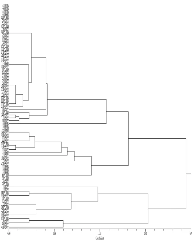
population of immunocompromised patients. Since pathogenicity and antifungal susceptibility often vary between strains, it is crucial to rapidly and accurately identify the strain causing infection so that the best treatment strategy is selected. The aim of the present work is to study a new microsatellite locus, CAIV, located inside IFF8 gene of Candida albicans. CAIV characterization is focused on its polymorphism, allelic frequencies, genotypic frequencies and discrimination power. This molecular marker will be used to assess strain relatedness of a group of C. albicans clinical isolates from several Portuguese health institutions as well as to compare strains collected from two biologic products, blood and vaginal exudates. A multilocus approach using CAI and CAIV molecular markers will be evaluated according to the discrimination power and ability to detect microevolutionary events when multiple isolates from the same patient are compared.
Materials
&
Methods
1. Origin and culture of the clinical isolates
A total of 97 Candida albicans clinical isolates from five different Portuguese institutions were studied. The isolates were obtained from one health centre, Centro de Saúde Carandá Braga (CSC), two oncology hospitals: IPO Porto (IPOP) and IPO Lisboa, Instituto Nacional Dr. Ricardo Jorge Lisboa (INSA), and Hospital São João Porto (HSJ). Approximately half of the strains were collected from blood cultures and the others were obtained from vaginal exudates, in a total of 78 independent patients. Considering the number of clinical isolates studied this means that, in some cases, more than one isolate from the same patient was examined.
All the isolates had been previously identified by conventional phenotypic methods like the ability to produce germ tubes and by rapid biochemical identification kits, such as API-32C (BioMérieux, St Louis Mo.) or by YBC Card (Vitek Systems Inc., Hazelwood Mo.), according to the clinical laboratories of origin. Isolates were named after their institution of origin followed by a number in order to preserve the anonymity of the patients.
Isolates were received in plates with Yeast Peptone Dextrose Agar (YPDA) medium or Sabouraud Dextrose Agar (SDA) medium, and were subjected to macroscopic analysis to confirm their purity. Pure cultures of each strain were then preserved at 4ºC in YPDA tubes. For long term analysis, they were also frozen at -80ºC in 1.5 ml of a 30% (w/v) glycerol solution.
2. Microsatellite selection and Primer design
A search in C. albicans genome sequences, available in databases from Stanford’s DNA Sequencing and Technology Center (http://www.sequence.stanford.edu/group/candida), was conducted for sequences containing microsatellite repeats. Previously, a search for repetitive sequences expected to have a very high degree of polymorphism was conducted based on two criteria: the number of simple repeat units (more than 20) and the location, outside a coding region. Following this search several microsatellites were
selected, characterized and used for C. albicans strain differentiation (Sampaio et
al., 2003; Sampaio et al., 2005).
According to assembly 20 of C. albicans genome sequencing project, it was found that some of the selected sequences containing repetitive regions were, in fact, located inside genes. A microsatellite consisting of trinucleotide repeats, located in
IFF8 gene, which codes for a GPI anchored protein, was then chosen for further
characterization in the present work and designated as locus CAIV.
To determine the chromosomal localization of the microsatellite the sequence selected was searched by BLAST against the latest release of C. albicans genome sequence, to give a location to a sequence contig (http://www-sequence.stanford.edu/group/candida).
Specific primers for the amplification of this locus were designed from the microsatellite flanking regions by using software Primer 3 available from www-genome.wi.mit.edu/cgi-bin/primer/primer3www.cgi. The conditions for primer design were that they should allow PCR amplification at an annealing temperature of 60ºC and that the amplified fragments should not exceed 500bp.
The forward primer was fluorescently labelled with FAM for detection in the automatic sequencer.
3. Characterization of the selected microsatellite locus 3.1. Genomic DNA isolation
The procedure followed was adapted from the method described by Kaiser et al., 1994. Cells were grown overnight in 15ml of YPD medium at 30ºC on a rotary shaker at 200rpm and then harvested by centrifugation at 5000rpm for 5 minutes at 4ºC. After ressuspending the cells in 1ml sorbitol buffer, 10µl of liticase were added. This mixture was incubated for an hour at 37ºC to break the cell wall. Another centrifugation at 5000rpm for 5 minutes was performed and spheroplasts in the pellet were ressuspended in 1ml of tris-EDTA. To break down the cell membranes, 30µl of 10% (v/v) SDS (sodiumdodecyl sulphate) solution was added. This suspension was heated at 65ºC for 30 minutes. After addition of 250µl of
potassium acetate (3M), the mixture was stored on ice for one hour for protein precipitation and centrifuged at 10000rpm for 10 minutes. To precipitate the nucleic acids an equal volume of cold isopropanol was added. The pellet thus obtained was subsequently washed twice with 70% ethanol (v/v) for 5 minutes. The dried pellet was finally ressuspended in 200µl of TE buffer (pH 7.4) and the DNA solution stored at 4ºC. From this solution, new ones were prepared with 25ng/µl for subsequent use in PCR reactions; these were stored at -20ºC.
3.2. PCR amplification
PCR reactions were performed in several independent isolates in order to evaluate the locus-specific amplification and its polymorphism.
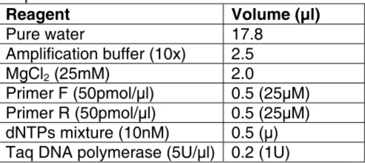
This locus was amplified using the reagent mixture presented in Table 1. While preparing this mixture, 10% excess of all components were added to ensure sufficient volumes. 1µl of each DNA template solution (total 25ng) isolated as described above was added to 24 µl of the mixture.
The sequences of the primers used were: F- 5’CCGCTTCTAATGATACTGGTGTT 3’ R- 5’TTTCCGTGGCATCAGTATCA 3’
Table 1. Reagents used to prepare the PCR amplification mixture. Reagent Volume (µl) Pure water 17.8 Amplification buffer (10x) 2.5 MgCl2 (25mM) 2.0 Primer F (50pmol/µl) 0.5 (25µM) Primer R (50pmol/µl) 0.5 (25µM) dNTPs mixture (10nM) 0.5 (µ) Taq DNA polymerase (5U/µl) 0.2 (1U)
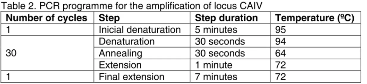
Amplification reactions were performed in the thermocycler (Icycler, BioRad) following the programme presented in Table 2.
Table 2. PCR programme for the amplification of locus CAIV
Number of cycles Step Step duration Temperature (ºC)
1 Inicial denaturation 5 minutes 95
Denaturation 30 seconds 94
Annealing 30 seconds 64
30
Extension 1 minute 72
1 Final extension 7 minutes 72
The annealing temperature of 64ºC was chosen after analysis of a range of values, from 58ºC to 66ºC.
3.3. Fragment size determination
3.3.1. Polyacrylamide gel electrophoresis
PCR products were analysed by electrophoresis in 6% (w/v) polyacrylamide gel under denaturing conditions provided by addition of 6.5% (w/v) urea. Buffer systems used were described by Gusmão et al. (1997) for the Multiphor II electrophoretic system, and consisted of gel buffer Tris-HCl (0.375M, pH 8.8) and loading buffer Tris-glicina (0.125M, pH 8.8). DNA fragments obtained were visualized by silver staining. Gels were washed with 10% (v/v) ethanol solution followed by a 1% (v/v) nitric acid solution. After rinsing with water, gels were incubated with a 0.2% (w/v) AgNO solution in the dark. They were then submerged in a 0.28M NaCO solution with 0.02% (v/v) formaldeid to reveal the DNA fragments, and finally fixed with 10% (v/v) acetic acid.
3.3.2. Genescan Fragment determination
When PCR conditions were optimized the genotyping of all the clinical isolates was performed by Genescan analysis. DNA samples were prepared by adding to 1µl of PCR products, 14.7µl of formamide with 0.3µl of molecular size marker
TAMRA GeneScanTM 500 (Applied Biosystems). These samples were denatured
by incubation at 96ºC for 5 minutes.
Samples were run in an ABI PRISM 310 genetic analyser (Applied Biosystems) and capillary electrophoresis was performed using POP4 polymer at 60ºC and
15Kv. Fragment size of PCR products was determined automatically using GeneScan 3.7 Analysis software (Applied Biosystems).
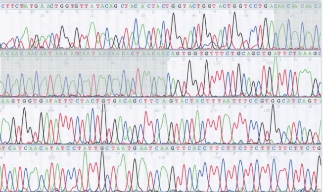
3.4. DNA sequencing 3.4.1. Allele sequence
The sequence of the alleles was determined in order to confirm the specific amplification of CAIV locus and to study the number of repetitions and the structure of the motif.
Single alleles were excised from polyacrylamide gels and eluted in 200µl of TE buffer, and subjected to three cycles of freezing (-20ºC) and towing (65ºC). These DNA fragments were reamplified in a PCR using the previous conditions and purified using Microspin S-300 HR (Amersham Biotech). 2µl of this product was added to 2µl of Mix BigDie Terminator v3.1 (Applied Biosystems) and 1µl of primer solution, either forward or reverse. The sequencing reaction took place in the thermocycler (Icycler, BioRad) following the programme described in Table 3.
Table 3. Programme used in the sequencing reaction of locus CAIV.
Number of cycles Step Step duration Temperature (ºC)
1 Inicial denaturation 10 minutes 96
Denaturation 10 seconds 94
Annealing 20 seconds 55
35
Extension 4 minutes 60
1 Final extension 10 minutes 60
These products were purified using AutoSeqTM G-50 columns (Amersham Biotech) according with the instructions. 20 µl of formamide were added and the solution was denatured at 96ºC for 3 minutes. These fragments were separated by capillary electrophoresis in an ABI PRISM 310 genetic analyser (Applied Biosystems) using POP4 polymer at 50ºC and 15Kv. Analysis of the sequence was performed with Sequence Analysis software 3.1 (Applied Biosystems).