FACULDADE DE MEDICINA
THE REPERTOIRE AND SPECIFICITY OF
FOLLICULAR REGULATORY T CELLS
Ana Raquel Maceiras de Oliveira
Orientador: Prof. Doutor Luís Ricardo Simões da Silva Graça
Tese especialmente elaborada para obtenção do grau de Doutor em Ciências Biomédicas no ramo de Imunologia
FACULDADE DE MEDICINA
THE REPERTOIRE AND SPECIFICITY OF
FOLLICULAR REGULATORY T CELLS
Ana Raquel Maceiras de Oliveira
Orientador: Prof. Doutor Luís Ricardo Simões da Silva Graça
Tese especialmente elaborada para obtenção do grau de Doutor em Ciências Biomédicas no ramo de Imunologia
Júri:
Presidente: Doutor José Augusto Gamito Melo Cristino, Professor Catedrático e Presidente do Conselho Científico da Faculdade de Medicina da Universidade de Lisboa.
Vogais: Doutor António Ângelo Bastos Alves de Freitas, Professor, Retired Scientist do Institut
Pasteur, Paris, França;
Doutor Manuel João Rua Vilanova, Professor Associado do Instituto de Ciências Biomédicas Abel Salazar da Universidade do Porto;
Doutora Jocelyne Danièle Michèle Demengeot, Investigadora Principal do Instituto Gulbenkian de Ciência;
Doutor Marc Veldhoen, Especialista de Reconhecido Mérito e Competência, Group Leader do Instituto de Medicina Molecular da Faculdade de Medicina da Universidade de Lisboa; Doutora Karine Serre, Especialista de Reconhecido Mérito e Competência, Investigadora do
Instituto de Medicina Molecular da Faculdade de Medicina da Universidade de Lisboa; Doutor Bruno Miguel de Carvalho e Silva Santos, Professor Associado com Agregação da
Faculdade de Medicina da Universidade de Lisboa;
Doutor Luís Ricardo Simões da Silva Graça, Professor Associado com Agregação da Faculdade de Medicina da Universidade de Lisboa; (Orientador).
Trabalho financiado pela Fundação para a Ciência e a Tecnologia no âmbito da bolsa SFRH/BD/88030/2012
ii
A impressão desta tese foi aprovada pelo Conselho Científico da Faculdade
de Medicina de Lisboa em reunião de 24 de Janeiro de 2017.
iii
As opiniões expressas nesta publicação são da exclusiva
v Ao meu pai que guardo com saudade
vii
Table of Contents
Index of Figures ... xi
Index of Tables ... xiii
Abbreviations ... xv Acknowledgments ... xix Abstract ... xxi Resumo ... xxiii I. General Introduction ... 1 Immune system ... 3
Innate immune system ... 3
Adaptive Immune system ... 6
B cells ... 7 Germinal centers ... 11 T cells ... 14 CD8 T cells ... 17 CD4 T cells ... 18 Th1 cells ... 19 Th2 cells ... 20 Th17 cells ... 21 Th9 cells ... 23 Th22 cells ... 23
Follicular helper T cells ... 24
Initial priming by dendritic cells ... 24
Full commitment to the Tfh program at the T-B border ... 26
Tfh function and maintenance at germinal centers ... 27
Additional transcription factors involved in Tfh differentiation ... 28
Tfh cells memory ... 29
Tfh cells in human beings ... 29
Regulatory T cells ... 30
viii
Peripherally induced Treg cells ... 32
Treg cells suppression mechanisms ... 32
Treg cell subsets ... 33
Foxp3- regulatory T cells ... 33
Follicular regulatory T cells ... 34
Tfr cell differentiation ... 35 GC regulation by Tfr cells ... 36 Tfr cells memory ... 37 GCs and autoimmunity ... 37 II. Objectives ... 39 III. Methods ... 43 Mice ... 45
Immunizations and cell transfers ... 45
Flow cytometry and cell sorting ... 45
In vitro cultures ... 46
RNA extraction, cDNA synthesis and CDR3 length analysis ... 47
RNA extraction, cDNA synthesis, TRA gene amplification, and deep sequencing ... 48
CDR3 data analysis ... 49
Deep sequencing data analysis ... 49
Statistical analysis ... 50
IV. Results ... 51
1. Tfr cells originate from thymic Treg cells ... 53
1.1. Introduction ... 55
1.2. Results ... 55
1.3. Discussion ... 57
2. Tfr cells are not specific for the immunizing antigen ... 59
2.1. Introduction ... 61
2.2. Results ... 61
2.3. Discussion ... 69
3. Tfr and Tfh populations have different TCR repertoires ... 73
ix 3.2. Results ... 75 3.3. Discussion ... 82 V. General Discussion ... 85 VI. References ... 91 VII. Appendix ... 115
xi
Index of Figures
Figure 1 – VDJ recombination process of BCR heavy chain. ... 8
Figure 2 – Class Switch Recombination process. ... 9
Figure 3 – GC initiation and development. ... 12
Figure 4 – V(D)J recombination of the TCR. ... 15
Figure 5 – Tfh differentiation steps. ... 25
Figure 6 – Tfh-B cell signals. ... 26
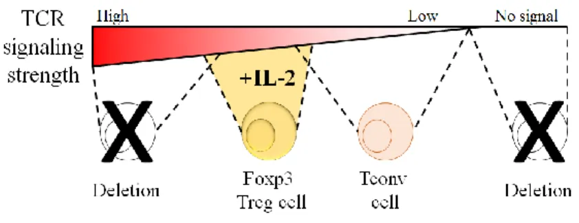
Figure 7 – TCR signaling strength determines Treg differentiation in the thymus. ... 31
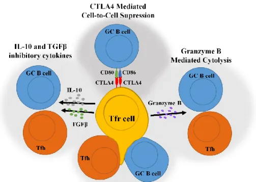
Figure 8 – Tfr cell suppression mechanisms. ... 36
Figure 9 – T follicular cells are virtually inexistent on popliteal LNs prior to immunization. ... 56
Figure 10 – Tfr cells do not differentiate from FOXP3- T cell precursors. ... 56
Figure 11 – Antigen-specific FOXP3+ tTreg cells can differentiate into Tfr cells. ... 57
Figure 12– Preferential accumulation of OVA-specific cells within Tfh, but not Tfr cells.62 Figure 13– Recruitment of OVA-specific cells as Tfr cells is independent of the immunizing antigen. ... 63
Figure 14– OT-II Treg cells specifically proliferate with OVA signals... 64
Figure 15– Recruitment of P25-specific and OVA-specific T cells into the Tfh pool of mice immunized with the corresponding antigens. ... 65
Figure 16– Tetramer+ cells are predominantly found among the Tfh population. ... 67
Figure 17– Tfr cells do not proliferate or survive with antigen signals. ... 68
Figure 18 – CDR3 spectratypes obtained from naïve CD4 T cells. ... 76
Figure 19 – Tfh cells present over-representations of specific CDR3 lengths. ... 77
Figure 20 – Analysis of CDR3 length distribution unveils a closeness between Tfr and Treg cells. ... 77
Figure 21 – 1D2β mice can mount specific responses against OVA. ... 79
Figure 22 – Tfr cells are oligoclonal and have more common clonotypes with Treg than Tfh cells. ... 80
Figure 23 – Tfr cells are closer to Treg cells than to any other population. ... 81
Figure 24 – Tfr cells origin and specificity. ... 88
Figure 25 – Speculative hypothesis on how Tfr cells prevent the selection of auto-reactive B cells. ... 89
xiii
Index of Tables
Table 1 – CD4 T cell subsets and its characteristics. ... 19
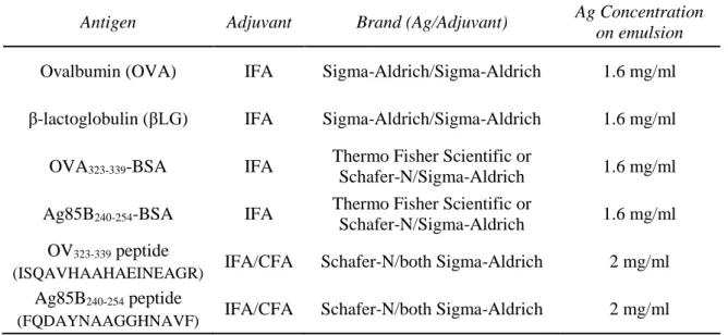
Table 2 – List of antigens and adjuvants used for immunization ... 46
Table 3 – Antibodies used for flow cytometry analysis ... 46
Table 4 – Primers used for CDR3 spectratyping/immunoscope analysis ... 48
Table 5 – Primers used for TCRα sequencing ... 49
Table 6 – Pairwise Multiple Comparison Analysis with Holm-Bonferroni Correction between Samples TRBV Perturbation Scoresa ... 78
xv
Abbreviations
ADCC - antibody-dependent cell cytotoxicity Ag – antigen
AHR – aryl hydrocarbon receptor AICD – activation-induced cell death
AID – activation-induced cytidine deaminase APC – antigen-presenting cell
ASC – antibody secreting cell
ASCL2 – achaete-scute homologue-2 AU – approximately unbiased
BATF – basic leucine zipper transcription factor BCL-6 – B cell lymphoma 6
BCR – B cell receptor BP – bootstrap probability bp – base pairs
BTLA – B and T lymphocyte attenuator cAMP – cyclic adenosine monophosphate CD – cluster of differentiation
CDR – complementarity-determining region CFA – Complete Freund’s adjuvant
CSF – colony-stimulating factor CSR – class switch recombination cTfh – circulating Tfh cells
CTLA-4 – cytotoxic T-lymphocyte-associated protein 4 CTV – CellTrace Violet
CXCL – C-X-C chemokine ligand CXCR – C-X-C chemokine receptor
DAMP – damage-associated molecular patterns DC – dendritic cell
DP – double positive DZ – dark zone
xvi
EAE – experimental autoimmune encephalomyelitis FACS – flow activated cell sorting
FASL – FAS ligand FcR – Fc receptor FcγR - IgG Fc receptor FcεR – IgE Fc receptor
FDC – follicular dendritic cell FOXP3 – forkhead box P3 FSC – forward scatter GC – germinal center
GFI-1 – growth factor independent-1
GITR – glucocorticoid-induced TNF receptor-related protein GM-CSF - granulocyte macrophage colony-stimulating factor hCD2 – human CD2
H-chain – immunoglobulin heavy-chain HVEM – herpesvirus entry mediator IBD – inflammatory bowel disease ICOS – inducible T cell co-stimulator ICOSL – ICOS ligand
IFA – Incomplete Freund’s adjuvant IFN – interferon
IL – interleukin
ILC – innate lymphoid cell
IPEX – immune dysregulation, polyendocrinopathy, enteropathy, X-linked syndrome IRF - interferon-regulatory factor
i.v. – intravenous
KLF2 – Krüppel-like factor 2
LAG-3 – lymphocyte-activation gene 3 L-chain – immunoglobulin light-chain LN – lymph node
LZ – light zone
MAC - membrane attack complex MCL1 – myeloid cell leukemia 1
xvii MS – multiple sclerosis
MZ – marginal zone
NFAT2 - Nuclear factor of activated T-cells 2 NK cell – natural killer cell
NKT cell – natural killer T cell
NT-ES – nuclear transfer - embryonic stem cell OVA – ovalbumin
PAMP – pathogen-associated molecular patterns PD-1 – programmed cell death 1
PD-L – PD-1 ligand
PI3K – phosphoinositide 3-kinase PRR – pattern recognition receptors PSGL-1 – P-selectin glycoprotein ligand 1 pTreg – peripherally-derived Treg cells RA – rheumatoid arthritis
RAG - recombination-activating gene
RORα – retinoic acid receptor-related orphan receptor alpha RORγt – retinoic acid receptor-related orphan receptor gamma-T RT – room temperature
SAP – SLAM-associated protein s.c. – subcutaneous
SHM – somatic hypermutation
SLAM – signaling lymphocytic activation molecule SLE – systemic lupus erythematosus
SP – single positive
SPF – specific pathogen free SSC – side scatter
STAT – signal transducer and activator S1PR1 – sphingosine-1-phosphate receptor 1 T-BET – T-box transcription factor
TCR – T cell receptor TD – T cell dependent
TdT – terminal desoxyribonucleotidyl transferase TI – T cell independent
xviii
TLR – Toll-like receptors TNF – tumor necrosis factor
TGFβ – transforming growth factor beta TRA/TCRα – T-cell receptor alpha chain TRAF3 – TNF receptor-associated factor 3 TRAV – V region of TRA
TRB/TCRβ – T cell receptor beta chain TRBV – V region of TRB
TRBVBJ – V and J regions of TRB Tconv – conventional T cells Tfh – follicular helper T cells Tfr – follicular regulatory T cells Treg – regulatory T cells
tTreg – thymic-derived Treg cells WT – Wild-type
xix
Acknowledgments
First, I want to thank my supervisor Luís Graça for giving me the opportunity to pursue a PhD in his group.
I would also like to thank everyone that was and still is in the lab during these 5 years, especially Sílvia, Marta Gomes, my homonymous Raquel, Andreia, and Patrícia.
To all the staff of the support units, especially flow cytometry, rodent facility, and bioimaging, thank you for sharing your precious time with me.
A special thanks to my thesis committee. I feel that you taught me some of the most important lessons and I will definitely and carefully keep them throughout my professional life.
And now in Portuguese:
Quero agradecer à minha família em especial à minha mãe, à minha tia Lucinda e à minha avó Gina. Obrigada, três Marias.
xxi
Abstract
Germinal centers (GCs) are key structures where B cells are selected to produce high affinity immunoglobulins of the adequate class. This selection is dependent of B cell interactions with specialized follicular helper T (Tfh) cells that provide help for class switch recombination, somatic hypermutation, and selection signals to GC B cells. It was recently described that the GC reaction is controlled by specialized Foxp3+ follicular regulatory T (Tfr) cells. These cells regulate the size and magnitude of the GC reaction, and have been also implicated in the prevention of autoimmunity.
This thesis aimed to answer two questions regarding Tfr cells biology. First, it was investigated whether Tfr cells derive from thymic-derived regulatory T (tTreg) cells. The second objective was to establish whether Tfr cells are specific for the non-self antigen driving the GC response, as Tfh cells, or if Tfr cells have a repertoire closer to Treg cells with specificity towards self-antigens.
It was found that Tfr cells originate exclusively from tTreg cells. Also, Tfr cells were not specific for the immunizing antigen. Indeed, as antigen-specific TCR-transgenic Tfr cells were not specifically recruited into the GC, nor could antigen-specific Tfr cells be detected using class-II MHC tetramers. Moreover, Tfr cells did not specifically proliferate in vitro when stimulated with the immunizing antigen. Lastly, repertoire analysis of Tfr, Treg and Tfh cells demonstrated that Tfh cells and Tfr cells have different repertoires with the latter retaining a repertoire closer to Treg cells.
Taken together, the presented results show a clear difference in the specificity and TCR usage by Tfh and Tfr populations from the same GCs. This distinct specificity is in line with different putative functions of the two populations: while Tfh cells promote antigen-specific B cell responses, Tfr cells prevent autoimmunity.
xxiii
Resumo
Os centros germinativos (GCs) são estruturas onde as células B sofrem hipermutação somática, seleção positiva e mudança de isotipo, de forma a produzirem anticorpos de alta afinidade e de isotipo adequeado à resposta imunitária em curso. No entanto, para que estes processos ocorram, as células B dos centros germinativos precisam de ajuda por parte de células T auxiliares foliculares (Tfh).
Recentemente, foi descrita uma nova população de células reguladoras FOXP3 positivas, as células T reguladoras foliculares (Tfr), que se encontram dentro dos GCs. Estas células estão envolvidas na regulação dos GCs, nomeadamente, na quantidade e na qualidade (afinidade) de anticorpos produzidos durante uma resposta imunitária. Para além disso, as células Tfr parecem também estar envolvidas na prevenção de doenças autoimunes.
As células Tfr partilham algumas características quer com as células T reguladoras (Treg) FOXP3 positivas, quer com as células Tfh. Tal como os linfócitos Treg, as células Tfr expressam FOXP3, CD25 e CTLA-4 e têm capacidade supressora. Por outro lado, assim como as células Tfh, as células Tfr expressam PD-1, ICOS, CXCR5 (que lhes permite migrar para o GC) e BCL-6, o principal factor de transcrição responsável pela diferenciação de células T CD4 foliculares. Tanto as células Tfr como as Tfh podem ser induzidas por imunização, já que esta induz GCs, num processo muito semelhante ao que é observado após vacinação.
O trabalho descrito nesta tese tinha dois objectivos principais. O primeiro era estabelecer qual a origem das células Tfr., isto é, se estas células se diferenciam a partir de células Treg originadas no timo ou a partir de células T CD4 convencionais (Tconv). O outro objectivo consistia em determinar a especificidade das células Tfr, ou seja, se estas células são específicas para o antigénio da imunização, ao qual as células B do GC estão a responder e a maturar a sua afinidade. Três linhas de trabalho foram estabelecidas para responder a estas questões.
Em primeiro lugar, efectuaram-se transferências de células Tconv específicas para um antigénio devido à expressão de um receptor de células T (TCR) transgénico. Foi verificado que estas células específicas, depois de uma imunização com o respectivo antigénio, não se diferenciavam em células Tfr. No entanto, quando uma população de células Treg específicas que se origina no timo está presente, estas dão origem a células Tfr. Assim, as células Tfr originam-se exclusivamente de células Treg que se originam no timo.
xxiv
Para estabelecer se as células Tfr, como as células Tfh, são específicas para o antigénio que induz a resposta do GC, foram realizadas experiências in vivo e in vitro. Primeiro, realizaram-se transferências de células transgénicas específicas para um antigénio, entre as quais uma população de células Treg também específicas. Após imunização com o respectivo antigénio (ou um antigénio controlo), verificou-se um recrutamento específico de células transgénicas como Tfh para o centro germinativo. No entanto, as células Treg específicas não foram preferencialmente recrutadas como células Tfr já que a mesma percentagem de células foi observada após imunização com o antigénio específico e com o antigénio controlo. Para confirmar os resultados anteriores, mas sem a utilização de células transgénicas, estudou-se a existência de células Tfr específicas após imunização com dois péptidos, utilizando para isso dois tetrâmeros de MHC classe II. Detectou-se uma população de células Tfh específicas (tetrâmero positivas) em imunizações contendo o péptido correspondente. Porém, não foram detectadas células Tfr específicas (tetrâmero positivas) em nenhuma das imunizações efectuadas. Verificou-se ainda que as células Tfr não reconheciam especificamente o antigénio da imunização que lhe deu origem, já que a sua proliferação in vitro foi igual independentemente do antigénio fornecido durante a cultura.
Por último, estudou-se o reportório das células Tfr, Tfh e Treg. O estudo do reportório, inicialmente feito através da determinação da distribuição do comprimento de CDR3 para cada segmento V da cadeia β do TCR, mostrou que as células Tfh apresentavam diferenças em relação à distribuição verificada para células T CD4 naïves. Estas diferenças consistiam principalmente em aumentos de utilização de comprimentos CDR3 específicos. A existência destes aumentos indica a presença de expansões clonais dentro da população de células Tfh. Os mesmos aumentos não foram observados nas células Tfr que, apesar de também apresentarem algumas diferenças em comparação com as distribuições das células T CD4 naïves, tinham o seu reportório próximo das células Treg. Para confirmar os resultados da análise de utilização de comprimentos de CDR3 e obter uma maior informação sem as limitações da técnica anterior, o reportório da cadeia α do TCR de murganhos 1D2β foi também sequencido. Este modelo de murganho, para além de ser repórter para FOXP3, expressa uma cadeia β do TCR transgénica (não variável) e tem apenas um dos alelos da cadeia α do TCR disponível para recombinação. Apesar disso, estes animais são capazes de montar respostas específicas contra um antigénio, visto que após imunização há formação de células T CD4 foliculares e as células Tfh destes animais conseguem reconhecer especificamente o antigénio correspondente em ensaios in vitro. A utilização deste modelo
xxv tem como vantagem obter a informação completa do reportório das populações em estudo sequenciando apenas a cadeia α do TCR. A análise do reportório das células Tfr, Treg e Tfh permitiu saber que, apesar de as células Tfr e Tfh serem oligoclonais, estas não têm clonótipos comuns. Por outro lado, a população celular que apresentava um reportório mais próximo das células Tfr era a população Treg, apesar desta última ser policlonal (sem expansões óbvias de clonótipos específicos). Estas observações permitem concluir que as células Tfr têm uma especificidade diferente das células Tfh, uma vez que o seu reportório é diferente, e que devem ser específicas para antigénios do próprio, tal como as células Treg de que se originam e com quem têm um reportório mais próximo.
Assim, os resultados permitem concluir que as células Tfr originam-se de células Treg e, não sendo específicas para o antigénio que induz a resposta do GC, têm um reportório próximo das células Treg, com especificidade para antigénios do próprio.
1
3
Immune system
The immune system is constituted by specialized cells and molecules which main role is to defend an organism from disease. Although immunity was first associated with protection against infectious disease, the immune system can also be activated by noninfectious foreign substances and damaged or altered cells of the self.
The immune system is divided into two fundamentally different parts, the innate and the adaptive immune systems, that interact and complement each other. The innate immune system consists of cellular and biochemical mechanisms that are already in place even before infection, and provides protection to the same extension independently of how many times it has encountered the same infectious agent. The adaptive immune system, on the other hand, takes longer to respond, but is characterized by the exquisite specificity for each infection and the ability to improve its response upon re-exposure to the same infectious agent.
Innate immune system
When the need for an immune response arises, the innate immune system is activated first to respond against the threat to the organism. In the case of an infection, the response is initiated within minutes through an inflammatory response.
The main activation mechanism of innate immune cells is through the recognition of a limited number of molecules called pathogen-associated molecular patterns (PAMPs) by pattern recognition receptors (PRRs). PRRs include Toll-like receptors (TLRs)1, C-type lectin receptors (like the mannose receptor2), scavenger receptors3, among others. These receptors, which can be present on the surface, in the endosomal vesicles, or in the cytoplasm of innate immune cells, bind different molecules from distinct pathogens, and consequently trigger different immune responses. TLRs, the most extensively studied PPRs, are divided in two groups based on their cellular localization: TLRs 1, 2, 4, and 5 are found in the cell surface and recognize bacterial and viral products on the extracellular space, while TLRs 3, 7, 8, and 9 are expressed mainly in endosomal vesicles and detect the presence of bacterial and viral nucleic acids. Furthermore, some PRRs also recognize cell damage signals by dying cells, called DAMPs (damage-associated molecular patterns), which allow the system to identify candidate cells for phagocytosis and initiate an inflammatory and immune response with these signals. Examples of DAMP receptors are TLR2 and TLR4 that bind host heat shock proteins4,5 and TLR9 that binds Chromatin-IgG complexes6. The activation
4
of PRRs cause cell activation, production of cytokines, and presentation of co-stimulatory signals that lead to activation of the immune system through an initial inflammatory response, recruitment of more cells to the local of the ongoing immune response, and further activation of specialized cell through cell-to-cell contact.
Cytokines are small molecules secreted by one or several cells to alter the behavior of itself (autocrine effect) or of other cells (paracrine effect if target cells in the vicinity or endocrine effect if the cytokine targets cells at long distances). Target cells receive the signal through receptors on their membrane that are specific for the cytokine which signaling will affect cell activation, division, apoptosis, or movement. While virtually all cells can produced cytokines, some immune cells, such as leukocytes, are specialized in that function. Cytokines are divided in subgroups depending on the function or target/producing cells: interleukins (IL) were initially described as being produced by, and affecting, white cells, but they have a very wide range of target cells and effects; chemokines induce cell migration by acting as chemoattractants; interferons (IFN) were initially associated with viral infections and their production mainly occurs in responses against intracellular pathogens; colony-stimulating factors (CSF) induce proliferation and differentiation of hematopoietic stem cells.
The innate immune system is composed by different types of cells with different functions, the complement system, and acute-phase proteins. Innate cells can be divided into three categories: cells that have high phagocytic capacity, like neutrophils, macrophages, and dendritic cells (DCs); basophils, mast cells, and eosinophils that act as producers of inflammatory mediators; and innate lymphoid cells (ILCs).
Macrophages and dendritic cells reside in different tissues and get activated upon onset of an immune response. Neutrophils, however, are the most abundant nucleated cells in the blood and are recruited into tissues due to local inflammatory signals. Macrophages and neutrophils are the main cells responsible for clearance of pathogens, infected cells, and immune complexes. Although DCs also have high phagocytic capacity, their main function is to connect the innate and adaptive immune system by, upon antigen uptake, migrating into the spleen and lymph nodes (LNs) where they act as antigen-presenting cells (APCs) to T cells. Other cell types can also act as APCs, as is the case of B cells and macrophages, but are not involved in the priming of T cells, a function mainly performed by DCs.
Eosinophils7 protect against parasitic infections, particularly infections by nematodes. As these parasites are large, eosinophils action is not through direct phagocytosis, but rather the release of large granules containing cytotoxic mediators on the surface of the parasitic
5 organism. Tissue resident mast cells and blood basophils7 release inflammatory meditators and induce inflammatory responses.
ILCs are divided in two main groups: cytotoxic ILCs and non-cytotoxic ILCs. The first comprises the long known natural killer (NK) cells, while the second is composed by group 1, 2, and 3 of ILCs (ILC1-3). All these cells have common characteristics with the adaptive immune T cells, however, they lack the capability of recognizing specific antigens8. NK cells recognize and kill microbial-infected or tumor cells and their surveillance is performed throughout the entire organism9. ILCs are common at sites of potential invasion or colonization by pathogens, such as barrier surfaces, and upon activation they rapidly initiate the release of cytokines. The type of cytokines release depends on the group of ILCs activated that in turn will depend on the kind of required response: while ILC1s produce IFNγ, ILC2s produce IL-4, IL-5, IL-9, and IL-13, and ILC3s produce IL-17, IL-22 (the same signature cytokines as Th1, Th2, and Th17 described later on the CD4 T cells subsection)8. Another important component of the innate immune system is the complement system10. This system is composed by a set of proteins (C1-C9) which bind to each other on a proteolytic cascade to generate pro-inflammatory mediators, pathogen opsonization, and lysis of the target cell through membrane-penetrating pores called membrane-attack complex (MAC). Although there are three activation pathways (the classical, the alternative, and the lectin pathways), all cascades converge in the generation of C3 convertase which cleaves C3 into the anaphylotoxin C3a, a peptide mediator of inflammation, and the opsonin C3b, the main effector molecule of the complement system.
The innate immune system has a major impact on the adaptive immune system, since the latter is dependent on antigen presentation by innate immune cells for full activation. Also, the ability to determine the necessary type of response by the innate immune system has an important role in directing the effector responses by the adaptive immune system (namely, Th1, Th2, or Th17). However, adaptive immune responses also have an extensive impact on innate capacity to fight infections: Fc receptors are present in most of innate immune cells and either facilitate phagocytose or lead to release of inflammatory molecules. Also, the classical activation pathway of the complement system is dependent on antibodies for activation, contrary to the other two pathways (alternative and lectin pathways) that rely on PAMP recognition.
6
Adaptive Immune system
The two main characteristics of the adaptive immunity that differentiates it from the innate immunity are the use of antigen-specific receptors and its ability to improve its response upon re-exposure to the same infectious agent.
T and B cells are the cell types that constitute the adaptive immune system. While B cells are associated with antibody production and consequently humoral immunity, T cells are responsible for cellular immunity through the action of CD8 T cells. However, both types of adaptive immunity require help from CD4 T cells.
One of the key points of these cells is their receptors, the T-cell receptor (TCR) in the case of T cells and the B-cell receptor (BCR) in the case of B cells. These receptors originate from the random recombination and mutation of multiple DNA segments that code for the antigen recognition area of the receptors, which in turn leads to the production of a very wide range of receptors: the T and B cell repertoires comprise over 108 different TCRs and over 1010 BCRs to guarantee the recognition of all pathogens that can ever be encountered. The other key point of T and B cells is their capability to acquire a memory phenotype during a primary immune response that allows more robust and rapid responses upon subsequent exposure to the same pathogen.
Both cell types derive from pluripotent stem cells, but, while B cells develop in the bone-marrow, T cell progenitors migrate into the thymus during their initial stages of development. As T and B development occurs in the thymus and the bone marrow, these organs are considered primary lymphoid organs. Both T and B cell development is dictated by the rearrangement steps of their antigen receptors and the ability to recognize antigens. However, while the BCR can recognize free antigens, T cells need another cell to process and present antigen peptides in major histocompatibility complex (MHC) molecules for TCR recognition. Once their development is complete, these cells migrate from the primary lymphoid organs into secondary lymphoid tissues, such as the spleen, LNs, and mucosa associated lymphoid tissues, where they wait the direct recognition or the presentation (in the case of B or T cells, respectively) of an antigen that their unique receptor can recognize in order to start an adaptive immune response.
7
B cells
The main function of B cells is the production of antibodies, which are key for the immune protection of the host against invading pathogens and are the basis for the vast majority of prophylactic vaccination strategies.
Although antibodies immune action is in their secreted form, they can also be produced as a membrane-bound receptor – the BCR. They are constituted by two heavy chains (H-chains) and two light chains (L-chains) bound by disulfide bonds (see Figure 1). The two recognition zones (bivalent), formed by N-terminal of each H-chain with an L-chain, bind the antigen through three hypervariable complementarity-determining regions (CDR1-3). B cells only express a functional immunoglobulin gene after successfully perform gene segments rearrangement, a process called V(D)J recombination11. There are four kind of gene segments (or regions): V (variable), D (diversity), J (joining), and C (constant) segments (see Figure 1). While the H-chain comprises the four segment types, the L-chain does not have D segments. On the other hand, there are two sets of VL, JL, and CL segments, κ and λ, that can be rearranged and originate a functional L-chain. During V(D)J recombination, segments are cut and spliced together by the action of several enzymes, such as recombination-activating gene 1 (RAG-1) and RAG-2. To further increase the diversity on the antigen specificity of these molecules, the splicing action of the enzymes is inaccurate, leading to frameshifts in the encoded base pairs, while the enzyme terminal desoxyribonucleotidyl transferase (TdT) can also insert nucleotides to change the coding sequence. As the CDR3 comprises the segment junctions, the same combination of V(D)J segments may have different CDR3 lengths (see Figure 1). As result, these processes remarkably increase the antibodies variability and can produce more than 1010 different immunoglobulins. There are also several C regions for the heavy chain that encode for different classes and subclasses of the antibody. In mouse there are five immunoglobulins classes and one of them with four subclasses: IgM, IgD, IgG3, IgG1, IgG2b, IgG2a, IgE, and IgA (coded by eight C segments: Cμ, Cδ, Cγ3, Cγ1, Cγ2b, Cγ2a, Cε, and Cα, respectively,
and ordered by gene order on the genome).
The recombination process takes place during B-cell development in the bone marrow or in the splanchnopleural region and fetal liver of the embryo prior to the bone marrow development. Upon BCR recombination, when a B cell successfully expresses an IgM molecule as BCR on its cell surface, auto-reactivity is tested in order to prevent the generation of harmful B cells that recognize and react against the self12. B cells are positively
8
selected and leave the bone marrow as immature B cells when BCRs mildly recognize and respond to self-antigens; if not, tolerance is achieved by clonal deletion of non-autoreactive or highly autoreactive B cells 13-18. When still available, secondary rearrangements of the non-rearranged L-chains can still rescue BCR specificity in a process denominated receptor editing19-21. Nevertheless, some self-reactive cells escape into the periphery22.
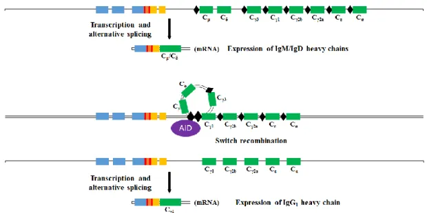
Only the H-chain C region recombination does not occur during the initial B cell development. B cells that have completed the initial development will only express IgM and IgD antibodies isotypes. For the other isotypes to be expressed by a B cell, class-switch recombination (CSR) has to occur. This process, mediated by the enzyme activation-induced cytidine deaminase (AID)23, involves the removal from the chromosome of portions of the antibody heavy chain locus, and the gene segments surrounding the deleted portion are rejoined to retain a functional antibody gene that produces antibody of a different isotype (see Figure 2). CSR occurs in secondary lymphoid organs upon activation and in the presence of specific cytokine signals.
Figure 1 – VDJ recombination process of BCR heavy chain.
The BCR is the result of a process denominated V(D)J recombination, where different gene segments are combined to obtain the final gene sequence. In the case of the heavy chain, a D segment is initially recombined with a J and the result is then recombined with a V segment. In the case of the light chain, there is only recombination of a J segment with a V segment (the light chain does not possess D segments). The recombination involves cut and splice in order to remove the unwanted gene sequences. Within the V(D)J rearranged sequences there are three complementarity-determining regions (CDR1-3) that are responsible for the direct recognition and binding of the antigen. These regions are hyper variable, especially in the case of the CDR3 that comprises the V(D)J segments junctions, which may suffer frameshifts in the encoded base pairs and insertion of nucleotides.
9 There are two subsets of B cells: B-1 and B-2 B cells. B-1 cells locate in peritoneal and pleural cavities and mucosal sites and are responsible for the production of natural IgM antibodies24.
B-2 cells develop in the bone-marrow, but their early differentiation is only terminated upon migration into B cell follicles in the spleen, where they differentiate into naïve follicular or marginal zone (MZ) B cells25. When their early development is completed, naïve follicular B cells can migrate into other secondary lymphoid organs such as LNs and Payer patches. MZ B cells, as B-1 cells, are also part of the first line of defense against blood-borne pathogens through T cell independent (TI) humoral responses26. On the first couple of days after the onset of an immune response, these cells rapidly differentiate into extrafollicular IgM-producing plasma cells, which secrete IgM as a decavalent pentamer that forms immune complexes with the pathogen27. However, since these cells differentiate independently of T cell help, they are short-lived and of low specificity as there is no affinity maturation. Follicular B cells are also able to respond to TI antigens; however they, seem to be more specialized in responding to antigens that also activate CD4 T cells, thus gaining the help of these cells and consequently mounting T cell dependent (TD) humoral responses. Follicular B cells get initially activated by recognition of antigen brought to lymphoid tissues. The recognized antigen is internalized and processed by the B cell to be presented on class-II
Figure 2 – Class Switch Recombination process.
In order for B cells to express antibodies of different classes, CSR needs to occur. This process involves somatic recombination through the action of the AID enzyme. The recombination involves the removal of a DNA fragment from the Cμ segment (if no recombination has happened yet) until the sequence before the wanted C segment. Specific sequence motifs on the DNA (black lozenges) indicate sites where recombination can occur. At the end of development in the bone marrow, and prior to CSR, B cells will only express IgM and IgD. CSR mainly occurs on germinal centers after B cell activation. The type of isotype to be expressed by the B cell is determined by the cytokines produced by CD4 helper T cells.
10
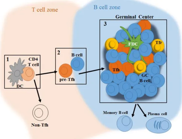
MHC (MHC-II) molecules to primed antigen-specific and specialized T cells. Thus, upon antigen receptor-dependent activation, B cells migrate to the border of the T cell zone and B cell follicle (T-B border), where they encounter pre-activated antigen-specific T cells (pre-follicular helper T cells or pre-Tfh cells). At this location, T and B cells form long-lived interactions, allowing B cells to proliferate and become fully activated (as well as leading to complete maturation of pre-Tfh cells into Tfh cells). Once a B cell becomes fully activated, it will reach a bifurcation between two fates: either join the extrafollicular response or the germinal center (GC) response.
In the extrafollicular response, B cells differentiate into unswitched IgM memory B cells28 or, after some rounds of division and in some cases CSR, into short-lived plasmablasts29. From the initial pool of activated B cells, cells with higher affinity to the antigen are the ones selected to become plasmablasts30,31; however, as they do not undergo affinity maturation, their antigen-specificity is rather low and unchanging.
B cells that commit to the GC fate migrate back to the center of the follicle where, with the help of Tfh cells and follicular dendritic cells (FDCs), establish a GC32,33. Within GCs, B cells undergo proliferation, affinity maturation, and CSR, leaving them as plasma or memory B cells with high affinity to the antigen.
Some published studies seem to indicate that the GC output is not plasma cells but rather plasmablasts not fully differentiated34,35. Then, these cells home mainly to the bone marrow, where they finally differentiate into long-lived plasma cells which have the capacity of sustaining a high level of antibody secretion for long periods of time36-38.
Memory B cells are characterized by their longevity and the capacity to rapidly and robustly respond to antigen re-exposure. These properties are one of the basis of vaccine success. IgM and IgG memory B cells, from the extrafollicular and GC responses, respectively, are the most studied populations within the memory B cell compartment. These two populations seem to have different properties and roles on secondary responses. IgM memory B cells, not having undergone affinity maturation and CSR, have a more diverse specificity, are able to persist longer in the organism, and are more prone to proliferate and join the new GC response. IgG+ cells memory B cells, with high affinity to the antigen, readily differentiate into antibody secreting plasmablasts, allowing a rapid response upon antigen rechallenge39,40.
11 Germinal centers
GCs are specialized structures where antibody affinity maturation and CSR occur41-45. The affinity maturation is the process that allows the improvement of antibody affinity over time during an immune response. This is accomplished through rounds of somatic hypermutation (SHM) of the V gene segment and selection of B cell clones with mutations that successfully improve their affinity to the antigen41,42,45.
As described before, follicular B cells migrate into the T-B border upon activation, where they engage in cognate interactions with pre-Tfh cells46,47 (Figure 3). These long-lived interactions ultimately lead to commitment to the GC pathway of the B cells with higher affinity to the antigen, and consequent changes in their transcriptional profile48-51. Genes like interferon-regulatory factor 4 (IRF4), MYC, B cell lymphoma 6 (BCL-6), and myeloid cell leukemia 1 (MCL1) are critical for GC B cells generation, and their upregulation occurs during the early initiation phase until day 3 of response. While some genes are key during early activation and/or late differentiation and their expression is only observed during that time (as is the case of IRF452,53), other genes need to be expressed throughout the whole GC reaction. One of them is BCL-6 that acts as a transcription repressor and is a key regulator of the GC B cell phenotype54-56: it is responsible for enabling the migration of B cells back to the follicle after commitment to the GC pathway by downregulation of Epstein-Barr virus-induced G-protein coupled receptor 2 (EBI2) and sphingosine-1-phosphate receptor 1 (S1PR1) that are responsible for localization at the T-B border32,33,57,58; BCL-6 induces an pro-apoptotic state, through the silencing of BCL-2, that will lead to the deletion of low affinity or autoreactive clones59,60; BCL-6 induces cell tolerance to DNA damage originated from AID activity and rapid proliferation by downregulating p53 and ATR61,62; BCL-6 regulates the expression of positive signaling mediators to allow a fine tune selection of high affinity BCRs63,64; and BCL-6 is involved in regulating plasma cell differentiation through the downregulation of BLIMP-163.
Thus, three to four days after initial activation by antigen encounter, GC B cell precursors, upon expression of BCL-6, are able to reverse their migratory properties and migrate back into the center of the B cell follicle. There, B cells form an early GC (already observable by microscopy) in-between a pre-existing network of C-X-C chemokine ligand 13 (CXCL13) secreting FDCs65,66. The GC B cell population has high expansion rates, and consequently a rapid increase in size of the GC occurs and leads to the formation of the B cell mantle compartment. This mantle is formed by the naïve B cells of the follicle that are pushed away by the GC67. Seven days after primary immunization, GCs have substantially increased in
12
size and are now denominated mature GCs67-69. Moreover, they can be easily divided in two regions: the light and dark zones (LZ and DZ, respectively)70. The light zone is populated by B cells, FDCs, macrophages, Tfh cells, follicular regulatory T (Tfr) cells, and other cell types68. FDCs are found not only on GCs, but also on primary follicles, and their main functions during a GC reaction are to present unprocessed antigen on their surface and secrete cytokines important for GC maintenance like IL-6 and the CXCL13 chemokine
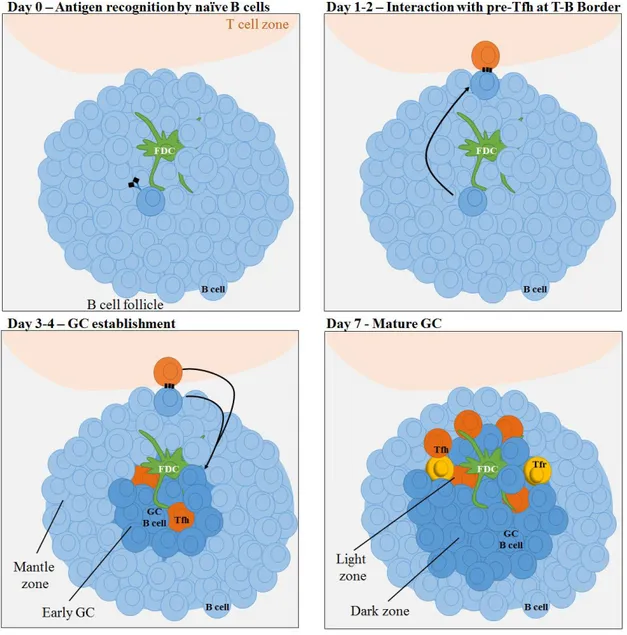
65,71-Figure 3 – GC initiation and development.
GC responses are initiated by antigen recognition by B cells within the follicle. Within 1-2 days after activation, B cells migrate into the T-B border where they engage in cognate interactions with pre-Tfh cells. Upon commitment to the GC program, Tfh and GC B cells migrate into the center of the follicle where an early GC establishes in between a network of FDCs. The formation of the GC pushes away pre-exiting naïve B cells which form the mantle zone. At day 7, the now mature GC has increased in size due to fast cell proliferation and it can be divided in two zones: the light zone composed by GC B cells, Tfh, Tfr, FDCs and other cell types, while the dark zone is mainly composed by rapidly dividing B cells.
13 73. Tingible-body macrophages are responsible for eliminating apoptotic B cells, thus preventing the accumulation of self-antigens which can lead to the generation and selection of autoreactive clones74-77. The dark zone is only densely populated with B cells among a network of CXCL-12 producing reticular cells (morphologically similar to FDCs)70.
During the maturation process undergone on the GC, B cells repeatedly migrate between dark and light zones. This migration is enabled by the chemokine receptors C-X-C chemokine receptor 4 (CXCR4) and CXCR5: centrocytes express CXCR5 and are found in the CXCL13 rich LZ, while centroblasts that also express CXCR4 are localized in the DZ where CXCL12 is abundant78. Within the DZ, GC B cells proliferate and undergo SHM that introduces point mutations in the rearranged V gene41,42,67,79. As for CSR, SHM is also mediated by the action of AID23. As result, a high number of cells with a vast range of affinities for the antigen arise. B cells then migrate into the LZ for selection by Tfh cells68. B cells that have successfully gained high affinity to the antigen receive positive signals, perform CSR, and differentiate into memory B cells or plasma cells before leaving the GC. B cells that have only moderately increased the affinity of their BCR to the antigen also receive positive signals, may also undergo CSR, and are able to migrate back into the DZ to undergo a new round of SHM to increase their affinity to the antigen. B cells that have lost their capability to recognize the antigen, and therefore are not able receive positive signals, are eliminated by apoptosis69.
The signals that trigger CSR are still poorly understood69. Nevertheless, the type of ongoing response and the signature cytokines of Th1, Th2, and Th17 responses determine the class of Ig to be produced80-82. IL-4 leads to the secretion of IgG1 and IgE, while IFNγ promotes IgG2a production. Published data so far favor the hypothesis that these cytokines are produced in situ by Tfh which can produce low levels of IL-4 or IFNγ83.
The affinity maturation process is dependent on Tfh signals79,84. High affinity B cells in the LZ capture more antigen from FDCs (compared to cells with low affinity BCRs), and consequently perform more MHC-TCR interactions with Tfh cells that ultimately lead to positive selection signals79,85. Moreover, from the pool of B cells that are selected to re-enter the DZ, the ones with higher affinities receive stronger signals from Tfh and present higher proliferation rates after migrating back into the DZ86. Additionally, the changes of antigen availability on FDCs during a GC reaction also ensure the increase in affinity of the B cells leaving the GC overtime87. Antibodies produced by plasma cells originated early from the GC will coat the antigen deposited in FDC. Thus, only GC B cells with higher affinity to the antigen will be able to overcome this competition and acquire antigen to present to Tfh cells.
14
Since antibodies with increasing affinities are gradually produced, the level affinity required also increases over time87. Although SHM allows the generation of high affinity antibodies, it may also give rise to GC B cells with self-reactive BCRs that must be deleted to prevent the onset of auto-antibody mediate autoimmunity88,89.
The signals that lead GC B cells to differentiate into memory B cells or plasma cells are still not very well understood. Nevertheless, differentiation of GC B cells into plasma cells or memory B cells seems to be time dependent90. Memory B cells, that present lower affinities and number of somatic mutation, are mostly generated first while differentiation into plasma only occurs at later stages, long after GCs peak in size.
T cells
T cells play a central role in adaptive immune responses since they are not only responsible for cytotoxic function, but are responsible for orchestrating the response by providing help to other cells. This functional duality is accomplished by the existence of two T cells populations: CD8 T cells and CD4 T cells, respectively. The designation “T cell” originated from the fact that its precursors leave the bone marrow and their differentiation occurs in the thymus.
As for B cells, a key feature of T cells is their specialized receptor that can recognize a wide range of antigens. However, the TCR is not able to bind and recognize free antigens in their natural structure, but rather small peptides presented on MHC molecules91. The TCR is constituted by two chains which are also product of somatic V(D)J recombination (see Figure 4)91. The majority of T cells expresses the αβ TCR that is composed by α and β chains. Nevertheless, there are also γδ T cells, which TCR is composed by γ and δ chains and have a more restrict repertoire, and natural killer T (NKT) cells that, although express an αβ TCR receptor, their TCR is considered invariant (Vα24Jα18 combines with a limited TCRβ repertoire in the case of type-1 NKTs) and recognizes hydrophobic antigens such as glycolipids91-93.
The TCR recombination process is performed in the same manner as the one described for the BCR recombination: a V, a D, and a J segments, in the case of the β chain, are cut and spliced together by the action of the same enzymes (RAG-1, RAG-2, and TDT); while in the case of the α chain, as there are no D segments, the recombination consists only in the junction of a V and a J segments (Figure 4). Again, besides the inaccuracy of the recombination process, the action of the enzyme desoxyribonucleotidyltransferase can insert
15 nucleotides in the coding sequence and further contributes for the high variability of the TCR repertoire.
Consequently, the CDR3, that comprises the segment junctions, may have different CDR3 lengths due to frameshift in the coding sequence. TCR specificity cannot be altered once the T cell finishes its receptor recombination, contrary to what is observed for the BCR that can improve its affinity to the antigen after the initial development by somatic hypermutation in GCs. Also, the TCR complex is not only composed by the αβ chains module (that recognize the peptides presented on MHC molecules) but also by invariant polypeptide chains responsible for intracellular signal-transmission, the CD3 module: CD3ε, CD3γ, CD3δ, and ζ.
Figure 4 – V(D)J recombination of the TCR.
As for the BCR, the TCR is also the result of V(D)J recombination. In the case of the α chain, there is the recombination of a J segment with a V segment, while for the β chain a D segment is recombined with a J followed by the recombination with a V. The three CDRs are the binding region of the TCR that not only recognize the peptide presented by a MHC molecule but also the MHC molecule itself. The three CDR3 regions are represented as spheres in violet (α chain CDRs) and blue (β chain CDRs) in the three dimensional representation in the box on the right.
16
There are several fate decision steps important in T cell development. The first occurs when committed lymphoid progenitors, which arise in the bone marrow, migrate into the thymus, lose the potential to develop as B cells or NK cells, and become committed T cells precursors. A second fate decision occurs when thymocytes start expressing a rearranged TCRβ chain and lose the potential to differentiate into γδ T cells.
Upon successful rearrangement of the α and β chains, thymocytes start co-expressing CD4 and CD8, thus becoming double positive (DP) thymocytes. As DP thymocytes already express a mature αβ TCR, the next steps of differentiation and fate decision involve the selection of the TCRs that, based on their affinity, can leave the thymus and are capable of participating in immune responses without triggering responses against the self. For that, the selection comprises 3 distinguishable processes: death by neglect, negative selection, and positive selection94. Thymocytes bearing TCRs that interact poorly with self-antigens-MHC complexes, and consequently are unable to induce intracellular signaling required for survival, suffer death by neglect. On the contrary, cells with TCRs that present very high affinity to self-antigens-MHC complexes are instructed to commit apoptotic cell death. This negative selection process prevents potential T cells that could cause autoimmune pathology from leaving the thymus, although some self-reactive T cells escape deletion95. DP thymocytes that generate intercellular signaling levels between death by neglect and negative selection initiate the multi-step positive selection.
The last important fate decision involves the CD4- or CD8-lineage commitment. This differentiation step comprises the passage from DP to single-positive (SP) state (CD4+CD8 -or CD8+CD4-) by silencing the transcription of one co-receptor locus96,97. CD8 and CD4 co-receptors are specific to class-I and class-II MHC molecules (MHC-I and MHC-II), respectively. Therefore, their expression restricts the interaction of the TCR to only one of the MHC molecules, and consequently establish the function of the cells when they leave the thymus. The selection of either CD4 or CD8 as co-receptor is determined by TCR affinity to self-peptides presented by either MHC-I or MHC-II molecules, as well as for the MHC molecule itself. This dichotomy on CD4 or CD8 expression, which results on T cell ability to recognize peptides presented by either MHC-I or MHC-II molecules and consequent associated function, is of high importance, since the two MHC molecules present peptides of different origins. MHC-I molecules, which are expressed by almost all nucleated cells, present peptides of intracellular (cytosolic and nuclear) origin to alert CD8 T cells for intracellular alterations and target cells for destruction. On the other hand, MHC-II molecules are only constitutively expressed on professional APCs (although their expression
17 can be induced in other cell types by IFNγ98-100) and present peptides derived from exogenous proteins degraded in the endocytic pathway. These peptides are then presented to CD4 T cells which in turn orchestrate intercellular immune responses. Nonetheless, there is a link between the two pathways (named cross-presentation) that allows exogenous peptides to be presented on MHC-I molecules and endogenous molecules on MHC-II molecules101,102. Taken all together, the commitment to CD4- or CD8-lineage depends on the capability of TCR to interact with MHC-II or MHC-I molecules in the thymus. This commitment then determines the function to be performed by the cell in the periphery. In the end of the development, mature CD4 and CD8 T cells, as well as γδ T cells and NKT cells, that have been positively selected, leave the thymus and migrate into secondary lymphoid organs.
CD8 T cells
CD8 T cells mediate cellular immunity by detecting infected or altered cells and inducing cell-mediated lysis, and these functions are primarily dependent on TCR recognition of antigens presented on MHC-I molecules.
Naïve CD8 T cells are initially primed on secondary lymphoid organs such as draining LNs and spleen. Priming is preferentially performed by DCs and occurs on peripheral regions enriched for antigens during early immune responses97,103. Upon activation, CD8 T cells with antigen-specific TCRs start producing IFNγ and rapidly expand104-106. This initial activation and expansion is not only dependent on TCR affinity but also on co-stimulatory signals (e.g., OX40 and CD27) and the presence of inflammatory cytokines like IL-12 and IFNα107,108. However, upon activation, antigen recognition by the TCR is sufficient to trigger CD8 T cells to kill altered cells. After activation in central lymphoid organs, CD8 T cells upregulate CXCR3 and migrate into site of ongoing response, where they continue to be stimulated by interacting with altered cells, resulting in cytolysis of those cells, and further inducing CD8 T cells proliferation109. Of note, in most types of responses, CD8 T cells initial activation by DCs and recruitment into infection site requires help from CD4 T cells110. Another important cytokine for CD8 T cells biology is IL-2. This cytokine, mainly produced by CD4 T cells, has not only an impact in the proliferation and cytolytic capacity of CD8 T cells but also regulates the formation of memory CD8 T cells111.
CD8 T cells use two different mechanisms to induce cell death on target cells, namely the perforin/granzyme-mediated apoptosis112 and activation-induced cell death (AICD)113, and
18
both are initiated upon antigen recognition and consequent TCR signaling. The first mechanism involves the secretion close to the target cell of granzymes and perforin that are stored in lysosomes within CD8 T cells. Perforin can either form pores on the plasma membrane of target cells or mediate endocytosis to allow granzyme entrance inside the cells and induce apoptosis. The AICD is dependent on the FAS ligand (FASL)-FAS interaction: the binding of the FASL on the CD8 T cell to the FAS receptor induces dead on the target cell by initiating a caspase dependent apoptosis process through the intracellular death domain of FAS.
Besides inducing cell death to target cells, CD8 T cells also produce cytokines like IFNγ and IL-10. IFNγ further induces CD8 T cells proliferation and function, acting a feed forward loop. On the contrary, the immune suppressive cytokine IL-10 has a regulatory function and is essential to avoid excessive tissue injury and consequently prevent autoimmune pathology during a viral infection114,115.
CD4 T cells
CD4 T cells are responsible to support immune responses by activating innate immune cells, B cells, CD8 T cells as well as non-immune cells. Moreover, CD4 T cells are also responsible for regulating and suppressing immune reactions. To be able to perform all these functions, naïve CD4 T cells can differentiate into different effector subpopulations depending on the signals provided by cells of the innate immune system at the time of T cell activation (See Table 1). These signals will in turn depend on the PRRs triggered on the DCs, and consequently the type of immune response that needs to be mounted. The duality of responses against intracellular and extracellular pathogens lead to the early discovery of type 1 T helper (Th1) and type 2 T helper (Th2) responses116. Since then, more differentiation subsets of CD4 T cells have been identified: Th17, Tfh, Th9, and Th22 cells that are involved in inflammatory responses, and forkhead box P3 positive (FOXP3+) regulatory T (Treg) and class 1 regulatory T (Tr1) cells that are engaged in immune suppression.
For a naïve CD4 T cell to differentiate into any of these subsets it is necessary an initial activation step that includes TCR recognition and co-stimulation signals. TCR recognition occurs when a naïve CD4 T cell is able to specifically recognize a peptide presented by an activated APC on a MHC-II molecule. The strength of the TCR stimulation depends on the affinity of the TCR to the molecule, and may also have an impact on the lineage commitment117,118. For a CD4 T cell to get fully activated, co-stimulation signals are also
19
Table 1 – CD4 T cell subsets and its characteristics.
CD4 Subset
Differentiation
signals Key regulators
Cytokines
produced Surface markers Th1 IL-12, IFNγ T-BET, STAT1, STAT4 IFNγ, Lfα, IL-2 CXCR3+IFNγR+
Th2 IL-4, IL-2 GATA3, STAT6, STAT5, STAT3, GFI-1, IRF4
IL-4, IL-5,
IL-13, IL-25 CCR4
+CCR6–ST2+
Th17 IL-6, TGFβ,
IL-21, IL-23 RORγt, RORα, STAT3
IL-17A, IL-17F,
IL-21, IL-22 CCR4
+CCR6+
Th9 IL-4, TGFβ IRF4, PU.1 IL-9 CCR4– CCR6+
Th22 IL-6, TNFα,
IL-1β AHR
IL-22, IL-13,
TNFα CCR4
+CCR6+CCR10+
Tfh IL-6, IL-21 BCL-6, ASCL2, c-MAF,
BATF, IRF4, STAT3, STAT1 IL-21, IL-4 CXCR5
+PD1+ICOS+
pTreg TGFβ, IL-2 FOXP3, Smad2, Smad3,
STAT5 IL-10, TGFβ
CD25+CTLA-4+
GITR+
Tr1 IL-27, IL-10,
IL-6 c-MAF, AHR IL-10 CD49b
+LAG-3+
required. These signals are provided by the DC that expresses CD80/CD86, inducible T cell co-stimulatory ligand (ICOSL), and OX40L, and provides positive signals through interaction with the CD4 T cells co-stimulatory receptors CD28, ICOS, and OX40, respectively. The last factors that determine the differentiation into a specific lineage are the cytokines produced by the DCs, which in turn depend on the PRRs triggered on the DC (see Table 1).
Th1 cells
Th1 cells are important for the protection against obligate intracellular pathogens, such as intracellular bacteria and viruses, as well as in immune response against tumors. Th1 differentiation is promoted by IL-12 and IFNγ, together with TCR signaling. While IL-12 is highly secreted by APCs upon activation through their PRR, IFNγ is produced by NK cells when exposed to IL-12119,120.
The master regulator of Th1 differentiation is the T-box transcription factor (T-BET), which activates genes necessary for the differentiation and is also capable of suppressing the differentiation into other cell lineages. T-BET expression is dependent on another transcription factor, the signal transducer and activator 1 (STAT1) which expression is upregulated by IFNγ121,122. Since T-BET induces high levels of IFNγ expression, a feed forward loop is established to ensure selective expansion and differentiation of Th1 cells123. Another important axis of Th1 differentiation is the IL-12-STAT4 pathway. IL-12 induces STAT4 which in turn activates IFNγ expression124. Since high levels of T-BET lead to the upregulation of IL-12 receptor β (IL-12Rβ) expression, Th1 differentiation is further
20
enhanced122. On the other hand, T-BET suppresses Th2 and Th17 lineages by impairing the function of their master regulators GATA3 and retinoic acid receptor-related orphan receptor gamma-T (RORγt), respectively125,126. Th2 development is further suppressed by inhibiting IL-4 expression127.
Upon full differentiation, Th1 cells main function is to produce cytokines that mount and boost an intracellular immune response. The two main cytokines produced are IFNγ that activates phagocytes, like macrophages, to increase their phagocytic activity, and IL-2 that, besides inducing proliferation and acquisition of cytolytic phenotype by CD8+ T cells, is required for the development of memory CD8+ T cells128.
Although the pro-inflammatory action of Th1 cells is of maximum importance for clearance of intracellular pathogens and tumors, their unregulated action can cause unwanted inflammatory diseases and self-reactivity. Th1 cells and their IFNγ production have been implicated in inflammatory diseases like inflammatory bowel disease (IBD), transplant rejection, graft-versus-host disease, and autoimmune diseases as type-1 diabetes and rheumatoid arthritis (RA)129-133.
Th2 cells
Th2 cells are involved in responses to extracellular parasites, including helminthes and nematodes, as well as in mucosal immunity of the lung, through the production of 4, IL-5, IL-9, IL-10, IL-13, IL-2IL-5, and amphiregulin.
Besides TCR recognition and initial activation, IL-4 and IL-2 are critical for a naïve CD4 T cell to follow the differentiation pathway to a Th2 cell. IL-4, the positive feedback cytokine for Th2 differentiation, induces STAT6 expression which in turn upregulates GATA3, the Th2 master regulator134-137. GATA3 is indispensable for Th2 response and it has been postulated that its action is mediated through three different mechanisms: (1) enhances Th2 cytokines production, (2) induces Th2 cells proliferation, and (3) suppresses Th1 differentiation by interacting with T-BET and suppressing STAT4 expression137,138. STAT6 is also important for the full Th2 differentiation as GATA3 does not directly regulate all Th2-realted genes139. On the other hand, STAT6 interaction with several of those genes loci is in turn dependent on the presence of STAT3140. STAT5 is another key transcription factor for this differentiation pathway. STAT5 is activated by IL-2 signaling and in coordinated activity with GATA-3 induces IL-4 expression by Th2 cells141,142. Growth factor independent-1 (GFI-1), a transcriptional repressor which expression is induced by TCR signaling alone or the IL-4-STAT6 pathway, selectively leads to Th2 expansion since it
21 induces proliferation on GATA3 high expressing cells143. IRF4 is another transcription factor that also has an impact on Th2 differentiation by activating the IL-4 promoter and upregulating GATA3 expression144,145.
During an ongoing immune response against extracellular pathogens, Th2 key effector cytokines act mainly upon innate cells. IL-4 induces the expression of IgE receptors: FcεRI on macrophages and B cells, while FcεRII expression is induced on basophils and mast cells146. Also, IL-4 induces lung mucus hypersecretion and secretion of inflammatory cytokines by lung fibroblasts147,148. Eosinophils and its precursors depend on IL-5 signaling for activation, recruitment, and apoptosis inhibition149,150. IL-9, firstly described as produced by Th2 cells, functionally activates several immune cell types such as B cells, neutrophils, eosinophils, and mast cells, while inducing mucin and chemoattractant factors production by airway epithelial cells151. IL-13 is involved in the elimination of gastrointestinal helminthes through the induction of mucus secretion and the increase of intestinal fluid content and intestinal contractility, and lung inflammatory responses by eosinophils activation and enhanced mucus secretion, as IL-4. Indeed, IL-4 and IL-13 seem to have redundant effects during Th2 responses. IL-25, a member of the IL-17 cytokines family, promotes Th2 responses by inducing IL-4, IL-5, and IL-13 production by a non-lymphocyte population152. Amphiregulin induces epithelial cell proliferation and is also involved in nematode expulsion153. Th2 cells also produce the anti-inflammatory cytokine IL-10 that, in this case, suppress Th1 proliferation and DC function154,155.
Moreover, and contrary to what is observed in Th1, Th2 responses are characterized by the production of high levels of specific immunoglobulins that neutralize foreign organisms. IL-4 and IL-13 production during this type of immune response have been implicated in inducing CSR and secretion of IgM, IgG1, IgE, and IgA antibodies.
Dysregulation of Th2 responses are responsible for allergic diseases, especially airway allergic diseases such as persistent asthma. IL-4, IL-13, and IL-9 have been implicated and play major roles in the induction and the immuno-pathogenesis of asthma.
Th17 cells
Th17 cells were the first CD4 T cells independent subset described after Th1 and Th2 discovery156. Th17 cells are involved in immune responses against fungi and extracellular bacteria and its action is mediated through the secretion of IL-17a, IL-17f, IL21, and IL-22. However, even before the discovery of Th17 cells, IL-17 production had already been