UNIVERSIDADE DE LISBOA
FACULDADE DE FARMÁCIA
Unravelling new ethnopharmacological roles
of Plectranthus species: biological activity
screening
Joana Eulália da Cruz Marçalo de Andrade
Dissertação de Mestrado
MESTRADO EM CIÊNCIAS BIOFARMACÊUTICAS
UNIVERSIDADE DE LISBOA
FACULDADE DE FARMÁCIA
Unravelling new ethnopharmacological roles
of Plectranthus species: biological activity
screening
Joana Eulália da Cruz Marçalo de Andrade
Dissertação de Mestrado orientada por:
Professora Doutora Patrícia Dias Mendonça Rijo
e co-orientada por:
Professora Doutora Célia Maria Cardona Faustino
MESTRADO EM CIÊNCIAS BIOFARMACÊUTICAS
2016
The presented thesis project was performed at the Food Sciences and Phytochemistry research group at the Research Center for Biosciences & Health Technologies (CBIOS) of Universidade Lusófona de Humanidades e Tecnologias (ULHT), under the supervision of Professor Doctor Patrícia Dias Mendonça Rijo, Ph.D and co-supervision of Professor Doctor Célia Maria Cardona Faustino, Ph.D.
Part of the research was developed under the Short-Term Scientific Mission of COST action CM1407 entitled “Natural diterpenoids as potential anti-tubercular drugs”, developed at the National Institute for the infectious Disease Lazzaro Spallanzani, Laboratorio di Biologia Cellulare e Microscopia Elettronica, Rome, Italy, under the supervision of Doctor Gian Maria Fimia, and co-supervision of Doctor Alessandra Romagnoli.
Additionally, some research was developed at the Centre for Marine Sciences (CCMAR), at the MARbiotech laboratories, University of Algarve, Faro, Portugal, under the supervision of Doctor Luísa Custódio.
i
Scientific production Papers
Marçalo J., Nicolai M., Reis C., Faustino C., Rijo P. (2016). Antioxidant, antityrosinase, antielastase, and anticollagenase activity of extracts and compounds from Plectranthus species.
Journal of Enzyme Inhibition and Medicinal Chemistry (IF 3.43), in preparation.
Marçalo J., Nicolai M., Reis C., Custódio L., Fimia G., Faustino C., Rijo P. (2016). Anti-inflammatory and anti-tubercular screening of diterpenes and triterpenoids from Plectranthus species. Journal of Ethnopharmacology (IF 3.05), in preparation.
Poster Communications National
Marçalo J., Roque L., Nicolai M., Rodrigues L.M., Reis C., Faustino C., Rijo P. PLGA nanoparticles with anti-inflammatory agents obtained from Plectranthus plants. III Symposium of Nanoscience and Biomedical Nanotechnology, Lisboa, Portugal, April 15th, 2016.
International
Faustino C., Marçalo J., Nicolai M., Rodrigues L.M., Reis C., Rijo P. In vitro efficacy assessment of extracts and compounds from Plectranthus species for skin delivery. International Society for Biophysics and Imaging of the Skin World Congress, Lisboa, Portugal, May 31st – June 3rd, 2016.
Marçalo J., Frias D., Nicolai M., Rodrigues L.M., Reis C., Faustino C., Rijo P. Antioxidant, antiacetylcholinesterase, antityrosinase, antielastase and anticollagenase activity of extracts and compounds from Plectranthus species. 6th International Congress of Aromatic and Medicinal Plants (CIPAM), Coimbra, Portugal, May 29th – June 1st, 2016.
Marçalo J., Custódio L., Rodrigues M.J., Nicolai M., Rodrigues L.M., Reis C., Faustino C., Rijo P. Exploring the biological activity potential of Plectranthus spp. diterpene derivates. Conference: 6th European Workshop in Drug Synthesis, Siena, Italy, May 15th – 19th, 2016.
Stanković T., Dinić J., Podolski-Renić A., Garcia C., Andrade J., Simões M.F., Rijo P., Pešić M. Diterpenoids isolated from Plectranthus spp. are selective towards human non-small cell lung carcinoma cells with P-glycoprotein overexpression. 2nd COST meeting. Centro de Investigaciones Biológicas (CIB-CSIC), Madrid, Spain, April 4th – 5th, 2016.
ii
Agradecimentos
Quero aproveitar esta oportunidade, para expressar a minha gratidão a todos aqueles que de algum modo fizeram parte da minha vida, e que por isso foram fundamentais para a construção desta tese.
Sempre em primeiro lugar, agradeço incondicionalmente à minha Mãe, pelo amor e presença sem condições e sem barreiras. Foi graças à sua força e dedicação que tudo foi possível atingir. Todos os objetivos e bandeiras hasteadas, foi graças à energia da minha Mãe que me fez seguir em frente. Foi este amor que se tornou mais forte e ajudou-me a ultrapassar todos os obstáculos. Com a minha Mãe partilho tudo e por isso, és a minha guerreira!
Este último ano não teria sido possível sem as minhas orientadoras. Agradeço do fundo do coração à minha querida orientadora Professora Dr.ª Patrícia Dias Mendonça Rijo, pela motivação, por todo o apoio, sabedoria, dedicação e oportunidades que me deu. Agradeço-lhe toda a paciência que teve comigo. Agradeço toda a motivação, abraços e sorrisos. Obrigado por acreditar e confiar em mim e me levar na Ciência pelo Mundo. Graças a si foi possível, é possível, será possível!
À minha co-orientadora Professora Dr.ª Célia Maria Cardona Faustino, um muito obrigado pela sua notável dedicação ao meu trabalho. O seu apoio foi fundamental na elaboração desta dissertação.
Agradeço também à Professora Dr.ª Marisa Nicolai, cuja sabedoria e empenho me auxiliou na elaboração dos protocolos enzimáticos. Um muito obrigado pela motivação que demonstrou no meu projeto, e por todas as horas dedicadas de apoio laboratorial, e também pessoal.
À minha família que foi sempre o meu porto de abrigo, de motivação e apoio. O meu irmão Francisco, a minha avó Manuela, a minha cunhada Soraia e a minha sobrinha Leonor, um enorme obrigado por estarem sempre presentes.
A todos os meus amigos, em particular às minhas incondicionais e intemporais amigas do “Norte”, Ana Rocha e Vera Macieira, agradeço pelo contacto apesar da distância, um muito obrigado à nossa amizade!
À minha amiga partner da Bioquímica, Sara Vicente pelo apoio e partilha. A sua amizade revelou toda a bondade e honestidade que muitos desejam, mas poucos têm. Pela sua ajuda nas crises, e pelo apoio em qualquer canto do Mundo: Grazie mille!
Quero também agradecer a todos os novos amigos e companheiros de viagem, que fiz ao longo deste ano, que tornaram os dias cansativos de laboratório muito mais alegres. A vossa amizade foi muito importante, um muito obrigado à Ana Mota, ao Luís Roque, à Catarina Garcia e ao Diogo Matias.
iii Como não podia deixar de ser, agradeço à minha querida amiga Íris Neto, que foi a melhor amizade que levo deste mestrado. A sua amizade incondicional ajudou-me em todas as fases, a conseguir atingir os meus objetivos. É uma amizade verdadeira que torna o Mundo muito mais bonito. Agradeço tudo, agradeço a sua presença e partilha nos bons e nos maus momentos, nos bons e nos maus resultados.
Agradeço ao Professor Dr. Gian Maria Fimia e à sua equipa de investigação, em particular à Dr.ª Alessandra Romagnoli por me terem aceite no seu laboratório, no “National Institute for
the Infectious Diseases Lazzaro Spallanzani” em Roma, e por me terem ajudado na
investigação. Agradeço o carinho e sabedoria partilhados.
Quero também agradecer à Dr.ª Luísa Custódio e à Dr.ª Maria João Rodrigues, do Centro de Ciências do Mar da Universidade do Algarve, por me terem aceite a desenvolver investigação. Não teria conseguido sem o vosso apoio e ajuda durante os ensaios.
Deixo o também meu agradecimento à Professora Dr.ª Catarina Reis pelo carinho, por me ajudar sempre que foi necessário e pela alegria. Ao Professor Dr. Amílcar Roberto agradeço o interesse e partilha de ideias.
Por fim, quero agradecer ao Universo por ter feito de mim uma guerreira, e me ter dado os melhores soldados de guerra que alguém podia pedir. A todas as batalhas que venci, e às ainda irei vencer, aqui estou. E estou, graças a todos os que já cá não estão, mas que estão sempre presentes em pensamento e no coração, e a quem dedico esta dissertação.
iv Abbreviations 1D One dimension 2D Two dimensions AA Arachidonic acid Abs Absorbance Roy 7α-acetoxy-6β-hydroxyroyleanone Ach Acetylcholine AChE Acetylcholinesterase
AChI Acetylcholine iodide
AChR Acetylcholine receptors
Ala Alanine
ANOVA Analysis of variance
AP-1 Activator protein-1
BHA Butylated hydroxyanisole
BSA Bovine serum albumin
cAMP Cyclic adenosine monophosphate
CBIOS Research Center for Biosciences & Health Technologies
CCMAR Center of Marine Sciences
CD14+ Cluster of Differentiation 14 monocytes
CFU Colony-forming units
ChC Collagenase
COSY Homonuclear correlation spectroscopy
COX Cyclooxygenase
Cyclo-abietene
(13S,15S)-6β,7α,12α,19-tetrahydroxy-13β,16-cyclo-8-abietene-11,14-dione
DeHRoy 6,7-dehydroroyleanone
DiHRoy 6β,7α-dihydroxyroyleanone
DMSO Dimethyl sulfoxide
DNA Deoxyribonucleic acid
DPPH 2,2-diphenyl-1-picrylhydrazyl
DTNB 5,5’-dithio-bis-(2-nitrobenzoic acid)
EC Enzyme Commission
ECM Extracellular matrix
EGCG Epigallocatechin gallate
v
ELISA Enzyme-linked immunosorbent assay
eNOS Endothelial nitric oxide synthase
et al. et alii / et aliae
ETAN Ethambutol
FAD Flavin adenine dinucleotide
FALGPA N-(3-[2-furyl]acryloyl)-Leu-Gly-Pro-Ala
FBS Fetal bovine serum
FMN Flavin mononucleotide
Forskolins 1,6-di-O-acetylforskolin:1,6-di-O-acetyl-9-deoxyforskolin
GADPH Glyceraldehyde 3-phosphate dehydrogenase
gHMBC Gradient selected phase-sensitive - Heteronuclear multiple
bond correlation
gHSQC Gradient selected phase-sensitive - Heteronuclear single
quantum correlation
Glu Glutamine
Halimane 11R*-acetoxy-halima-5,13E-dien-15-oic acid
Halimane Bu (11R*,13E)-15-butyryloxyhalima-5,13-dien-11-ol
Halimane Diol (11R*,13E)-halima-5,13-diene-11,15-diol
Halimane Ester (11R*,13E)-11-acetoxyhalima-5,13-dien-15-oic methyl ester
HEPES 4-(2-hydroxyethyl)-1-piperazineethanesulfonic acid
HPLC High performance liquid chromatography
HSD Honest significant difference
IC50 Drug concentration causing 50% inhibition
IFN-γ Interferon-γ
IL Interleukin
INH Isoniazid
iNOS Inducible nitric oxide synthase
IR Infrared
KOJ Kojic acid
LC3 Microtubule-associated protein 1A/1B-light chain 3
LDH Lactate dehydrogenase
L-DOPA L-3,4-dihydroxyphenylalanine
L-NAME ω-Nitro-L-arginine methyl ester hydrochloride
LPS Lipopolysaccharide
MAPK Mitogen-activated protein kinase
vi
MIC Minimum Inhibitory Concentration
MMP Metalloproteinase
MOI Multiplicity of infection
mRNA Messenger ribonucleic acid
Mtb Mycobacterium tuberculosis
MTT 3-(4,5-dimethylthiazol-2-yl)-2,5-diphenyltetrazolium bromide
NADPH Nicotinamide adenine dinucleotide phosphate
NED N-(1-naphthyl)ethylenediamine
NF-κB Nuclear factor-κB
NMR Nuclear magnetic resonance
nNOS Neuronal nitric oxide synthase
NO Nitric oxide
NOESY Nuclear overhauser spectroscopy
NOS Nitric oxide synthase
NSAID Nonsteroidal anti-inflammatory drug
NT Not tested
p62 Nucleoporin p62
Parvifloron D 11-hydroxy-2α-(4-hydroxybenzoyloxy)-abieta
5,7,9(11),13-tetraen-12-one
PBMC Peripheral blood mononuclear cell
PBS Phosphate-buffered saline
PE Plectranthus ecklonii Benth.
PG Plectranthus grandidentatus Gürke
PGE2 Prostaglandin E2
PGG2 Prostaglandin G2
PGH2 Prostaglandin H2
PLA2 Phospholipase A2
PLC Phospholipase C
PM Plectranthus madagascariensis (Pers.) Benth
PN Plectranthus neochilus Schltr.
PO Plectranthus ornatus Codd.
PP Plectranthus porcatus van Jaarsv. & P.J.D. Winter
PPP Plectranthus prostratus Gürke
PS Plectranthus saccatus Benth.
ROS Reactive oxygen species
vii
RPMI Roswell Park Memorial Institute
SANA N-succinyl-(Ala)3-p-nitroanilide
SAR Structure-activity relationships
SCF Supercritical fluid
SD Standard deviation
SIRS Systemic inflammatory response syndrome
Smad Mothers against decapentaplegic homolog
SOD Superoxide dismutase
SQSTM1 Sequestosome 1/p62
STS Staurosporine
TB Tuberculosis
TDR Totally-drug resistance
TGF-β Transforming growth factor-β
TMPD N,N,N',N'-tetramethyl-p-phenylenediamine
TNF-α Tumor necrosis factor-α
TOCSY Total correlation spectroscopy
TPP Tuberculosinyl diphosphate
Tris-HCl Tris(hydroxymethyl)aminomethane hydrochloride
Tyr Tyrosinase
U Enzyme unit
URS Ursolic acid
UV Ultraviolet
v/v Volume/volume
WHO World Health Organization
viii
Abstract
This dissertation focused on the biological activity screening of several Plectranthus spp. plants, aiming to unravell novel ethnopharmacological roles and further research, of several extracts (methanol, ethyl acetate, acetone and water) and isolated compounds. Previous evidences on interesting biological activities of Plectranthus spp. constituents, directed the study for antioxidant, anti-skin ageing, anti-inflammatory, and anti-mycobacterial activities.
Antioxidant results revealed an increased activity of the methanol extracts (20-76%), due to the presence of polyphenols widely known as antioxidants. Moreover, P. grandidentatus (62.3 ± 0.43%) and P. ecklonii (55.5 ± 1.66%) registered a high scavenging activity in the ethyl acetate extract, in comparison with quercetin (89.0 ± 2.5%), most likely due to the presence of abietane diterpenes.
Acetylcholinesterase (AChE) was studied in vitro to evaluate the enzymatic inhibition, due to recent discoveries on non-neuronal cholinergic system in the skin. This assay showed that the Plectranthus spp. organic extracts did not significantly inhibited AChE.
Concerning the tyrosinase inhibition assay, it was observed a high inhibition for the P.
ecklonii methanol (65.9 ± 3.42%), P. grandidentatus acetone (67.9 ± 3.55%), and P. saccatus
acetone (56.5 ± 5.68%) organic extracts. On the other hand, the aqueous extract of P. porcatus was the only one that showed enzymatic inhibition (65.0 ± 8.67%). From the tested isolated compounds, abietane diterpenes, mainly present in the organic extracts of P. grandidentatus,
P. madagascariensis, and P. ecklonii were highly active against tyrosinase, in more than 46%
and up to 75%, especially compared to kojic acid (92.9 ± 7.28%).
In the collagenase assay, all tested extracts and compounds showed a high enzymatic inhibition which was in the range of 28-76%. The higher inhibition results were obtained from
P. neochilus methanol extract (76.4 ± 2.09%), P. ecklonii aqueous extract inhibited (75.59 ±
6.5%) and rosmarinic acid (44.78 ± 4.53%), in comparison with epigallocatechin gallate (93.1 ± 5.27%).
In contrast to the results of the previous enzymatic assays, the anti-elastase assay revealed that in general the extracts did not decrease elastase activity (30-42%). Nonetheless, the isolated compounds were tested, and they were able to highly inhibit elastase activity. Particularly, the oleanolic:ursolic acids mixture (1:4) with 63.4 ± 2.56% and Parvifloron D with 52.8 ± 3.76%, were the most efficient in elastase inhibition specially compared with ursolic acid used as positive control (69.9 ± 3.65%).
The anti-inflammatory assay was performed by the quantification of NO production using the Griess reaction. The non-cytotoxic isolated compounds revealed to be unable to reduce NO production, after LPS stimulated inflammation (ranging from 16-23 µM), in
ix comparison with the normal quantities of NO production within the cells (17.7 ± 0.67 µM), and with the positive control L-NAME that decreased NO until reaching 3.9 ± 0.24 µM.
Finally, the preliminary results of Mycobacterium tuberculosis H37Rv growth assay, revealed low CFU/mL (dilution 10-3) especially regarding one halimane diterpene compound
(2.1×10⁵ CFU/mL), similar to the positive controls isoniazid (1.2×10⁵ CFU/mL), and ethambutol
(2.0×10⁵ CFU/mL), suggesting a potential alternative for further anti-tubercular studies.
Overall, according to the references of this study, this was the first report on Plectranthus spp., concerning the assays of skin-related enzymatic inhibition in vitro, anti-inflammatory assay, and Mycobacterium tuberculosis H37Rv growth, with a preliminary scientific validation upon known ethnopharmacological uses.
KEYWORDS: Plectranthus spp.; Anti-inflammatory; Antioxidant; Skin enzymatic
x
Resumo
As plantas do género Plectranthus L’Héritier (família Lamiaceae) são selecionadas em várias investigações científicas devido aos vários usos etnofarmacológicos pelas populações indígenas, e facilidade de crescimento em zonas temperadas. Estas plantas são compostas por cerca de 350 espécies reconhecidas pelos seus óleos essenciais e compostos terpénicos.
Nesta dissertação pretendeu-se desenvolver e complementar o screening de várias atividades biológicas, a partir de extratos e compostos isolados de sete espécies de
Plectranthus (P. grandidentatus Gürke, P. ecklonii Benth., P. ornatus Codd., P.
madagascariensis (Pers.) Benth., P. porcatus van Jaarsv. & P.J.D.Winter, P. neochilus Schltr.,
e P. prostratus Gürke). Estes estudos biológicos envolveram a pesquisa de atividades antioxidante, anti-envelhecimento da pele, anti-inflamatória e anti-micobacteriana.
Foram obtidos vinte e oito extratos (sete aquosos e vinte e um orgânicos, nomeadamente de acetona, metanol e acetato de etilo) de todas as plantas em estudo, obtendo-se os resíduos secos (mg de extrato/g de planta).
Nos resultados preliminares da atividade antioxidante executada pelo método da recaptação do radical DPPH, verificou-se que os extratos de metanol detiveram os valores mais elevados de recaptação de radicais livres (20-76%), e, portanto, de maior atividade antioxidante. Concluiu-se que esta atividade poderá estar relacionada com as elevadas quantidades em polifenóis nos extratos metanólicos, amplamente citados como antioxidantes. Adicionalmente, foi também registada uma elevada atividade antioxidante para os extratos de acetato de etilo de P. ecklonii (55.5 ± 1.66%) e P. grandidentatus (62.3 ± 0.43%), muito provavelmente devido à presença de diterpenos abietânicos. Na verdade, estes foram os resultados que demonstraram maior atividade antioxidante comparável ao controlo positivo usado, quercetina (89.0 ± 2.5%).
A acetilcolinesterase (AChE, EC 3.1.17), conhecida pela sua ação na terminação do impulso nervoso pela hidrólise da acetilcolina, produzindo colina, foi estudada in vitro usando o método de Ellman, de modo a avaliar a inibição enzimática pelos extratos e/ou compostos isolados. Este ensaio foi realizado com base nos dados recentes da literatura, evidenciando um sistema colinérgico não-neuronal pela presença de acetilcolina na pele (humana). Com base nos resultados obtidos, foi possível concluir que nenhum dos vinte e um extratos orgânicos estudados inibiram a AChE no ensaio enzimático in vitro.
Relativamente aos ensaios in vitro de inibição enzimática relacionada com a pele, o ensaio de inibição da tirosinase (EC 1.14.18.1), uma monooxigenase de cobre que catalisa a síntese de melanina pela produção de L-DOPA a partir de L-tirosina, revelou que os extratos orgânicos de P. ecklonii metanol (65.9 ± 3.42%), P. grandidentatus acetona (67.9 ± 3.55%), e P. saccatus acetona (56.5 ± 5.68%), diminuíram significativamente a atividade enzimática da tirosinase.
xi Por outro lado, o extrato aquoso de P. porcatus foi o único a exibir inibição enzimática da tirosinase (65.0 ± 8.67%). De modo a confirmar a atividade registada para os extratos, foram testados os compostos isolados. De facto, os compostos abietânicos (maioritariamente presentes nos extratos orgânicos de P. grandidentatus, P. madagascariensis, e P. ecklonii) demonstraram forte atividade contra a tirosinase, em mais de 46% e até 75%, especialmente em comparação com o ácido kójico (92.9 ± 7.28%).
Relativamente ao ensaio enzimático de inibição da colagenase (EC 3.4.24.3, metaloproteinase envolvida na clivagem do colagénio), todas as amostras inibiram, de um modo geral, a atividade enzimática (28-76%). Na verdade, o extrato metanólico de P.
neochilus inibiu a colagenase em 76.4 ± 2.09%, bem como o extrato aquoso de P. ecklonii
(75.6 ± 6.5%) e o composto ácido rosmarínico em 44.78 ± 4.53%. Estes resultados preliminares sugerem que a presença de compostos polifenólicos e diterpenos abietânicos nos extratos de Plectranthus, têm uma elevada eficiência inibitória da colagenase semelhante à epigallocatequina galhato (93.1 ± 5.27%).
Em contraste com o que foi obtido nos ensaios enzimáticos da tirosinase e da colagenase, no ensaio enzimático de inibição da elastase (EC 3.4.21.36, protease de serina envolvida na quebra da elastina) não foi observada a diminuição da atividade enzimática após tratamento com os vinte e oito extratos de Plectranthus (30-42% de inibição enzimática). No entanto, os compostos isolados testados demonstraram uma elevada inibição. Particularmente, esta ação foi observada para a mistura de ácidos oleanólico:ursólico 1:4 (63.4 ± 2.56%) e para a Parviflorona D (52.8 ± 3.76%), em comparação com o ácido ursólico usado como controlo positivo (69.9 ± 3.65%).
De um modo geral, os extratos e/ou compostos isolados das sete espécies de
Plectranthus, revelaram uma elevada capacidade, como redutores da híper pigmentação da
pele (provocada pela atividade excessiva da tirosinase), e na manutenção da integridade dérmica, através da manutenção de colagénio e elastina, pela inibição da colagenase e da elastase. Assim, em conjunto com a atividade antioxidante contra a produção de ROS, e na inibição de enzimas relacionadas com a integridade dérmica e pigmentação, será possível o desenvolvimento de uma aplicação cosmética em ensaios futuros.
Por outro lado, a capacidade anti-inflamatória dos compostos foi avaliada através da inibição da produção de NO, usando a reação de Griess. Assim, após a avaliação da citotoxicidade dos compostos testados (em várias concentrações) através do ensaio MTT, foi medida a inflamação em células de macrófagos RAW 264.7. Após estimulação da inflamação celular com LPS, e tratamento com os compostos não tóxicos, este ensaio revelou que os compostos testados não foram capazes de reduzir a produção de NO (16-23 µM), em comparação com a quantidade de NO produzida naturalmente pelas células (17.7 ± 0.67 µM) e com o L-NAME, usado como controlo positivo (3.9 ± 0.2 µM). Uma vez que a inflamação
xii não é apenas proporcional à produção de NO, mas também de outros mediadores inflamatórios, será necessário expandir a investigação desta atividade biológica. De modo a compreender, se de facto, os compostos isolados de Plectranthus são anti-inflamatórios por outros mecanismos, como por exemplo, por inibição enzimática da ciclooxigenase-2 (COX-2).
Finalmente, foi avaliado o crescimento de Mycobacterium tuberculosis (Mtb) H37Rv após a infeção de macrófagos, derivados de células mononucleares do sangue periférico (PBMC). Numa primeira abordagem, o crescimento bacteriano foi monitorizado pela contagem de unidades formadoras de colónias (UFC), nas primeiras horas de infeção, e 13 dias após infeção e tratamento com os compostos selecionados. Após a normalização dos valores de UFC/mL, os resultados revelaram uma diminuição substancial de colónias de Mtb (na diluição 10-3), apenas após o tratamento com um composto halimano obtido por hemi-síntese (2.1×10⁵
UFC/mL).
Este resultado foi muito semelhante ao dos controlos positivos aplicados, isoniazida (1.2×10⁵ UFC/mL) e etambutol (2.0×10⁵ UFC/mL), sugerindo a possível aplicabilidade deste
tipo de compostos em estudos futuros de terapia anti-tuberculose. Dado o resultado deste halimano na notável diminuição de colónias de Mtb, será relevante prosseguir na possível avaliação do mecanismo de ação que conduziu à inibição do crescimento bacteriano. Para tal, poder-se-ão aplicar técnicas de Western blot e imunofluorescência usando marcadores de autofagia, e avaliar a biogénese do fagolisossoma pelos macrófagos infetados.
No geral, de acordo com as referências consultadas, são aqui reportados os primeiros estudos em ensaios de inibição de enzimas relacionadas com a pele, na atividade anti-inflamatória por via do NO, e na atividade anti-tuberculose de extratos e compostos isolados de Plectranthus. Os resultados obtidos nesta dissertação fornecem uma validação científica preliminar, sobre os usos amplamente conhecidos e reportados, por estas plantas medicinais. Na verdade, são necessários mais estudos na pesquisa de novos alvos terapêuticos mais específicos e com menos efeitos adversos, a partir das atividades etnofarmacológicas de
Plectranthus reportadas até agora. Assim como validações científicas no uso de produtos à
base de plantas usados no tratamento, cura e prevenção de doenças.
Uma vez que os produtos naturais representam uma fonte única de compostos protótipo no desenvolvimento de novos fármacos, os metabolitos secundários isolados de Plectranthus revelam neste estudo, o seu potencial no design de novos medicamentos.
PALAVRAS-CHAVE: Plectranthus spp.; Anti-inflamatório; Antioxidante; Inibição enzimas da pele; Mycobacterium tuberculosis.
xiii Table of contents Scientific production ... i Papers ... i Poster Communications ... i Agradecimentos ... ii Abbreviations ... iv Abstract ... viii Resumo ... x
I. INTRODUCTION AND OBJECTIVES ... 1
I.1 Overview of natural products from medicinal plants ... 2
I.2 Plectranthus plants L’ Héritier genus (Lamiaceae) ... 3
I.2.1 Isolated compounds from Plectranthus plants ... 5
I.2.1.1 Diterpenes ... 7
I.2.1.1.1 Abietane diterpenes ... 8
I.2.1.1.2 Labdane diterpenes ... 9
I.2.1.1.3 Halimane diterpenes ...10
I.2.1.2 Triterpenes and phytosterols ...11
I.2.1.3 Polyphenolic compounds ...12
I.3 Biological activities of Plectranthus natural products ...13
I.3.1 Antioxidant activity ...14
I.3.2 Inhibition of skin-related enzymes ...15
I.3.2.1 Enzymes in skin pigmentation and ageing – Tyrosinase, elastase, and collagenase ...16
I.3.2.2 Acetylcholinesterase in the skin – Non-neuronal cholinergic system ....18
I.3.3 Anti-inflammatory activity ...20
I.3.4 Anti-Mycobacterium tuberculosis activity ...22
I.4 Objectives ...25
II. EXPERIMENTAL SECTION ...26
II.1 Reagents and materials ...27
II.2 Equipment ...27
II.3 Plant material and preparation of extracts ...28
II.4 Isolated compounds ...28
II.5 Antioxidant activity - Radical scavenging assay with 2,2-diphenyl-1-picrylhydrazyl (DPPH) ...29
II.6 In vitro inhibition of skin-related enzymes ...30
xiv
II.6.2 Anti-tyrosinase enzymatic assay ...31
II.6.3 Anti-collagenase enzymatic assay ...31
II.6.4 Anti-elastase enzymatic assay ...32
II.7 Anti-inflammatory activity ...33
II.7.1 Cytotoxicity assay with 3-(4,5-dimethylthiazol-2-yl)-2,5-diphenyltetrazolium bromide (MTT) ...33
II.7.2 Nitric oxide quantification in LPS-stimulated RAW 264.7 cells treated with non-cytotoxic compounds ...33
II.8 Mycobacterium tuberculosis H37Rv growth assay ...34
II.8.1 Peripheral blood mononuclear cell (PBMC) isolation ...34
II.8.2 Lactate dehydrogenase (LDH) cytotoxicity assay ...35
II.8.3 Macrophage infection with Mycobacterium tuberculosis, and treatment with compounds ...36
II.9 Statistical analysis ...37
III. RESULTS AND DISCUSSION...38
III.1 Plectranthus spp. extracts ...39
III.2 Organic extracts antioxidant activity by scavenging of DPPH radical ...40
III.3 Skin-related enzymatic inhibitions in vitro ...41
III.3.1 Organic extracts AChE inhibition in vitro ...41
III.3.2 Plectranthus spp. extracts and isolated compounds in tyrosinase inhibition in vitro ...43
III.3.3 Plectranthus spp. extracts and isolated compounds in collagenase inhibition in vitro ...46
III.3.4 Plectranthus spp. extracts and isolated compounds in elastase inhibition in vitro ...50
III.4 Anti-inflammatory study on isolated compounds by NO quantification with Griess assay ...53
III.4.1 MTT cytotoxicity assay of isolated compounds on RAW 264.7 cells ...53
III.4.2 NO production upon inflammation on RAW 264.7 cells after treatment with non-cytotoxic isolated compounds ...55
III.5 Mycobacterium tuberculosis H37Rv growth with CFU assay ...57
III.5.1 LDH release (cytotoxicity assay) on macrophages derived from PBMC...57
III.5.2 Colony-forming units assay for Mycobacterium tuberculosis H37Rv growth . ...58
IV. CONCLUSIONS AND FUTURE PERSPECTIVES ...61
REFERENCES ...64
xv Appendix I – Cytotoxicity evaluation using the MTT assay: Graphical results for the tested compounds concentrations ...81 Appendix II – Anti-inflammatory assay: Cyclooxygenase-2 enzymatic inhibition in
1
2
I.1 Overview of natural products from medicinal plants
Natural products from medicinal plants have been used by indigenous populations for thousands of years, as traditional medicines, remedies, potions, and oils for the treatment of many ailments (Rijo et al., 2014a; Dias et al., 2012). For their vast ethnomedicinal and ethnopharmacological applications which inspired current research in drug discovery, natural products provide new and important leads against various pharmacological targets (Butler, 2004; Dias et al., 2012). In addition, it is well known that many of the drugs in the market have been developed from natural products (Rijo et al., 2013a; Pereira et al., 2015; Balunas and Kinghorn, 2005).
The 20th century was known for the intensive search for new compounds using chemical
synthesis from most of multinational pharmaceutical companies. Natural products operations were dismissed or scaled down, despite the significant number of natural product-derived drugs, that reached phase III clinical trials (Butler, 2004; Dias et al., 2012; Mishra and Tiwari, 2011). However, the chemical synthesis approach did not fulfill the high expectations concerning the discovery of new lead alternatives, thus there has been a renewed interest on natural products, regarding their unique structural diversity (Butler, 2004; Cragg et al., 2008; Dias et al., 2012; Mishra and Tiwari, 2011; Rey-Ladino et al., 2011).
The most ancient records of natural products go back to the “Ebers Papyrus” dated 2900 B.C., the “Chinese Planta Medica” from 1100 B.C., and “Ayurveda Pharmacopoeia” from India more than 5000 years ago (Cragg and Newman, 2013; Samuelsson and Bohlin, 2010). The first was an Egyptian pharmaceutical document recording over 700 plants many still used today to alleviate cough, inflammation, and colds. These plants were to be used as gargles, infusions, ointments, and even pills. The second, documented over 100,000 prescriptions, plants practice, raw material, and most are still used today in Chinese Traditional Medicine (Cragg et al., 2008; Dias et al., 2012).
According to the World Health Organization (WHO), by 2012 about 80% of people still depended on traditional medicines as primary health care, and 80% of the 122 plant-based drugs were being used according to their ethnopharmacological use (Cragg and Newman, 2013; Dias et al., 2012). Medicines considered basic and essential by WHO are about 250, 11% of which are exclusively of vegetable origin. The vast majority of synthetic drugs originate from natural precursors, and over 60% of antitumor and anti-infective drugs are based on plants natural metabolites (Bandaranayake, 2006; Robbers et al., 1996; Rout et al., 2009)
.
Two well-known examples of drug discovery from natural products are acetylsalicylic acid (aspirin), obtained from salicin, a natural product isolated from Salix alba L. (Dias et al., 2012; Mishra and Tiwari, 2011), and morphine isolated from Papaver somniferum L. (Cragg and Newman, 2013; Cragg et al., 2008; Dias et al., 2012). Thus, plants can have an important
3 role on pharmacological research and drug development, not only when the bioactive compounds are directly used as therapeutic agents, but also when they are used as raw material for drug synthesis, or as a base model for new biologically active compounds (Mendonça-Filho, 2006; Swain, 1972).
However, validating and using plants as a phytopharmaceutical requires careful and exhaustive applied research, to set this resource at the same level of conventional pharmaceutical products (Batanouny et al., 1999). In order to keep up with the drug discovery market, the research on natural products needs to improve screening, isolation, and chemical characterization (Butler, 2004; Dias et al., 2012).
Since less than 10% of the world’s biodiversity has been evaluated, many potential natural lead compounds await discovery of their biological activity (Dias et al., 2012). Thus, it is imperative to improve medicinal plants study, and learn from the traditional health practitioners that bear the knowledge of many generations of trial and error.
I.2 Plectranthus plants L’ Héritier genus (Lamiaceae)
Plants from the Plectranthus L’ Hérit. genus belong to the Lamiaceae family, and are also known as spur flowers (Rice et al., 2011). The Lamiaceae is well known for its biologically active essential oils, common to many family members, its ornamental and culinary herbs such as basil, lavender, mint, rosemary, sage, and thyme (Naghibi et al., 2005; Wagstaff et al., 1998).
Being easy to grow, there are about 350 species of Plectranthus widely distributed across the warm and tropical areas of Africa, Asia and Oceania. These plants were probably brought to the Mediterranean areas due to the 16th century Portuguese maritime discoveries
(Rijo et al., 2014a; Rosa et al., 2015).
The genus Plectranthus was first described in 1788 by Charles L’ Héritier a keen French botanist who named these plants after the Latin “plecton” meaning spur, and “Anthos” for flower (Rijo et al., 2013b). This particular genus is widely known for their biologically active essential oils regarding its aromatic nature (Rice et al., 2011), and so it is commonly used in traditional medicine (see Table 1). In fact stems, leaves, roots, and tubers are used to treat around thirteen categories of ailments as described in Economic Botany Data Collection Standard (Cook, 1995).
Some traditional application include uses as vermicides, antiseptics, purgatives, pain relief, and nausea (Rijo et al., 2005), making the Plectranthus plants the most recurrent medicinal plants cited in literature (Gaspar-Marques et al., 2006; Lukhoba et al., 2006). Generally, Plectranthus species are rich in essential oils having more than 0.5% of volatile oil on a dry weight basis, composed mainly of mono- and sesquiterpenes (Abdel-Mogib et al.,
4 2002). Also these plants are notorious as household (culinary, fly repellent, aromatic) ornament and for horticulture (gardens, bonsai, rockeries), since they are very aromatic, produce stunning flowers, and are plague resistant (Lukhoba et al., 2006; Rice et al., 2011).
Table 1 Traditional medicinal uses of the studied Plectranthus spp. plants
Plectranthus spp. Ethnobotanical uses Reference
P. madagascariensis (Pers.) Benth.
Cough; colds; asthma; scabies; small wounds; skin ailments;
infection
(Lukhoba et al., 2006)
P. grandidentatus Gürke Blood circulation; infection (Lukhoba et al., 2006) P. neochilus Schltr. Hepatic insufficiency; dyspepsia;
anti-helmintic; infection
(Gabriel et al., 2016; Lukhoba et al., 2006)
P. ecklonii Benth.
Nausea; meningitis; tuberculosis; antifungal; headache; allergic rhinitis; skin ailments; gastrointestinal infections;
malaria
(Rice et al., 2011)
P. porcatus van Jaarsv.
& P.J.D.Winter Infections (Lukhoba et al., 2006)
P. prostratus Gürke Genito-urinary system problems (Lukhoba et al., 2006)
P. ornatus Codd.
Digestive tract (stomach and liver complications); diuretic; pain; fever;
inflammations; infections
(Lukhoba et al., 2006)
P. saccatus Benth. Infections; fungicide; insect anti-feedant
(Rijo, 2011; Wellsow
et al., 2006)
In Africa and Asia, Plectranthus are mostly used for digestive, pain, skin, and infections complaints whereas Caribbean populations exploited them for epilepsy symptoms relief (Lukhoba et al., 2006).
Plectranthus plants are very difficult to catalogue because of the ambiguous
morphology criteria, resulting in copious taxonomic setback. Due to these issues, one
Plectranthus plant can be recorded with many names thus preventing suitable research on
ethnobotanical uses (Lukhoba et al., 2006). Helpful information about the medicinal uses of most Plectranthus plants can be found in online databases such as NAPRALERT, CAB-direct, SEPASAL and Kew Library Catalogue.
The worldwide interest in the use of medicinal plants has been growing, with its beneficial effects being rediscovered for the development of new drugs. Therefore, new therapeutic agents with specific targets and less adverse side effects, supports the need to extend the studies on Plectranthus plants.
5
I.2.1 Isolated compounds from Plectranthus plants
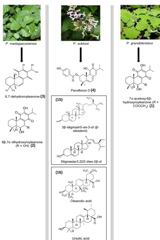
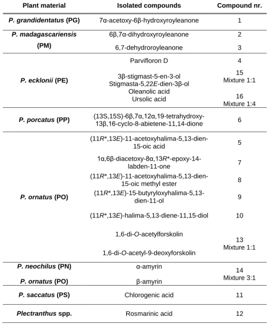
Several phytochemical studies on Plectranthus plants have described a great number of isolated compounds, and Figure 1 depicts the link between Plectranthus species and isolated compounds, with chemical structures and compounds number reported in this thesis.
Figure 1 Studied Plectranthus plants species and related isolated compounds with internal identification
numbers. Compounds (15) were studied in a 1:1 mixture and (16) in a 1:4 mixture. Plants pictures were obtained from (Van Jaarsveld E., 2006).
(1) (3) (4) (2) (15) (16)
6 R1=Ac; R2= COOH (11R*,13E)-11-acetoxyhalima-5,13-dien-15-oic acid R1= OH; R2= CH2OCOCH2CH2CH3 (11R*,13E)-15-butyryloxyhalima-5,13-dien-11-ol R1=OH; R2= CH2OH (11R*,13E)-halima-5,13-diene-11,15-diol 1α,6β-diacetoxy-8α,13R*-epoxy-14-labden-11-one R1=Ac; R2=COOMe
(11R*,13E)-11-acetoxyhalima-5,13-dien-15-oic methyl ester
R1=R2=Ac; R3=OH 1,6-di-O-acetylforskolin R1=R2=Ac; R3=H 1,6-di-O-acetyl-9-deoxyforskolin α-amyrin β-amyrin P. saccatus Chlorogenic acid (PS) Rosmarinic acid (Plectranthus spp.) Chlorogenic acid
Figure 1 (Cont.) Studied Plectranthus plants species and related isolated compounds with internal
identification numbers. Compounds (14) were studied in a 1:1 mixture and (14) in a 3:1 mixture. Plants pictures were obtained from (Van Jaarsveld E., 2006).
(6) (5) (9) (10) (8) (7) (14) (14) (11)
7 The main compounds found in Plectranthus plants are abietane, phyllocladane, kaurane, clerodane and labdane diterpenoids (Figure 1), together with long-chain alkylphenols, aristolane sesquiterpenes, ursane, oleane, and lupine triterpenois, flavonoid, and phenolic compounds (Naghibi et al., 2005; Rijo et al., 2005).
Terpenes are usually present in essential oils and resins, which include over 10,000 compounds and are divided into mono-, di-, tri- and sesquiterpenes, depending on the number of carbon atoms and isoprene (C5H8) groups (Doughari, 2012; Lovkova et al., 2001). Lamiaceae is one of the families where diterpenes have been found, and they can be divided
in 91 skeletons, although the majority belong to the abietane skeleton (Rijo, 2011).
Isolation of diterpenes usually requires organic extracts together with maceration, ultrasound, or super critical fluid extraction methods (Rijo et al., 2012). For higher isolation efficacy, there should be an adequate selection of solvents with different polarities, according to the desired compounds (usually with greater scientific relevance like phenols, terpenes and flavonoids).
The most common organic solvents have intermediate polarity such as acetone, dichloromethane and ethyl acetate, or high polarity like methanol (Rijo et al., 2005, 2014a, 2014b; Waksmundzka-Hajnos and Sherma, 2011).
The often used maceration technique is long-lasting, environmentally unfriendly, hazardous and with some associated human toxicity. However, other extraction methodologies can overcome these problems and simultaneously extract the decomposable highly oxygenated terpenes contained in some Plectranthus plants (Marques et al., 2002).
For natural products extraction and isolation, supercritical fluid extraction (SCF), especially employing supercritical CO2, has become a widespread choice. Cutting-edge
technologies allow precise regulation of changes in temperature and pressure, and thus manipulation of the solvating property of the SCF. These settings enhance the quality of extraction of a wide range of natural products (Bernardo-Gil et al., 2011).
Structural identification of the metabolites is based on physicochemical and spectroscopic data, typically UV, IR, 1H and, 13C NMR spectra. Usually NMR can be further
completed with 2D COSY, TOCSY, gHSQC, gHMBC, and 1D NOESY spectra, mass spectra, and elemental analysis (Maria Fátima Simões, 2010; Rijo et al., 2005).
I.2.1.1 Diterpenes
Diterpenes have a wide structural variability due to their singular roles in plant growth development, and in the resistance to environmental stress (Rijo et al., 2013b). These compounds have a hydrocarbon skeleton, mostly cyclic, and highly oxygenated with hydroxyl, carbonyl, and carboxylic groups in an aliphatic or aromatic framework (Rijo et al., 2014b; Maria Fátima Simões, 2010; Coutinho et al., 2009; Abdel-Mogib et al., 2002). It is the cyclization
8 method of the carbonated skeleton allied with the oxygenated groups that gives diterpenes the wide range of pharmacological abilities (Rijo et al., 2013b). Some of the most common diterpene skeletons can be found in Figure 2.
Due to their high boiling point, diterpenes are not considered as essential oils, but rather as a resin, the remaining material after steam distillation (Wang et al., 2005).
I.2.1.1.1 Abietane diterpenes
Abietane diterpenes (mainly isolated from P. madagascariensis, P. grandidentatus and
P. ecklonii, are the skeleton with the highest occurrence and most widespread in Lamiaceae.
Indeed the royleanone structure of abietane diterpenes, as well as its derivatives, are already extensively known for their antimicrobial, cytotoxic, antiproliferative, and antifungal activities (Rijo et al., 2013a, 2013b; Pereira et al., 2015; Rijo et al., 2014b; Burmistrova et al., 2015, 2013; Rijo et al., 2012).
Two of most the promising metabolites are Parvifloron D and 7α-acetoxy-6β-hydroxyroyleanone. Because of the oxidized abietane and phenol groups, these compounds show a wide range of biological activities (Figure 3). Biological activity has been improved by their derivatization into more bioactive compounds such as 6β,7α-dihydroxyroyleanone or ester derivatives (Rijo, 2011).
Parvifloron D, isolated from P. ecklonii is widely known for its toxicity, but has also been reported with high antioxidant power at in vitro assays, suggesting an interesting efficacy in converting free radicals into more stable products. However, these do not take into account the compound bioavailability or toxicity at in vivo systems (Rosa et al., 2015).
Figure 2 Pathway of the halimane, labdane, pimarane, beyerane, clerodane, abietane and kaurane
diterpenoid skeletons, widely isolated from Plectranthus species, showing the numeration of the hydrocarbon skeleton. Adapted from (Rijo et al., 2013b).
9 The quinone moiety present in abietane diterpenes compounds has been associated with antimycobacterial, antimicrobial, and antitumoral activities (Gaspar-Marques et al., 2006; Rijo, 2011; Rijo et al., 2010, 2014b). Often found in nature, quinones represent important features at stabilizing free radicals in many biological systems suffering from oxidative stress, protein inactivation through irreversible reactions enhancing antimicrobial activity, and intermediate melanin synthesis pathway in human skin (Rijo, 2011; Riley, 1997; Shadyro et
al., 2002).
Figure 3 Structural representation of three abietane diterpenes, Parvifloron D (A),
7α-acetoxy-6β-hydroxyroyleanone (B), and 6β,7α-di7α-acetoxy-6β-hydroxyroyleanone (C). Here are also depicted some of the most important structural characteristics granting a wide biological activity. Adapted from (Rijo, 2011).
Structure activity relationships (SAR) analyses have shown that due to their amphipathic character, these diterpenes can damage bacteria cytoplasmatic membrane. Also, they can increase lipophilicity and/or promote hydrogen-bonding of the hydrophilic moiety (Rijo et al., 2014b). The cytotoxic and antiproliferative abilities against human tumor cells are probably due to their alkylating properties (Burmistrova et al., 2013, 2015).
Since royleanones can be present in many traditional remedies, more in vitro and in vivo studies should be performed, to understand their medicinal value consistent with the ethnopharmacological uses.
I.2.1.1.2 Labdane diterpenes
By 2005 the isolation of new diterpenes concerning the labdane skeleton was reported (Rijo et al., 2005). These diterpenes had a forskolin-like backbone (isolated from P. ornatus at ethyl acetate extract, see Figure 1), one of the most interesting compounds originally obtained from Plectranthus barbatus Andr. (also known as Coleus forskohlii Briq.) (Rijo et al., 2005, 2013b).
Forskolin compounds are specially notorious for several biological activities, regarding their cardiotonic, platelet aggregation inhibition, and anti-inflammatory properties (Klatz and Goldman, 2003; Rijo et al., 2005). Also, forskolin (Figure 4) has a catecholamine-like action by increasing intracellular cyclic adenosine monophosphate (cAMP), through the activation of adenylate cyclase. Previous studies suggest that this activation occurs in synergy with a wide
10 range of hormones and neurotransmitters (Bone and Mills, 2013). This interesting bioactivity leads to vasodilatory properties, and improved fat loss in double-blind clinical trials (Bone and Mills, 2013; Hayashida et al., 2001; Pizzorno and Murray, 2013).
Regarding the presence of non-volatile compounds, 1,6-di-O-acetylforskolin (Figure 4B) is obtained by acetylation of forskolin, whereas 1,6-di-O-acetyl-9-deoxyforskolin (Figure 4C) is an intermediate of forskolin, starting with 9-deoxyforskolin. Although the potential therapeutic benefit of forskolin-like compounds, there is a concern about the use of these products on drug metabolizing enzymes in the liver (Lukhoba et al., 2006).
Another labdane diterpene is Plectrornatin C depicted in Figure 4D, that was isolated from the acetone extract of P. ornatus. This compound was previously demonstrated to have moderate antifungal activity (Rijo et al., 2002), and cyclooxygenase-2 (COX-2) inhibition by 59% at a concentration of 0.6 mM (Rijo, 2011).
Figure 4 Resemblance of the chemical structures of some labdane diterpenes with relevance for Forskolin (A),
1,6-di-O-acetylforskolin (B), 1,6-di-O-acetyl-9-deoxyforskolin (C), and Plectrornatine C (D). Here are also depicted the flavanone ring (in red) granting the labdanes a wide biological activity. Adapted from (Rijo, 2011).
As seen in Figure 4, some labdane diterpenes (A-D) seem also to be related with flavanones. Flavanones have been a promising source for lead compounds as the focus of many researches for biologically active components. Indeed, they were reported to have significant cytotoxic activity against leukemia and lung carcinoma cells, senescence, and cardiovascular diseases (Ketabforoosh et al., 2014; Shi et al., 2010). However, they are rarely present in natural sources, which limits the flavanone biological applications, and realizing their existence in Plectranthus spp. plants could bring a new light into labdane pharmacological activities.
I.2.1.1.3 Halimane diterpenes
Diterpenes with a halimane skeleton are mainly isolated from the acetone extract of P.
ornatus (see Figure 1Figure ), and still have some unknown pharmacological properties.
Considering halimane biosynthesis, they appear to be related to the labdanes by a series of methyl and hydride shifts (Silva et al., 2011).
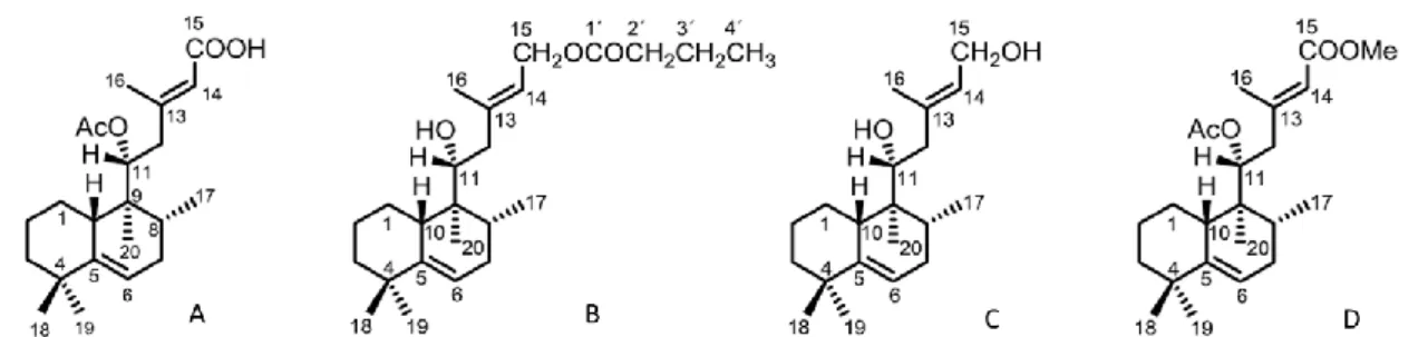
Unfortunately, studies on halimane biological activities are scarce. In 2011 some halimane derivatives derived from the halimane diterpene A (Figure 5), namely halimane diterpenes B-D, were reported to be strong inhibitors of COX-2 in a range of 59 to 79% (Rijo, 2011).
11 More recently, new findings regarding tuberculosis, namely the pathogenic strain of
Mycobacterium tuberculosis, reported the existence of tuberculosinols (Nakano et al., 2011),
very similar in structure to halimane diterpenes. Tuberculosinols have been reported to suppress phagosome maturation, and inhibit phagocytosis by macrophage cells (Hoshino et
al., 2011). More details will be discussed in Section I.3.4.
Figure 5 Chemical structures of some halimane diterpenes. (11R*,13E)-11-acetoxyhalima-5,13-dien-15-oic acid
(A), (11R*,13E)-15-butyryloxyhalima-5,13-dien-11-ol (B), (11R*,13E)-halima-5,13-diene-11,15-diol (C), and (11R*,13E)-11-acetoxyhalima-5,13-dien-15-oic acid methyl ester (D). Compounds B-D are derivatives from A. Adapted from (Rijo, 2011).
I.2.1.2 Triterpenes and phytosterols
Triterpenes and sterols are two types of compounds that genetically derive from the same squalene precursor, whose cyclic structures comprise trans or cis cyclohexane and cyclopropane units (Wink, 2010). Distinguishing triterpenes from sterols can be hard since they share common structural characteristics, therefore one possible way is by identifying their synthetic routes (Wink, 2010). Whereas sterols can be present in the Animalia, triterpenes are unique of the Plantae kingdom (Gabay et al., 2010).
Pentacyclic triterpenes such as ursolic and oleanolic acids (Figure 6A and 6B), α- and β-amyrin (Figure 6C and 6D), can be isolated from P. neochilus, P. ornatus, and P. ecklonii, using acetone or hexane extracts (Gabriel et al., 2016; Rijo et al., 2005; Simões et al., 2010). Oleanolic and ursolic acids are usually isolated simultaneously and are isomers that only differ on the methyl group at C-29 as depicted in Figure 6.
Figure 6 Chemical structure of ursolic (A) and oleanolic (B) acids and their characteristic difference at C-29,
α-amyrin (C) and β-α-amyrin (D). Adapted from (Rijo, 2011).
The triterpenoids α- and β-amyrins are known for their antinociceptive properties, that have been demonstrated in several in vivo models (Gabriel et al., 2016; Rijo et al., 2005). Although the mechanisms of action are not yet fully understood, it is considered that they are independent from important endogenous systems (like opioidergic, serotoninergic and
12 noradrenergic), stabilizing the action of mast cell membranes involved in inflammation process (Backhouse et al., 2008).
Oleanolic and ursolic acids have proven efficacy as antioxidant regarding radical scavenging and superoxide decrease (Ghimeray et al., 2015; Hwang et al., 2014), antimicrobial activities (Maria Fátima Simões, 2010), anti-inflammatory, antitumor including inhibition of skin tumorigenesis (Cha et al., 1998), and as potent elastase and collagenase inhibitors (Lee et al., 2001; Thring et al., 2009; Tu and Tawata, 2015; Ying et al., 1991).
Phytosterols are notorious for their anti-inflammatory properties (Backhouse et al., 2008). These compounds are synthesized by the mevalonate pathway of terpenoid formation, and can be regarded as direct precursors of steroid saponins, alkaloids, pregnanes, androstanes, that share the same backbone (Wink, 2010).
Two examples of these compounds that can be isolated from Plectranthus plants, usually from P. ecklonii (Figure 1), are β-sitosterol and stigmasterol. These have been validated for anti-inflammatory and analgesic purposes (Backhouse et al., 2008; Ghimeray et al., 2015), although it was previously though that stigmasterol could not be considered as major anti-inflammatory agent (García et al., 1999). Nonetheless, both compounds distinctly reduce myeloperoxydase activity influenced by the neutrophil migration inhibition into inflamed tissue (De la Puerta et al., 2000; García et al., 1999; Villaseñor et al., 2002, 2004)
Furthermore, β-sitosterol was proven to inhibit human prostate cancer cells growth, revealing an effectiveness in apoptosis induction regarding prostaglandin production (Awad et
al., 2005). According to the investigation held by Nashed et al. (Nashed et al., 2005),
phytosterols are strongly associated with reduced plasma cholesterol concentrations and atherosclerosis lesions by 20% and 60% respectively, together with lowering of pro-inflammatory cytokines (Ghimeray et al., 2015).
I.2.1.3 Polyphenolic compounds
Besides diterpenes, Plectranthus plants are rich in phenolic compounds (mainly isolated from aqueous extracts, see Figure 1). Polyphenols are chemical compounds primarily responsible for the fruit coloring (Doughari, 2012). These are classified as phenolic acids, flavonoids and non-flavonoid (Doughari, 2012). In addition to their antioxidant properties, they play a very important role in the plant defenses against herbivores, pathogens and predators; therefore they have an application in the control of human agents infections (Doughari, 2012). Polyphenols, such as rosmarinic acid ubiquous to all Plectranthus spp., and chlorogenic acid obtained mainly from P. saccatus (Rijo et al., 2014a), are present in aqueous fractions using decoction, infusion microwave or ultrasound extraction methods (Rijo et al., 2012, 2014a). Overall polyphenols are present in the diet and are known for their antioxidant,
13 antimicrobial, and anti-inflammatory properties (Ghimeray et al., 2015; Pereira et al., 2015; Sergent et al., 2010).
I.3 Biological activities of Plectranthus natural products
In line with the literature, the most common compounds in Plectranthus are pimarane, labdane, neoclerodane, halimane, and abietane diterpenes, and they have been extensively studied for their biological activity (Rijo et al., 2013b).
Previous studies on the secondary metabolites samples used in this project, were reported in partial studies regarding their medicinal use, although the pharmacological properties of some compounds remain unknown (Rijo et al., 2013c).
Although many compounds seem to have great potential according to their biological activities as described in Table 2, research focused on the screening and scientific validation of Plectranthus plants are still half-way.
The next sections will review in more detail the state of the art concerning the studied biological activities of Plectranthus secondary metabolites within this thesis.
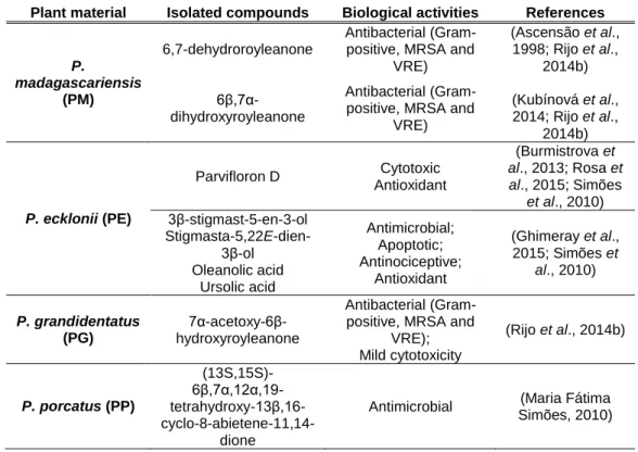
Table 2 Terpene, phytosterols, and polyphenols isolated from the described Plectranthus spp. plants, and their
main biological activities.
Plant material Isolated compounds Biological activities References
P.
madagascariensis (PM)
6,7-dehydroroyleanone
Antibacterial (Gram-positive, MRSA and
VRE) (Ascensão et al., 1998; Rijo et al., 2014b) 6β,7α-dihydroxyroyleanone Antibacterial (Gram-positive, MRSA and
VRE) (Kubínová et al., 2014; Rijo et al., 2014b) P. ecklonii (PE) Parvifloron D Cytotoxic Antioxidant (Burmistrova et al., 2013; Rosa et al., 2015; Simões et al., 2010) 3β-stigmast-5-en-3-ol Stigmasta-5,22E-dien-3β-ol Oleanolic acid Ursolic acid Antimicrobial; Apoptotic; Antinociceptive; Antioxidant (Ghimeray et al., 2015; Simões et al., 2010) P. grandidentatus (PG) 7α-acetoxy-6β-hydroxyroyleanone Antibacterial (Gram-positive, MRSA and
VRE); Mild cytotoxicity (Rijo et al., 2014b) P. porcatus (PP) (13S,15S)- 6β,7α,12α,19- tetrahydroxy-13β,16- cyclo-8-abietene-11,14-dione
Antimicrobial (Maria Fátima
14
Table 2 (Cont.) Terpene, phytosterols, and polyphenols isolated from the described Plectranthus spp. plants, and
their main biological activities.
Plant material Isolated compounds Biological activities References
P. ornatus (PO) (11R*,13E)-11- acetoxyhalima-5,13-dien-15-oic acid Antimicrobial (MRSA); Antimycobacterial (Layre et al., 2014; Mann and Peters, 2012; Nakano et al., 2011; Rijo et al., 2011) (11R*,13E)-15- butyryloxyhalima-5,13-dien-11-ol (11R*,13E)-halima-5,13-diene-11,15-diol (11R*,13E)-11- acetoxyhalima-5,13-dien-15-oic methyl ester
1α,6β-diacetoxy-
8α,13R*-epoxy-14-labden-11-one
Antibacterial (Rijo et al., 2013a)
1,6-di-O-acetylforskolin
Anti-inflammatory (Rijo et al., 2005)
1,6-di-O-acetyl-9-deoxyforskolin P. neochilus (PN) P. ornatus (PO) α-amyrin β-amyrin Cytotoxic; Anti-inflammatory; Analgesic (Backhouse et al., 2008; Gabriel et al., 2016; Rijo et al., 2005)
P. saccatus (PS) Chlorogenic acid
Antioxidant; Antimicrobial Anti-acetylcholinesterase; tyrosinase; collagenase; elastase; Anti-inflammatory (Falé et al., 2012, 2011; Ghimeray et al., 2015; Pereira et al., 2015)
Plectranthus spp. Rosmarinic acid
Antioxidant; Antimicrobial; Anti-acetylcholinesterase; Anti-inflammatory (Falé et al., 2012, 2011; Ghimeray et al., 2015; Pereira et al., 2015)
I.3.1 Antioxidant Activity
Reactive oxygen species (ROS) are mainly produced at mitochondrial oxidative phosphorylation, in cellular response to xenobiotics, cytokines, and bacterial attack. Therefore, oxidative stress refers to the imbalance of ROS over the cell ability to have an effective antioxidant response (Ray et al., 2012).
Although a natural physiological process in biologic systems, the consequences of oxidative stress are present in many pathologies such as cancer, inflammation, and neurodegenerative disorders, renewing the interest to discover new antioxidant molecules (Rosa et al., 2015). When ROS are extensively generated, such as superoxide anion (O2●-),
hydrogen peroxide (H2O2) and hydroxyl radical (HO●), redox-active metal ions like Fe2+ or Cu2+
originate harmful products from reacting rapidly with lipids, DNA and proteins and thus resulting in mutagenesis and enzymatic denaturation (Dhawan, 2014; Ray et al., 2012; Thring
15 free radicals and chelating catalytic metals, or by acting as oxygen scavengers (Nsimba et al., 2008). However, the most used antioxidants such as butylated hydroxyanisole (BHA) or propyl gallate, are suspected to induce liver damage and carcinogenesis in animals (Nsimba et al., 2008). Therefore, it is widely important to discover new and eventually more powerful and less cytotoxic antioxidant agents from natural sources, such as Plectranthus plants.
Polyphenols are powerful ROS scavengers, therefore they work as the most powerful antioxidants (Wahab et al., 2014) and they can be found in many plants, including Plectranthus (Rijo et al., 2012; Rosa et al., 2015). These plant-derived compounds exhibit a wide range of biological effects as antioxidants, such as anti-ageing, vitamin C protection and reduction of α-tocopheryl radical (Wahab et al., 2014; Rijo et al., 2014b).
The phenol groups present in rosmarinic and chlorogenic acids, can form relatively stable phenoxyl radicals, thereby disrupting chain oxidation reactions in cellular components (Pandey and Rizvi, 2009).
Also, abietane diterpenes, Parvifloron D and 7α-acetoxy-6β-hydroxyroyleanone have been proved to have the capacity to chelate the 2,2-diphenyl-1-picrylhydrazyl (DPPH) radical. Parvifloron D (IC50 0.11 ± 0.018 mM) was found to be a much stronger antioxidant than
7α-acetoxy-6β-hydroxyroyleanone (IC50 1.85 ± 0.071 mM), when compared to the positive control
quercetin (IC50 0.0075 ± 0.0010 mM) (Rijo, 2011; Rosa et al., 2015). Besides, Parvifloron D
can protect DNA from oxidative stress by plasmid DNA cleavage assay in the presence of H2O2 and Fe (II) (Rosa et al., 2015).
Since one of the antioxidant mechanisms is the capacity to scavenge the free radicals that are formed during carcinogenesis, neurodegeneration, diabetes, inflammation, and ageing processes, an antioxidant agent can be useful as an adjuvant therapy (Falé et al., 2011; Ray
et al., 2012).
Also, compounds associated with reduced levels of oxidative stress, are supposed to have skin-protective properties, through the inhibition of natural skin-related enzymes such as tyrosinase, elastase, and collagenase (Chen et al., 2015; Ghimeray et al., 2015). Further elucidation will be given throughout section I.3.2.
I.3.2 Inhibition of skin-related enzymes
The skin is one of the most important organs of the body, since it provides protection from the external environment, and from any damage caused by the UV-light, chemicals, and microorganisms (Moon et al., 2010; Thring et al., 2011).
There are many forms of skin ageing, but one the most common stress-inducing factor is exposure to UV irradiation, that disturbs the intrinsic pathways involving elasticity and pigmentation changes (Wahab et al., 2014; Thring et al., 2011; Moon et al., 2010). Solar
16 exposure causes ultimately the formation of lipid peroxides, and ROS that can overcome the endogenous cellular antioxidant system, thus induce proteinases capable of changing the matrix (Thring et al., 2009). Moreover, skin disorders are associated with increased oxidative stress, that enhances enzymatic activity such as tyrosinase, elastase, and collagenase (Ghimeray et al., 2015; Vallisuta et al., 2014). Therefore, there is an interest to identify natural antioxidants that can be inhibitors of these enzymes to fight against skin ageing and hyperpigmentation (Popoola et al., 2015; Tu and Tawata, 2015).
I.3.2.1 Enzymes in skin pigmentation and ageing – Tyrosinase, elastase, and collagenase
Tyrosinase (EC 1.14.18.1) is a bi-nuclear copper monooxygenase that catalyzes the first two stages of melanin biosynthesis. The copper ions are coordinated with the histidine residues on the active site, and are critical for catalytic activity (Chen et al., 2015; Popoola et
al., 2015). In the initial step, L-tyrosine is hydroxylated to L-3,4-dihydroxyphenylalanine (also
known as L-DOPA), that is further oxidized to the corresponding dopaquinone. The oxidative polymerization of several dopaquinone derivatives originates melanin (Masuda et al., 2009; Tu and Tawata, 2015), thus tyrosine is considered to be the rate-limiting regulator (Tu and Tawata, 2015).
Melanin is an important cellular component responsible for skin color, and has photo-protective properties against harmful effects of UV radiation in normal skin pigmentation. In contrast, abnormal pigmentation caused by formation of excessive ROS can lead to senile lentigines, feckles, and melasma (Chen et al., 2015; Tu and Tawata, 2015).
Many tyrosinase inhibitors such as hydroquinone, kojic acid (IC50 0.03 mM (Masuda et
al., 2009)), azelaic acid, electron-rich phenols, and arbutin (IC50 83.3 mM (Masuda et al.,
2009)), have been reported for their capability of inhibiting melanin overproduction (Chen et
al., 2015). However, due to their adverse side effects, low formulation stability, and poor skin
penetration their use is limited, hence the search for new agents from natural products. In fact, quercetin, a strong antioxidant compound, has been reported to highly inhibit tyrosinase activity (IC50 0.1 mM (Masuda et al., 2009)).
Besides hyperpigmentation, UV irradiation can also cause changes in the extracellular matrix (ECM) by the breakdown of collagen, elastin, proteoglycans, and fibronectin, thus damaging skin collagenous tissues (Jung et al., 2014). It is believed that ROS can upregulate expression of several matrix metalloproteinases (MMP), and decrease the synthesis of ECM proteins such as collagen and elastin. In Figure 7 two signaling pathways are outlined according to previous studies.