Article
Printed in Brazil - ©2015 Sociedade Brasileira de Química0103 - 5053 $6.00+0.00
A
*e-mail: [email protected]
Abietane Diterpenes from
Hyptis crassifolia
Mart. ex Benth. (Lamiaceae)
Karísia S. B. Lima,a Maria L. S. Guedesb and Edilberto R. Silveira*,a
aDepartamento de Química Orgânica e Inorgânica, Centro de Ciências,
Universidade Federal do Ceará, CP 12.200, 60021-940 Fortaleza-CE, Brazil
bDepartamento de Botânica, Instituto de Biologia, Universidade Federal da Bahia,
40170-290 Salvador-BA, Brazil
O estudo fitoquímico do extrato etanólico das raízes de Hyptis crassifolia Mart. ex Benth. (Lamiaceae) levou ao isolamento e elucidação estrutural de nove diterpenos identificados como 11,12,15-tri-hidroxi-8,11,13-abietatrien-7-ona, 6α ,11,12,15-tetra-hidroxi-8,11,13-abietatrien-7-ona, 11,12,16-tri-hidroxi-17(15→16)-abeo-abieta-8,11,13-trien-7-ona, (16S)-12,16-epoxi-11,14-di-hidroxi-17(15→16)-abeo-abieta-8,11,13-trien-7-ona, incanona, ferruginol, sugiol, óxido de 11-oxomanoíla e óxido de 11β-hidroximanoíla. Os compostos 11,12,16-tri-hidroxi-17(15→16)-abeo-abieta-8,11,13-trien-7-ona e6α,11,12,15-tetra-hidroxi-8,11,13-abietatrien-7-ona são inéditos na literatura, a 11,12,15-tri-hidroxi-8,11,13-abietatrien-7-ona está sendo relatada pela primeira vez como um novo diterpeno abietano natural, enquanto que para a (16S)-12,16-epoxi-11,14-di-hidroxi-17(15→16)-abeo-abieta-8,11,13-trien-7-ona,isoladaanteriormente de Teucrium divaricatum Subsp. villosum, propõe-se uma revisão dos dados de ressonância magnética nuclear (NMR) 1H e 13C. A determinação estrutural de todos os constituintes foi realizada através de
técnicas espectroscópicas como espectrometria de massas de alta resolução (HRMS), infravermelho (IR), NMR de 1H e 13C, incluindo sequências de pulsos uni e bidimensionais, e comparação com
dados descritos na literatura.
The phytochemical study of the ethanol extract from roots of Hyptis crassifolia Mart. ex Benth. (Lamiaceae) led to the isolation and structure elucidation of nine diterpenes identified as 11,12,15-trihydroxy-8,11,13-abietatrien-7-one, 6α ,11,12,15-tetrahydroxy-8,11,13-abietatrien-7-one, 11,12,16-trihydroxy-17(15→16)-abeo-abieta-8,11,13-trien-7-one, (16S)-12,16-epoxy-11,14-dihydroxy-17(15→16)-abeo-abieta-8,11,13-trien-7-one, incanone, ferruginol, sugiol, 11-oxomanoyl oxide and 11β-hydroxymanoyl oxide. Compounds 11,12,16-trihydroxy-17(15→16)-abeo-abieta-8,11,13-trien-7-oneand6α ,11,12,15-tetrahydroxy-8,11,13-abietatrien-7-one are new, 11,12,15-trihydroxy-8,11,13-abietatrien-,11,12,15-tetrahydroxy-8,11,13-abietatrien-7-one is being reported for the first time as a new natural abietane diterpene, while for (16S)-12,16-epoxy-11,14-dihydroxy-17(15→ 16)-abeo-abieta-8,11,13-trien-7-one, previously isolated from Teucrium divaricatum Subsp. villosum, a revision of the 1H and 13C nuclear magnetic resonance (NMR) data previously reported is proposed.
Structure determination of all constituents was performed by mean of spectroscopic techniques such as high resolution mass spectrometry (HRMS), infrared (IR), 1H and 13C 1D and 2D NMR
and comparison with literature data.
Keywords: Hyptis crassifolia, Lamiaceae, abietane diterpenes, rearranged abietane diterpenes, labdane diterpenes
Introduction
Lamiaceae consists of about 258 genera and 6970 species distributed around the world,1 mainly centered in
the Mediterranean region despite reports of their occurrence in Australia and South America.2 The Hyptis genus includes
around 400 species distributed throughout the Americas, West Africa, Fiji Island (Oceania), and western India.3
They are found in Northern and Northeastern Brazil, especially in the cerrado.4 Plants from this genus are used
Previous studies on the chemical constitution of Hyptis
species report the isolation of triterpenoids,5 flavonoids,6,7
lignans,7,8 α-pyrone derivatives9,10 and diterpenoids,11-14
namely labdanes and abietanes. The abietane diterpenes have attracted particular attention, due to their biological activities, in particular as antioxidant,15-17 antibacterial,18-20
cytotoxic,21-24 antiviral25,26 and antimalarial.27 Among the
diterpenes that exhibit these activities, were observed several highly oxygenated compounds that can present either the C-ring saturated or unsaturated, aromatic, and also with an ortho or para-naphtoquinone pattern. Previous works carried out on plants of the genus Hyptis from the Northeast of Brazil flora, reported the isolation of many abietane diterpenes, for example those isolated from roots of H. martiusii,11H. platanifolia12 and H. carvalhoi.28
In this paper, it is reported the phytochemical analysis of the ethanol extract from roots of Hyptis crassifolia Mart. ex Benth., a small shrub that grows in abundance in the state of Bahia to which no phytochemical investigation has been reported so far. This study led to the isolation and structure elucidation of nine diterpenes: four abietanes (1, 2,
6 and 7), three rearranged abietanes (3-5) and two labdanes (8 and 9). Compounds 2 and 3 have not yet being reported in the literature, whereas 1 is being reported for the first time from a natural source. A revision of the previously published nuclear magnetic resonance (NMR) data in the literature29 is being proposed for compound 4.
Experimental
General procedures
Melting points were measured on a digital Marconi MA-381 apparatus and are uncorrected. Optical rotations [α]D20 were determined with a Jasco P-2000 digital
polarimeter, operating with tungsten lamp at a wavelength of 589 nm at 20 °C. Infrared (IR) spectra were recorded using a Perkin-Elmer FTIR 100 spectrometer using the universal attenuated total reflectance accessory (UATR). High-resolution electrospray ionization mass spectra (HR-ESI-MS) were performed with a SHIMADZU LCMS-IT-TOF (225-07100-34) equipped with a Z-spray ESI (electrospray) source operating either in the negative or positive mode. 1H and 13C NMR (1D and 2D) spectra were
performed on Bruker Avance DPX-300 and/or DRX-500 spectrometers equipped with 5 mm direct probe or inverse detection Z-gradient probe, respectively. 1H NMR (300.13
and 500.13 MHz) and 13C NMR (75.47 and 125.75 MHz)
spectra were measured at 25 °C. Chemical shifts (d), expressed in parts per million (ppm), are referenced by the signal of the residual non-deuterated hydrogens
(1H NMR) and the central peak of carbon-13 (13C NMR)
of the deuterated solvents. Flash column chromatography and column chromatography (CC) were carried out on silica gel 60 A (Whatman, 70-230 mesh) and silica gel 60 A (Acros Organic, 0.035-0.070 mm), respectively. Thin layer chromatography (TLC) was performed on precoated silica gel polyester sheets (Kieselgel 60 F254, 0.20 mm, Merck)
by detection with a spray reagent of vanillin/perchloric acid/EtOH solution followed by heating at 100 °C. Normal phase semi-preparative HPLC separations were performed with a Shimadzu LC-20AT pump, UV-PDA (SPD-M20A) detector and WATERS-1525 pump, UV-PDA (WATERS 2996) detector, a Phenomenex Silica column (10 × 150 mm, 5 µm particle size), with a flow rate of 4.72 mL min−1 and
column oven temperature 40 °C, monitoring at 273.8 nm.
Plant material
The roots of Hyptis crassifolia Mart. ex Benth. were collected in July 2010, at Mucujê county (Diamantina Plateau, Bahia State). The plant material was identified by Prof Maria Lenise Silva Guedes of Instituto de Biologia, Departamento de Botânica, Universidade Federal da Bahia. A voucher specimen (No. 95.183) is deposited in the Herbário Alexandre Leal Costa of the Departamento de Botânica, Universidade Federal da Bahia.
Extraction and isolation
The roots (875 g) of Hyptis crassifolia Mart. ex Benth., dried and crushed were macerated with EtOH (3 × 6.0 L), after extraction with hexane, at room temperature after standing for 72 h. The ethanol solution was concentrated under reduced pressure at 50 °C to yield the respective extract (26.0 g). The EtOH extract (22.0 g) was coarsely fractionated over a silica gel column using hexane/CH2Cl2
(1:1), CH2Cl2, CH2Cl2/EtOAc (1:1), EtOAc and MeOH
as eluents. The hexane/CH2Cl2 (1:1) fraction (674.4 mg)
was chromatographed on a silica gel column with hexane, hexane/CH2Cl2 (9:1, 7:3, 1:1), CH2Cl2 and MeOH to
give 36 fractions (10 mL each), that after TLC analysis were pooled to 9 fractions (F1HD-F9HD). F4HD (52.8 mg, 7:3
hexane/CH2Cl2) was separated through HPLC using a 9:1
hexane/EtOAc as eluent (v/v), with an injection volume of 200 µL, under isocratic conditions to yield compound 6
(6.0 mg), [α]D20 +25.7 (c 0.2, CHCl3) {lit. [α]D20 +42.9°
(c 0.2, CHCl3)}.30 F6HD (60.2 mg, hexane/CH2Cl2 7:3) after
successive recrystallizations in MeOH yielded 18.5 mg of 8 in the form of colorless crystals, mp 70.7-73.4 °C, [α]D20−33.8° (c 0.2, CHCl3) {lit. mp 96-97 °C, [α]D20−103.2°
was obtained from F8HD (85.0 mg, 1:1 hexane/CH2Cl2),
after semi-preparative HPLC analysis (mobile phase 98:2 hexane/isopropanol). The CH2Cl2 fraction (1.26 g) was
chromatographed over silica gel by elution hexane/CH2Cl2
(1:1, 3:7), CH2Cl2, CH2Cl2/EtOAc (1:1), EtOAc and MeOH
to give 32 fractions (10 mL each), that were combined into nine resulting fractions after TLC analysis (F1D-F9D). F4D
(110.5 mg, 1:1 hexane/CH2Cl2) was submitted to
semi-preparative HPLC analysis, using hexane/EtOAc, 8:2 (v:v) as mobile phase to yield 7 (19.0 mg) in the form of yellow crystals, mp 283.4-285.4 °C, [α]20D +26.1 (c 0.1, CHCl3)
{lit. mp 282-285 °C, [α]D25 +12.3° (c 0.1, CHCl3)}.32 F6D
(195.8 mg, 3:7 hexane/CH2Cl2), was rechromatographed
on silica column by elution with hexane, hexane/CH2Cl2
(9:1, 8:2, 7:3, 1:1), CH2Cl2 and MeOH to yield 9 (47.3 mg)
in the form of yellow crystals, mp 78.9-79.9 °C, [α]20D +30.2°
(c 0.1, CHCl3) {lit. mp 106-107 °C and [α]D25 +12.3° (c 0.1,
CHCl3)}.33 Compound 5 (4.6 mg) was obtained as a yellow
resin, [α]D20 +15.7° (c 0,095, CHCl3) {lit. [α]25D +140.8° (c 0.1,
CHCl3)},32 from F7D (33.7 mg, 3:7 hexane/CH2Cl2), after
successive recrystallization with CH2Cl2/MeOH, (1:1). F8D
(479.7 mg, CH2Cl2) was chromatographed on silica column
with CH2Cl2:MeOH (98:2, 95:5 and 1:1) to give 22
sub-fractions (10 mL each), that were pooled to 6 sub-sub-fractions (F8D1-F8D6), after TLC analysis. F8D5 was subjected to HPLC
analysis, using hexane/EtOAc, 8:2 (v:v) as mobile phase to yield compounds 1 (4.0 mg), 2 (3.2 mg) and 3 (3.7 mg).
11,12,15-trihydroxy-8,11,13-abietatrien-7-one (1)
Yellowish resin; [α]D20 +17.13° (c 0.0975, CHCl3; IR
vmax/cm−1 3385, 2927, 2865, 1726, 1669, 1564, 1459, 1311;
HR-ESI-MS m/z 333.2062 [M + H]+, (calc. for C
20H29O4,
333.2060); 1Hand 13C NMR spectral data, see Table 1.
6α,11,12,15-tetrahydroxy-8,11,13-abietatrien-7-one (2)
Yellowish resin; [α]D20−18.15° (c 0.074, CHCl3); IR
vmax/cm−1 3436, 2928, 2869, 1720, 1675, 1611, 1566,
1463, 1372, 1309, 1298, 1128; HR-ESI-MS m/z 349.2007 [M + H]+ (calc. for C
20H29O5, 349.2010); 1H and 13C NMR
spectral data, see Table 1.
11,12,16-trihydroxy-17(15→16)-abeo
-abieta-8,11,13-trien-7-one (3)
Yellow resin; [α]D20 +57.92° (c 0.08, CHCl3); IR,
vmax/cm−1 3401, 2927, 2861, 1722, 1671, 1609, 1564, 1461,
1368, 1316; HR-ESI-MS m/z 333.2064 [M + H]+ (calc.
for C20H28O4, 333.2060); 1Hand 13C NMR spectral data,
see Table 1.
(16S)-12,16-epoxy-11,14-dihydroxy-17(15→16)-abeo
-abieta-8,11,13-trien-7-one (4)
Yellow solid; mp 195.7-197.7 °C; [α]D20 +26.79° (c 0.24,
CHCl3); IR vmax/cm−1 3382, 2926, 2865, 1618, 1459, 1352,
1253, 1016, 807; HR-ESI-MS m/z 331.1889 [M + H]+
(calc. for C20H27O4, 331.1909); 1Hand 13C NMR spectral
data, see Table 1.
Results and Discussion
The EtOH extract from roots of Hyptis crassifolia
Mart. ex Benth. was fractionated by silica gel column chromatography after elution with pure or binary mixtures of hexane, CH2Cl2, EtOAc and MeOH. Thehexane-CH2Cl2
(1:1) and CH2Cl2 fractions were subjected to various
chromatographic procedures leading to the isolation of nine compounds (1-9, Figure 1), whose structures were elucidated by spectroscopic methods, such as IR, high resolution mass spectrometry (HRMS) and particularly,
1H and 13C NMR (1D and 2D).
Compound 1 was obtained asa yellowish resin. The IR spectrum exhibited absorption bands for hydroxyl group at 3385 cm−1, Csp3-H groups at 2927 and 2865 cm−1, skeletal
bands of benzene ring at 1609 and 1564 cm−1, conjugated
C=O at 1669 cm−1 and C–O of phenol and alcohols at
1254 and 1145 cm−1, respectively. The molecular formula
C20H28O4 was determined by HR-ESI-MS, based on the
quasi-molecular ion at m/z 333.2062 [M + H]+ (calcd.
for C20H29O4 m/z 333.2060). The CPD and DEPT 135° 13C NMR spectra displayed 20 and 11 signals, respectively,
one from a conjugated aryl ketone carbonyl (dC 199.4),
six sp2 carbons of a benzene ring (d
C 116.5-147.6), one
non-hidrogenated sp3 carbon bearing an oxygen (d C 76.9),
and 12 non-functionalized sp3 carbons (d
C 18.1-50.5;
two quaternary, one methine, four methylenes and five methyls) (Table 1). The 1H NMR spectrum showed five
characteristic singlets of methyl groups attached to non hydrogenated carbons at dH 0.95 (Me-18), 0.98 (Me-19),
1.40 (Me-20), 1.68 (Me-17) and 1.70 (Me-16). A signal at dH 7.49 (H-14) was ascribed to one proton of
a pentasubstituted benzene ring. In addition, a pair of double doublets at dH 2.53 (J 17.0, 14.4 Hz, H-6b) and 2.63
(J 17.0, 2.8 Hz, H-6a) were observed, corresponding to one methylene group coupled to a methine at dH 1.85 (dd,
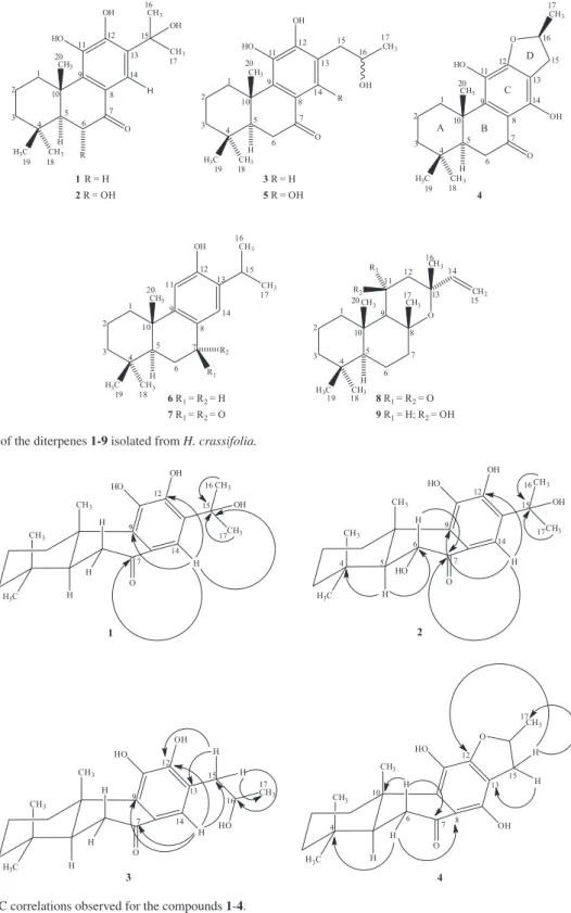
J 2.8, 14.4 Hz, H-5). The presence of a hydroxyl group at C-15 was accomplished by the HMBC spectrum analysis, showing long-range correlations of the methyl groups at dH 1.70 (Me-16) and 1.68 (Me-17) with the oxygenated
carbon at dC76.9 (C-15, 2J) (Figure 2). Furthermore, the
was determined by correlations of the benzene proton at dH 7.49 (H-14) with the oxygenated carbon at dC76.9 (C-15, 3J), with the sp2 carbons at d
C 147.6 (C-12, 3J) and 139.4
(C-9, 3J) and with the carbonyl group at d
C 199.4 (C-7, 3J)
(Figure 2). Then, compound 1 was characterized as the 11,12,15-trihydroxy-8,11,13-abietatrien-7-one, a diterpene previously synthesized during the structural determination
of callicarpone by Kawazu et al.,34 but that is being reported
for the first time in the literature as a new natural abietane. Compound 2 was also obtained as a yellowish resin. The molecular formula C20H28O5 was determined by
HR-ESI-MS, based on the quasi-molecular ion at m/z 349.2007 [M + H]+ (calcd. for C
20H29O5 m/z 349.2010). The IR
spectrum showed stretching bands consistent with the
Table 1. 1H and 13C NMR data (d in ppm, J in Hz) for compounds 1-5
1b 2b 3b 4a 5a
C dC dH dC dH dC dH dC dH dC dH
1 36.4 3.24 (bd,
J 13.5 Hz)
36.6 3.21 (bd,
J 12.8 Hz)
36.4 3.29 (bd,
J 13.3 Hz)
37.8 3.47 (bd,
J 14.3 Hz)
38.1 3.44 (bd,
J 14.0 Hz)
1.44 (m) 1.41 (m) 1.44 (dd,
J 3.6, 13.3 Hz)
1.39 (overlap) 1.34 (m, 1H)
2 19.2 1.77 (td,
J 3.2, 6.4 Hz)
19.1 1.70 (overlap) 19.2 1.80 (td,
J 3.6, 7.0 Hz)
20.2 1.55 (bd,
J 14.2 Hz)
20.3 1.76 (dd, J 2.8, 14.2 Hz)
1.60 (m) 1.55 (m) 1.60 (td,
J 3.6, 7.0 Hz)
1.79 (m) 1.56 (dt,
J 3.6, 14.2 Hz)
3 41.5 1.50 (bd,
J 13.5 Hz)
42.6 1.57 (m) 41.5 1.50 (bd,
J 13.3 Hz)
42.5 1.48 (overlap) 42.6 1.50 (bd,
J 12.6 Hz)
1.30 (m) 1.36 (m) 1.32 (m) 1.33 (m) 1.29 (dd,
J 4.0, 14.0 Hz)
4 33.7 − 34.3 − 33.4 − 34.5 − 34.5 –
5 50.5 1.85 (dd,
J 2.8, 14.4 Hz)
55.6 1.81 (d,
J 13.5 Hz)
50.6 1.88 (dd,
J 2.9, 14.3 Hz)
51.9 1.75 (dd,
J 2.5, 14.7 Hz)
51.7 1.76 (dd,
J 2.7, 14.4 Hz)
6 35.8 2.63 (dd,
J 2.8, 17.0 Hz)
73.4 4.60 (d,
J 13.5 Hz)
35.8 2.63 (dd,
J 1.9, 14.8 Hz)
36.4 2.63 (d,
J 14.7 Hz)
36.5 2.66 (dd,
J 2.7, 16.0 Hz)
2.53 (dd,
J 14.4, 17.0 Hz)
2.54 (dd,
J 14.8, 17.0 Hz)
2.51 (dd,
J 2.0, 16.9 Hz)
2.53 (dd,
J 2.7, 16.0 Hz)
7 199.4 − 200.4 − 199.7 − 206.4 − 206.3 −
8 124.6 − 121.6 − 125.3 − 111.3 − 109.8 −
9 139.4 − 138.9 − 139.2 − 142.3 − 138.5 −
10 40.3 − 41.4 − 40.3 − 42.2 − 41.7 −
11 142.8 − 142.9 − 143.4 − 133.6 − 137.4 −
12 147.6 − 148.3 − 148.1 − 158.9 − 159.2 −
13 128.6 − 129.1 − 122.9 − 112.2 − 112.1 −
14 116.5 7.49 (s) 116.7 7.44 (s) 122.4 7.43 (s) 156.6 − 155.4 −
15 76.9 − 76.8 − 40.2 2.90 (dd,
J 1.9, 14.8 Hz)
35.0 3.27 (overlap) 32.6 2.87 (dd,
J 3.5, 14.3 Hz)
2.78 (dd,
J 7.4, 14.8 Hz)
2.75 (dd,
J 7.3, 15.3 Hz)
2.78 (dd,
J 7.0, 14.3 Hz)
16 30.6 1.70 (s) 30.6 1.71 (s) 71.3 4.31(m) 83.9 5.08 (bq, J 6.9 Hz) 69.9 4.09 (m) 17 30.8 1.68 (s) 30.8 1.69 (s) 23.5 1.29 (d, J 6.2 Hz) 22.3 1.48 (d, J 6.2 Hz) 22.9 1.17 (d, 6.2) 18 33.4 0,95 (s) 35.9 1.21 (s) 33.7 0,95 (s) 34.5 0.96 (s) 33.8 0.96 (s)
19 21.8 0.98 (s) 22.8 1.25 (s) 21.8 1.00 (s) 22.2 0.99 (s) 22.2 0.99 (s) 20 18.1 1.40 (s) 19.1 1.55 (s) 18.1 1.40 (s) 18.2 1.39 (s) 18.3 1.38 (s)
OH 6.09 (s) 6.14 (s) 6.19 (s) 3.51 (s, 1H) 4.58 (s, 1H)
OH 9.86 (s) 9.94 (s) 4.61 (s, 1H) 5.49 (s, 1H)
aIn 500/125 MHz, CD
presence of hydroxyl groups at 3436 cm−1 and stretching
bands of Csp3-H groups at 2928 and 2869 cm−1, and as for
compound 1, a conjugated aryl ketone group at 1675, 1611 and 1566 cm−1. Stretching bands at 1258 and 1080 cm−1
were consistent with the presence of C–O group of phenol and alcohols,respectively. Comparative analysis of 1H and 13C NMR data of the compounds 1 and 2 revealed several
similarities. The major diferences in the 13C NMR spectrum
were the disappearance of a methylene at dC 35.8 for 1
and the appearance of an oxymethine at dC 73.4 for 2. The
deshielding of C-5 (dC 50.5 in 1) to dC 55.6 and the shielding
of C-8 (dC 124.6 in 1) to dC 121.6 are in accordance with
β and γ effects, respectivelly, of the hydroxyl positioned at C-6. The stereochemistry of the hydroxyl at C-6 was
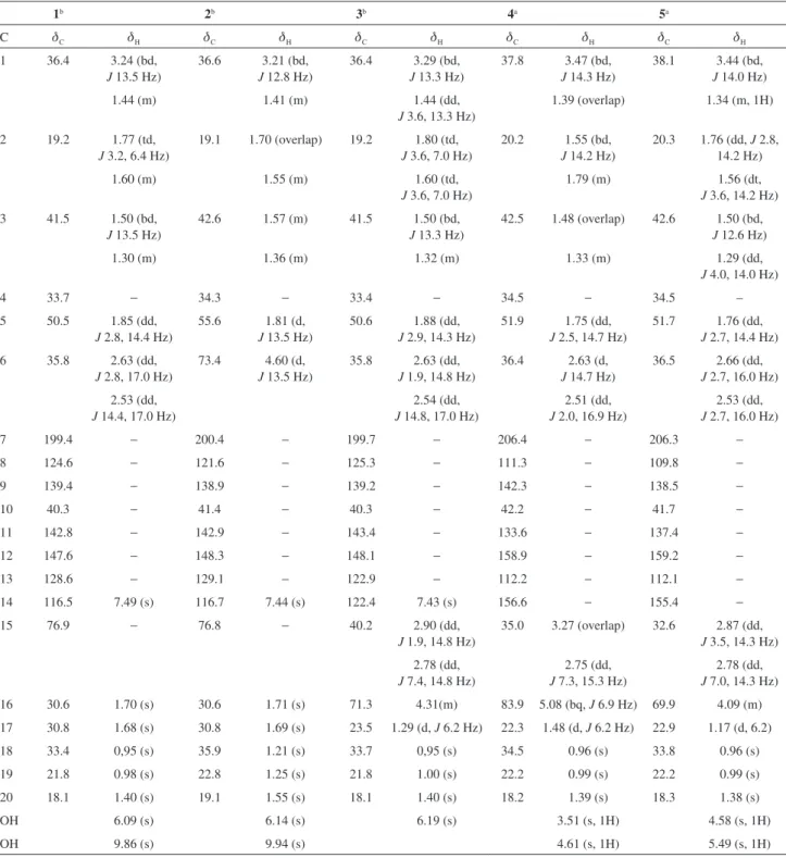
Figure 1. Structures of the diterpenes 1-9 isolated from H. crassifolia.
H3C
CH3 CH3 H O H HO OH CH3 CH3 OH R 1 2 3 4 5 6 7 8 9 10 11 12 13 14 15 16 17 18 19 20
1R = H
2R = OH
H3C
CH3 CH3 H O R HO OH CH3 OH 1 2 3 4 5 6 7 8 9 10 11 12 13 14 15 16 17 18 19 20
5R = OH 3R = H
O
H3C
CH3 CH3 H O OH HO CH3 1 2 3 4 5 6 7 8 9 10 11 12 13 14 15 16 17 18 19 20 4 A B C D
H3C
CH3 CH3 H OH CH3 CH3 R1 R2
6R1= R2= H
7R1= R2= O 1 2 3 4 5 6 7 8 9 10 11 12 13 14 15 16 17 18 19 20
H3C
CH3 CH3 H CH2 O CH3 CH3 R1 R2
8R1= R2= O
9R1= H; R2= OH 1 2 3 4 5 6 7 8 9 10 11 12 13 14 15 16 17 18 19 20
Figure 2. Key HMBC correlations observed for the compounds 1-4.
O
H3C
CH3 CH3 H HO H H H OH 1 CH3 CH3 OH 7 9 12 14 15 16 17 O
H3C
CH3 CH3 H HO H H HO OH CH3 CH3 OH 2 4 5 6 7 9 12 14 15 16 17 O
H3C
CH3 CH3 H HO H H H 3 OH CH3 HO H H 7 9 12 13 14 15 16 17 O H3C
assigned as α-equatorial on the basis of the coupling constants of H-5 [dH 1.81 (d, J 13.5 Hz)] and H-6 [dH 4.60
(d, J 13.5 Hz)], a typical axial-axial hydrogen coupling of cyclohexane rings. The changes in the 1H NMR spectrum
of 2, relatively to 1, are all in agreement with the above discussion. The axial oxymethine hydrogen attached to the
α-carbon to the carbonyl appeared at dH 4.60 (d, J 13.5 Hz),
while all methyls attached to quaternary carbons underwent a light deshielding. Accordingly with the afore mentioned spectral data, the structure of 2 was characterized as the new diterpene 6α ,11,12,15-tetrahydroxy-8,11,13-abietatrien-7-one.
Compound 3 was obtained as a yellow resin. The molecular formula C20H28O4 was determined by HR-ESI-MS,
based on the quasi-molecular ion at m/z 333.2064 [M + H]+
(calcd. for C20H29O4 m/z 333.2060). The NMR data of
compound 3 showed remarkable similarities with those of compound 1 (see Table 1). The major diferences account for an oxymethine at C-16 (dC 71.3; dH 4.31 m) and an
extra diastereotopic methylene at C-15 [dC 40.2; dH 2.90
(dd, J 1.9, 14.8 Hz, H-15a) and dH 2.78 (dd, J 7.4, 14.8 Hz,
H-15b)]. The singlets at dH 1.70 (Me-16) and 1.68 (Me-17)
correspondent to the geminal methyls of the benzene side chain of 1 are missing, while a doublet for a primary methyl C-17 [dC 23.5; dH 1.29 (d, J 6.2 Hz)] appears on
the 1H NMR. This information is in agreement with a
rearrangement of the aromatic side chain of compound 1. The 13C NMR data is totally in accordance with the
suggested change, appearing the extra methylene (C-15) at dC 40.2, the oxymethine (C-16) at dC 71.3, and the
methyl (C-17) at dC 23.5. The other expected changes were
the shielding of C-13 (dC 122.9) due to the replacement
of the deshielding β-hydroxy and β-methyl effect on compound 1, for the shielding γ-effect of both groups on compound 3. The release of the crowding steric hindrance of the branched side chain on compound 1 through the rearrangement for a normal chain on compound 3 also affects the chemical shift of C-14 (dC 122.4), now with a
remarkable deshielding effect (∆dC = 5.9). The spectral data
discussed above are consistent with a 17(15→16)-abeo -abietane diterpenoid.32,35 The skeleton of 3 was confirmed
through a detailed analysis of the HMBC spectrum by long-range correlations of the aromatic proton at dH 7.43
(H-14) with the carbons at dC 148.1 (C-12, 3J), 139.2 (C-9, 3J) and 40.2 (C-15, 3J), and with the carbonyl carbon in
dC 199.7 (C-7, 3J) (Figure 2). Thus, compound 3 was
identified as a new 17(15→16)-abeo-abietane diterpene, the 11,12,16-trihydroxy-17(15→16)-abeo -abieta-8,11,13-trien-7-one.
Compound 4 [α]D20 +26.79º (c 0.24, CHCl3) was isolated
as a yellow solid (mp 195.7-197.7 °C). The molecular
formula, C20H26O4, was established after HR-ESI-MS
analysis based on the quasi-molecular ion at m/z 331.1889 [M + H]+ (calcd. C
20H27O4m/z 331.1909). Compound 5,
already known, showed very similar spectroscopic data to those of compound 4 (see Table 1). Its HR-ESI-MS
quasi-molecular ion at 347.1879 [M − H]−, 18 Da higher
than that of compound 4, suggested that compound 4 could be a dehydrated derivative. The IR spectrum of 4 exhibited absorption bands for hydroxyl groups at 3382 cm−1 and
a conjugated ketone carbonyl group at 1639 cm−1. The 13C CPD NMR spectrum exhibited 20 signals, that after
the 1H,13C-HSQC spectrum analysis allowed to determine
the presence of an aryl ketone carbonyl, chelated to the hydroxyl at C-14, at dC 206.4, six sp2 carbons at the region
of dC111.3-158.9, one very deshielded oxymethine carbon
at dC 83.9, and 12 sp3 saturated carbons (dC 18.2-51.9)
(Table 1). Analysis of the 1H NMR spectrum did not
show any aromatic proton, but three singlets of methyl groups attached to quartenary carbons at dH 0.96 (Me-18),
0.99 (Me-19) and 1.39 (Me-20), and a methyl doublet at dH 1.48 (d, J 6.2 Hz, Me-17). In addition, an oxymethine
proton atdH 5.08 (bq, J 6.9 Hz, H-16) and a diastereotopic
methylene at dH 3.27 (H-15a); 2.75 (dd, J 15.3, 7.3 Hz,
H-15b) have been observed. The main difference was the deshielding of C-16 (dC 69.9 in 5) to dC 83.9 due the
formation of an α-methyldihydrofuran ring condensed with the fully substituted benzene ring at the positions C-12 and C-13. The appearance of a chelated hydroxy at dH 13.44
in the 1H NMR spectrum (CDCl
3) of compound 4 (see
Supplementary Information (SI) section) evidenced that the ring closure has been done through the C-12 hydroxy. The substitution pattern of the C aromatic ring was definitively established from the HMBC analysis by long-range correlations of the methylene protons at dH 3.27 (H-15a)
and 2.75 (H-15b) with the carbons at dC 158.9 (C-12, 3J), 112.2 (C-13, 2J) and 22.3 (C-17, 3J). In addition, the
correlations of the methylene protons at dH 2.63-2.51 (H-6)
with the carbons at dC 34.5 (C-4, 3J), 206.4 (C-7, 2J), 111.3
(C-8, 3J) and 42.2 (C-10, 3J) (Figure 2) were also observed.
To the C-16 stereogenic center was attributed the same relative stereochemistry as that in either teuvincenone A or E (αH, βMe), taking in consideration the similar chemical shifts and coupling constant values of the 2H-15, H-16 and Me-17 protons,35 as well as the carbon-13 chemical
Ulubelen et al.29 to which the structure suggested is the
same asthe one proposed for 4, did not show a good matching. For instance, the carbonyl chemical shift at dC 185.6 is not compatible with the C-7 aryl ketone carbonyl
of compound 4 (dC 206.4), once other aryl ketones described
in the literaturehave carbon ressonances of approximately dC 205.0.32,36,37 It appears that the carbonyl chemical shift at
dC 185.6 previously reported,29 is more compatible with a
cross conjugated aryl ketone (ca. dC 188.0)36 or α-hydroxy
cross conjugated aryl ketones (ca. dC 183.0).29,36 In addition,
several other inconsistencies can be pointed out, for example, the 13C NMR chemical shifts of C-4 (d
C 37.2),
C-6 (dC 29.7), C-8 (dC 107.6) and C-10 (dC 35.4).29 Thus,
the NMR data assignment previously published for villosin A should be revised, or an alternative structure should be considered. According to the analysis described above, the structure of 4 was assigned as (16S)-12,16-epoxy -11,14-dihydroxy-17(15→16)-abeo-abieta-8,11,13-trien-7-one.
The remaining compounds were characterized as incanone (5),32 ferruginol (6),30,38 sugiol (7),39
11-oxomanoyloxide (8)40 and 11β-hydroxymanoyloxide
(9),33 by extensive 1D and 2D NMR spectroscopy analyses
and by comparison of the spectral data with those reported in literature.
Conclusions
The unprecedent phytochemical analysis of
Hyptis crassifolia, a herb wildly dispersed through the neighborhood of Mucujê, a small town at Chapada Diamantina, BA, Brazil, has led to the isolation of four abietane diterpenes, two of which are new, 1 and 2. This is in agreement with the chemotaxonomic profile of the genus Hyptis already reported in the literature. On the other hand, rearranged abietanes, two of which are new (3 and 4), are being reported for the first time from Hyptis. The known labdane diterpenes 8 and 9, isolated from other genera like Salvia33 and Kyllinga,40 have not been reported
from Hyptis previously. In addition, the NMR data of the rearranged abietane 4, previously identified as villosin A,29
was reassigned.
Supplementary Information
Supplementary data are available free of charge at http://jbcs.sbq.org.br as PDF file.
Acknowledgments
The authors are grateful to CNPq, CAPES, PRONEX, FUNCAP, FINEP for the fellowships and financial support.
References
1. Judd, W. S.; Campbell, C. S.; Kellogg, E. A.; Stevens, P. F.;
Plant Systematics: a Phylogenetic Approach, 2nded.; Sinauer
Associates Inc:Sunderland, 1999.
2. Falcão, D. Q.; Menezes, F. S.; Rev. Bras. Farmacogn.2003, 84, 69.
3. Trindade, R. C.; Barbosa Júnior, A. M.; Santos, P. O.; Costa, M. J. C.; Alves, J. A. B.; Nascimento, P. F. C.; Melo, D. L. F. M.; Blank, A. F.; Arrigoni-Blank, M. F.; Alves, P. B.; Nascimento, M. P. F.; Quim. Nova 2008, 31, 1648.
4. Harley, R. M.; Vanzolini, P. E.; Heyer, W. R.; An. Acad. Bras. Cienc.1988, 71.
5. Deng, Y. E.; Balunas, M. J.; Kim, J.; Lantvit, D. D.; Chin, Y.; Chai, H.; Sugiarso, S.; Kardono, L. B. S.; Fong, H. H. S.; Pezzuto, J. M.; Swanson, S. M.; Carcache, E. J. B.; Kinghorn, A. D.; J. Nat. Prod.2009, 72, 1165.
6. Pereda-Miranda, R.; Delgado, G.; J. Nat. Prod.1990, 53, 182. 7. Novelo, M.; Cruz, J. G.; Hernandez, L.; Pereda-Miranda, R.;
Chai, H.; Mar, W.; Pezzuto, J. M.; J. Nat. Prod.1993, 56, 1728. 8. Kuhnt, M.; Rimpler, H.; Heinrich, M.; Phytochemistry1994,
36, 485.
9. Pereda-Miranda, R.; Hernandez, L.; Villavicencio, M. J.; Novelo, M.; Ibarra, P.; Chai, H.; Pezzuto, J. M.; J. Nat. Prod. 1993, 56, 583.
10. Boalino, D. M.; Connolly, J. D.; McLean, S.; Reynolds, W. F.; Tinto, W. F.; Phytochemistry2003, 64, 1303.
11. Araújo, E. C. C.; Lima, M. A. S.; Silveira, E. R.; Magn. Reson. Chem.2004, 42, 1049.
12. Araújo, E. C. C.; Lima, M. A. S.; Nunes, E. P.; Silveira, E. R.;
J. Braz. Chem. Soc.2005, 16, 1336.
13. Porter, R. B. R.; Biggs, D. A. C.; Reynolds, W. F.; Nat. Prod. Commun.2009, 4, 15.
14. Ohsaki, A.; Kishimoto, Y.; Isobe, T.; Fukuyama, Y.; Chem. Pharm. Bull.2005, 53, 1577.
15. Kabouche, A.; Kabouche, Z.; Ozturk, M.; Kolak, U.; Topçu, G.; Food Chem.2007, 102, 1281.
16. Lee, W. S.; Kim, J.; Han, J.; Jang, K. C.; Sok, D.; Jeong, T.;
J. Agric. Food Chem.2006, 54, 5369.
17. Kolak, U.; Kabouche, A.; Öztürk, M.; Kabouche, Z.; Topçu, G.; Ulubelen, A.; Phytochem. Anal.2009, 20, 320.
18. Yang, Z.; Kitano, Y.; Chiba, K.; Shibata, K.; Kurokawa, H.; Doi, Y.; Arakawa, Y.; Tada, M.; Bioorg. Med. Chem.2001, 9, 347. 19. Tada, M.; Kurabe, J.; Yoshida, T.; Ohkanda, T.; Matsumoto, Y.;
Chem. Pharm. Bull.2010, 58, 818.
20. Smith, E. C. J.; Wareham, N.; Zloh, M.; Gibbons, S.; Phytochem. Lett.2008, 1, 49.
21. Yang, J.; Du, X.; He, F.; Zhang, H.; Li, X.; Su, J.; Li, Y.; Pu, J.; Sun, H.; J. Nat. Prod.2013, 76, 256.
Henriques, J. A. P.; Pessoa, C.; Costa-Lotufo, L. V.; Food Chem.
Toxicol.2008, 46, 388.
23. Li, S.; Wang, P.; Deng, G.; Yuan, W.; Su, Z.; Bioorg. Med. Chem.
Lett.2013, 23, 6682.
24. Wang, W.; Xiong, J.; Tang, Y.; Zhu, J.; Li, M.; Zhao, Y.; Yang, G.; Xia, G.; Hu, J.; Phytochemistry2013, 89, 89. 25. Yang, L.; Li, L.; Huang, S.; Pu, J.; Zhao, Y.; Ma, Y.; Chen, J.;
Leng, C.; Tao, Z.; Sun, H.; Chem. Pharm. Bull.2011, 59, 1102. 26. Araújo, E. C. C.; Lima, M. A. S.; Montenegro, R. C.;
Nogueira, M.; Costa-Lotufo, L. V.; Pessoa, C.; Moraes, M. O.; Silveira, E. R.; Z. Naturforsch.2006, 61c, 177.
27. Achenbach, H.; Waibel, R.; Nkunya, M. H. H.; Weenen, H.;
Phytochemistry1992, 31, 3781.
28. Lima, K. S. B.; Pimenta, A. T. A.; Guedes, M. L. S.; Lima, M. A. S.; Silveira, E. R.; Biochem. Syst. Ecol.2012, 44, 240. 29. Ulubelen, A.; Topcu, G.; Olçal, S.; Phytochemistry1994, 37,
1371.
30. Marcos, I. S.; Beneitez, A.; Moro, R. F.; Basabe, P.; Díez, D.; Urones, J. G.; Tetrahedron2010, 66, 7773.
31. Gabetta, B.; Zini, G.; Danieli, B.; Phytochemistry1989, 28, 859.
32. Gao, J.; Han, G.; Phytochemistry1997, 44, 759.
33. Topçu, G.; Tan, N.; Ulubelen, A.; Sun, D.; Watson, W. H.;
Phytochemistry1995, 40, 501.
34. Kawazu, K.; Inaba, M.; Mitsui, T.; Agr. Biol. Chem.1967, 31, 498.
35. Rodríguez, B.; Bruno, M.; La Torre, M. C.; Savona, G.; Prozzr, F.; Phytochemistry1990, 29, 2710.
36. Cañigueral, S.; Iglesias, J.; Sánchez-Ferrando, F.; Virgili, A.;
Phytochemistry1988, 27, 221.
37. Rodríguez, B.; Robledo, A.; Pascual-Villalobos, M. J.; Biochem. Syst. Ecol.2009, 37, 76.
38. Carvalho, M. S.; Baptistella, L. H. B.; Imamura, P. M.; Magn. Reson. Chem.2008, 46, 381.
39. Bajpai, V. K.; Kang, S. C.; J. Food Saf.2011, 31, 27. 40. Mahmout, Y.; Bessiere, J.; Dolmazon, R.; J. Agric. Food Chem.
1993, 41, 277.
Submitted: May 8, 2014