Influence of aerobic fitness on age-related lymphocyte DNA
damage in humans: relationship with mitochondria
respiratory chain and hydrogen peroxide production
Maria Paula Mota&Francisco M. Peixoto&Jorge F. Soares&Pedro A. Figueiredo& José C. Leitão&Isabel Gaivão&José A. Duarte
Received: 28 July 2009 / Accepted: 22 February 2010
# American Aging Association 2010
Abstract The aim of this study was to analyze the influence of aerobic fitness (AF) on age-related lym-phocyte DNA damage in humans, giving special attention to the role of the mitochondrial respiratory chain and hydrogen peroxide production. Considering age and AF (as assessed by VO2max), 66 males (19–59 years old) were classified as high fitness (HF) or low fitness (LF) and distributed into one of the following groups: young adults (19–29 years old), adults (30–39 years old), and middle-aged adults (over 40 years old). Peripheral lymphocytes obtained at rest
were used to assess DNA damage (strand breaks and formamidopyrimidine DNA glycosylase (FPG) sites through the comet assay), activity of mitochondrial complexes I and II (polarographically measured), and the hydrogen peroxide production rate (assayed by fluorescence). Results revealed a significant interaction between age groups and AF for DNA strand breaks (F=8.415, p=.000), FPG sites (F=11.766, p=.000), mitochondrial complex I activity (F=7.555, p=.000), and H2O2 production (F=7.500, p=.000). Except for mitochondrial complex II activity, the age variation of the remaining parameters was significantly attenuated by HF. Considering each AF level, an increase in DNA strand breaks and FPG sites with age (r=0.655, p=0.000, and r=0.738, p=0.000, respectively) was only observed in LF. Moreover, decreased mitochondrial complex I activity with age (r=−.470, p=.009) was reported in LF. These results allow the conclusion that high AF seems to play a key role in attenuating the biological aging process.
Keywords DNA damage . FPG sites . Oxidative stress . Mitochondrial respiratory chain . Aging . Exercise
Introduction
It is widely accepted that oxidative stress results in molecular damage, some of which accumulates with age, causing progressive physiological attrition with DOI 10.1007/s11357-010-9138-8
M. P. Mota (*)
:
J. F. Soares:
J. C. Leitão University of Trás-os-Montes and Alto Douro, Centro de Investigação em Desporto, Saúde e Desenvolvimento Humano, Vila Real, Portugale-mail: mpmota@utad.pt F. M. Peixoto
:
I. GaivãoUniversity of Trás-os-Montes and Alto Douro, Centro de Ciência Animal e Veterinária, Vila Real, Portugal
P. A. Figueiredo
Instituto Superior da Maia,
Centro de Investigação em Actividade Física, Saúde e Lazer,
Porto, Portugal J. A. Duarte
Faculty of Sports, Centro de Investigação em Actividade Física, Saúde e Lazer,
an increased susceptibility to disease and risk of death (Agarwal and Sohal 1994; Figueiredo et al. 2008; Lenaz et al. 2000; Ventura et al. 2002). Indeed, the age-related increased concentration of DNA-oxidized bases (Agarwal and Sohal 1994; Randerath et al. 1996) clearly connects oxidative stress with the aging process and supports the concept of the existence of DNA mutation accumulation throughout life in organs and systems (Beckman and Ames 1998a; Lin and Beal 2006), including the immune system (Miller 1996). Although different origins and mechanisms have been proposed to explain the occurrence of DNA mutations, it is possible that mitochondria play an important role in age-related immunodeficiency (Ross et al.2002). The supporters of this theory state that age-related oxidative damage to mitochondria results in a progressive reduction in mitochondrial bioenergetic capacity, leading to cellular energy deficits that compromise overall cellular functionality (Figueiredo et al. 2008; Judge and Leeuwenburgh 2007; Lenaz et al. 2000; Wallace 2005) and to an increase in reactive oxygen species (ROS) formation with oxidative stress and damage to cell macro-molecules such as DNA (Beckman and Ames 1998b; Wallace2005).
Apart from the age influence itself, it is widely described that chronic exercise reduces oxidative stress and damage, both by decreasing ROS produc-tion and increasing antioxidant capacity, and that it improves mitochondria efficiency in several organs and systems (Ascensão et al.2007; Starnes and Taylor 2007). Bearing this in mind, it is expected that exercise may counteract some of the undesirable effects of the aging process on the cardiovascular system as described by Ascensão et al. (2007), on skeletal muscle as described by den Hoed et al. (2008), and on the brain as described by Navarro and Boveris (2007). However, in regard to the immune system, little is known about the hypothetical influ-ence of chronic physical exercise on the age-related oxidative stress condition and associated mitochon-drial functionality in immune cells. Therefore, with this study, we searched for age and fitness-related changes in DNA damage and mitochondrial function-ality in the circulating lymphocytes of healthy sub-jects. To reach this purpose, we evaluated in males of different ages (from 19 to 59 years old) and with distinct levels of aerobic fitness (AF; estimated through maximal oxygen uptake) the degree of
DNA damage (through the comet assay), the rate of mitochondria ROS production (estimated by the rate of hydrogen peroxide production, H2O2), and the mitochondrial functionality (assessed by respiratory chain complex I and II activity). Based on data provided by other studies using different organs and tissues, we hypothesized that the fittest subjects should present lymphocytes with lower levels of ROS production and DNA damage paralleled with enhanced mitochondrial functionality.
Materials and methods
Subjects
Sixty-six healthy male volunteers between the ages of 19 and 59 years who were nonsmokers and who had different habitual physical activity habits (ranging from sedentary to highly and frequently active) participated in this study. Considering their chrono-logical age, subjects were included in one of the following groups: young adults (YAd, n=21, aged between 19 and 29 years), adults (Ad, n=18, aged between 30 and 39 years), and middle-age adults (MAAd, n=27, aged between 40 and 59 years). Based on individual maximal oxygen uptake (VO2max) and on the expected variation of VO2max with age, in each group subjects were further classified as high fitness (HF: n=13 YAd, n=9 Ad, and n=14 MAAd) or low fitness (LF: n=8 YAd, n=9 Ad, and n=13 MAAd), following the cut-off points proposed by Shvartz and Reibold (1990; those subjects with VO2max values considered good or better integrated the HF, while the remaining ones were included in the LF). Before participating in the study, all subjects were informed about the protocol, which was ap-proved by the local ethical committee, and signed an informed consent. Afterwards, the consenters under-went a medical examination in order to exclude pathologies or diseases.
Maximal oxygen uptake
To assess the AF of all subjects, VO2max was determined 1 week before blood sample collection via an open-circuit spirometry using a treadmill incremental exercise following the Bruce protocol (Bruce et al. 1973). The test started at 2.74 km h−1
(1.7 mph) at a gradient of 10%. At 3-min intervals, the incline of the treadmill increased by 2%, and the speed increased. At least three of the following criteria were attained to establish individual maximal oxygen consumption: (1) a plateau in oxygen con-sumption with increasing exercise intensity, (2) a respiratory exchange ratio of 1.10, (3) achievement of age-predicted maximal heart rate, and (4) perceived exertion of 18 on the Borg scale (Midgley et al. 2007). For safety reasons, heart rate was monitored during all exercise testing.
Blood collection and lymphocyte isolation
For the comet assay (single cell gel electrophoresis), between the hours of 9:00 and 10:00 a.m., ∼30 µl of capillary blood was taken from a finger prick of each subject and added to 1 ml of phosphate buffer solution (PBS) in a 1.5-ml Eppendorf tube. After gentle mixing, 100 µl of Histopaque 1077 (Sigma) was underlaid using a pipette. The sample was then centrifuged at 200×g for 3 min at 4°C. Lymphocytes were retrieved in 100 µl from just above the boundary between PBS and Histopaque using a pipette. One milliliter of PBS was added, and the sample was spun again. The supernatant was then removed as much as possible using a pipette, being cells on the pellet re-suspended in 280 µl of 1% low melting point agarose (Gibco) in PBS at 37°C.
To assess mitochondrial functionality and the rate of H2O2 production, 20 ml of peripheral venous blood was collected between 9:00 and 10:00 a.m. Blood lymphocytes were isolated following routine proce-dures of successive centrifugation in a Ficoll’s gradient, and the final protein concentration was quantified according to Bradford's method (Bradford1976).
DNA damage estimation with the comet assay
DNA strand breaks were measured using the comet assay, or the single cell gel electrophoresis assay. The suspended cells in low melting point agarose were transferred as two 70-µl drops to slides previously precoated with 1% normal melting point agarose (Gibco) in H2O to aid attachment of the gels. Two slides per person were made, one to measure basal DNA strand breaks and the other to identify 8-oxoguanines and additional altered purines through incubation with formamidopyrimidine DNA
glycosy-lase (FPG). This is a lesion-specific enzyme that detects this type of damage and creates a strand break during incubation after lysis, which increases its sensitivity as well as its specificity. Each drop on a slide was covered with an 18×18-mm cover slip and left at 4°C for 5 min. Cover slips were then removed from the slides and placed in a vertical jar with 1 ml Triton X-100 to 100 ml of lysis solution (2.5 M NaCl, 0.1 M EDTA, 10 mM Tris, pH 10, 4°C) and kept in the dark for 1 h. Slides were then washed in three changes of enzyme reaction buffer (40 mM HEPES, 0.1 MKCl, 0.5 mM EDTA, 0.2 mg/ml BSA, pH 8.0 with KOH, 4°C) in a staining jar for 5 min each time. After removing slides from the last wash, the excess liquid was dabbed off with absorbent paper. Fifty microliters of enzyme solution (or buffer alone, as a control) was placed onto gel and covered with a 22× 22-mm cover slip. Slides were put into a moist box (to prevent desiccation) and incubated at 37°C for 30 min for FPG activity. After this incubation, the slides were removed and gently placed (minus cover slips) on platforms in an electrophoresis tank, forming 1 or 2 complete rows, and immersed in an electro-phoresis solution (0.3 M NaOH and 1 mM EDTA) for 40 min (4°C). Then, electrophoresis was performed for 30 min at 25 V (constant voltage setting). After that, the slides were washed three times with neutralizing buffer (0.4 M Tris, pH to 7.5 with
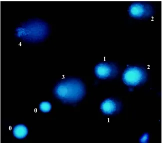
0 1 2 4 3 0 1 2
Fig. 1 Fluorescent micrograph of comet assay from lympho-cytes stained with ethidium bromide showing the different classes (0–4) of DNA damage
concentrated HCl) in a staining jar for 5 min at 4°C. The slides were then stored at room temperature. Slides were stained with ethidium bromide (2 µg/ml) immediately before visualization of comet DNA using a Carl Zeiss fluorescent microscope. Tail intensity was measured in each cell by the visual score method, classifying them into five categories representing different degrees of DNA damage, ranging from 0 (cell without observable migration and so without damage) to 4 (cell with intense observable migration and so intense damage; Fig.1). In each drop, 100 cells were observed; since each slide had two drops, the average of both drops was calculated, with the scores ranging from 0 (undamaged) to 400 (completely damaged; Collins 2004). The comet assay classifica-tion was performed in a blind fashion by an experienced researcher in the field. After at least a month, 9% of the slides (equivalent to six subjects) were visualized once again to test–retest reliability. Mitochondria oxidative activity
Oxygen utilization by intact lymphocytes (equivalent to 135 µg of protein) was measured polarographically in 0.5 ml of standard medium (pH 7.4) containing 0.3 M mannitol, 10 mM KCl, 5 mM MgCl2, 10 mM KH2PO4, and 0.25 mg bovine serum albumin with a Clark electrode in a water-jacketed cell at 37°C (Hansatech Instruments, Norfolk, UK). Intact cell oxidative activity was determined according to Miro et al. (1999) and Rustin et al. (1994). Oxidative rates (expressed as nanomoles oxygen/minute/milligrams lymphocyte protein) were assessed after permeabili-zation of the cellular membrane with digitonin (1%) and the addition of the following substrates: pyruvate (5 mM; complex I substrate) plus malate (1 mM), and succinate (20 mM; complex II substrate) in the presence of rotenone (4 µM) and ADP (0.4 mM) and malonate (20 mM). After the subtract addiction, the rate of oxygen consumption was measured for 6 min as the maximal oxygen consumption per minute registered for further data treatment. To quantify the specific contribution for oxygen con-sumption of electron transport chain complexes I and II, inhibitors for each substrate were further used (4 µM rotenone for pyruvate oxidation and 20 mM malonate for succinate oxidation) to determine the oxygen consumption measured during 6 min and the lowest average value per minute registered. The real
contribution of electron transport chain complexes I and II activity was then calculated by subtracting the values of oxygen consumption per minute obtained in the presence of the specific inhibitor from the rate of oxygen uptake assessed without complex inhibition.
Hydrogen peroxide assay
Hydrogen peroxide release from lymphocyte mitochon-dria into the overlying medium was assayed using a modification of the method described by Valletta and Berton (1987). This is based on the conversion of homovanillic acid (HVA) to its fluorescent dimer in the presence of H2O2and horseradish peroxidase (HRP). To measure mitochondrial H2O2production, the assay was performed in permeabilized lymphocytes, with mitochondrial respiration substrates for complex I (pyruvate/malate) and for complex II (succinate) in the presence of ADP. Assays were performed in the absence of mitochondrial respiration inhibitors and in the presence of inhibitors (1 µM rotenone, 20 mM malonate, and 10 µM antimycin A). Both reactions were performed in a reaction mixture containing 800 mM HVA and 6 U/ml HRP at 37°C.
After 15 min, the reaction was stopped with 2 ml of cold glycine NaOH (pH 12) containing 25 mM EDTA. The lymphocyte suspensions were then centrifuged at 5,000 rpm for 10 min at 4°C. The fluorescence of supernatants was measured at 312 nm as excitation and 420 nm as emission wavelengths in a spectrofluorimeter (Jasco FP 777). In each experiment, incubation was also performed with a control sample containing the reaction mixture alone (i.e., without lymphocytes) to correct for any spontaneous dimerization of HVA. The H2O2 concentrations used to establish standard curves were prepared by diluting a 30% H2O2 solution. Final dilutions were made in a glycine NaOH buffer. Peroxide generation was calculated using a standard curve of H2O2. Extracellular H2O2 levels were obtained as nanomoles H2O2/15 min/mg protein and were expressed as the difference of H2O2produced in the presence of inhibitors and the H2O2 produced in the absence of inhibitors.
Statistical analysis
Data were processed using SPSS software. After verifying the normality of the studied variables with the Kolmogorov–Smirnov test, means and standard
deviation (±SD) were used for further data analysis. An intraclass correlation coefficient was used to observe comet assay test–retest reliability, revealing a high reliability coefficient (r=0.93). Age group and AF effects for the quantified biomarkers in lympho-cytes were tested using a two-way factorial analysis of variance. The statistical significance for all multi-ple comparisons was adjusted with respect to the Tukey method. Pearson's r was used to find correla-tion between variables and age for each level of AF. p values less than 0.05 were considered significant.
Results
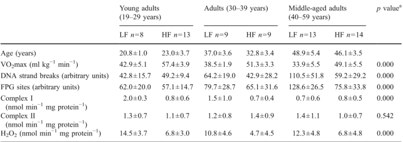
Data of age, VO2max, and quantified biomarkers in lymphocytes from subjects with either LF or HF in the three age groups are displayed in Table1.
As expected, the comparison between groups points out that VO2max decreases with age in both LF and HF subjects, as shown by the significant interaction between AF and age group (F=44.355, p=.000). Post hoc comparison revealed differences between YAd and Ad (p=0.022) as well as between YAd and MAAd (p=0.001).
With regard to DNA damage, the interaction between AF and age group was significant for both markers (F=8.415, p=.000 and F=11.766, p=.000 for DNA strand breaks and FPG sites, respectively).
Post hoc comparison revealed differences between MAAd and YAd (p = 0.002) as well as between MAAd and Ad (p=0.028). In the YAd group, the DNA strand breaks were somewhat higher in the HF subjects. However, in the two older groups, this variable was increasingly higher with age in the LF subjects. Our results also give evidence that DNA damage induced by oxidative stress (FPG sites) is higher in MAAd compared to YAd (p=0.000) and to Ad (p=0.027). In all age groups, FPG sites were also higher in subjects with lower AF.
The results of mitochondria complex I activity in LF and HF subjects evidenced a different variation between age groups: the interaction of AF and age group significance (F=7.555, p=.000). While in LF subjects higher mitochondrial complex I activity was observed in YAd—decreasing afterward with age until MAAd—in the HF group, it remained fairly stable across different age groups. In YAd and Ad groups, mitochondrial complex I activity was higher in LF subjects compared to HF subjects. Post hoc comparison revealed differences between YAd and MAAd (p=0.019). No significant effect of age group, AF, or interception of both factors in mitochondrial complex II activity was observed.
Our results revealed that mitochondria H2O2 production changed significantly with age and AF (F=7.500, p=.000), as evidenced by the higher values observed in the LF subjects of all age groups.
Table 1 Summary data of age, VO2max, and quantified biomarkers in lymphocytes from 66 men with low aerobic fitness (LF) and high aerobic fitness (HF) in different age groups
Young adults (19–29 years)
Adults (30–39 years) Middle-aged adults (40–59 years)
p valuea
LF n=8 HF n=13 LF n=9 HF n=9 LF n=13 HF n=14
Age (years) 20.8±1.0 23.0±3.7 37.0±3.6 32.8±3.4 48.9±5.4 46.1±3.5
VO2max (ml kg−1min−1) 42.9±5.1 57.4±3.9 38.5±1.9 51.3±3.3 33.9±5.5 49.1±5.5 0.000 DNA strand breaks (arbitrary units) 42.8±15.7 49.2±9.4 64.2±19.0 42.9±28.2 110.5±51.8 59.2±29.2 0.000 FPG sites (arbitrary units) 62.0±20.0 57.1±14.7 79.7±28.7 65.1±31.6 128.6±26.5 75.8±33.8 0.000 Complex I
(nmol min−1mg protein−1)
2.0±0.3 0.8±0.6 1.5±1.0 0.7±0.4 0.7±0.6 0.8±0.5 0.000 Complex II
(nmol min−1mg protein−1)
1.3±0.7 1.1±0.7 1.2±0.8 1.4±0.9 1.4±1.1 1.0±0.7 0.542 H2O2(nmol min−1mg protein−1) 14.5±3.7 6.8±3.0 10.8±4.6 4.7±4.5 12.3±4.8 6.8±4.8 0.000 Values are means ± SD
a
Relationship between variables and age according to aerobic fitness
In the complete sample, the association between age and the studied variables was analyzed for the LF and HF subjects independently.
VO2max was inversely correlated with age in both AF levels (r=−.663, p=.000 and r=−.578, p=.000 for LF subjects and HF subjects, respectively), as shown by the linear regression analysis depicted in Fig.2.
Bearing in mind age-related changes either in total lymphocyte DNA damage or oxidative DNA damage, our results pointed out that only in LF subjects did both variables increase significantly with age (r=.655, p=.000 and r=.738, p=.000, respectively). The linear
regression analysis of DNA damage markers with age can be seen in Figs.3and 4.
The results also revealed that mitochondrial com-plex I activity was inversely correlated with age in LF subjects, as shown by the respective linear regression analysis depicted in Fig.5. No correlation was found between age and mitochondrial complex II activity or H2O2production in subjects with different AF.
Discussion and conclusion
The present study reports an age-related increase in total DNA lymphocyte damage as well as oxidized DNA sites. Moreover, our results suggest that this
60 50 40 30 20 10 Age (yrs) 70,00 60,00 50,00 40,00 30,00 20,00 VO2max (ml/kg/min) R2=0.457 R2=0.334
Fig. 2 Linear regression between VO2max and age for both low fitness (circle, broken line) and high fitness (asterisk, solid line) subjects
60 50 40 30 20 10 Age (yrs) 200,00 150,00 100,00 50,00 0,00
DNA strand breaks (arbitr
ary units)
R2=0.024
R2=0.433
Fig. 3 Linear regression between DNA strand breaks and age for both low fitness (circle, broken line) and high fitness (asterisk, solid line) subjects
age-related DNA damage seems to be attenuated in subjects with higher AF, in which lymphocytes are characterized by a reduced ROS production.
The increased DNA damage with age observed in the present study is in accordance with other data obtained from different tissues in laboratory animals (Agarwal and Sohal 1994; Chen et al. 2007; Kregel and Zhang2007; Nakamoto et al.2007) and humans (Krajcovicova-Kudlackova et al.2008). Furthermore, the observed increase of FPG sites with age strongly suggests the contribution of oxidative stress to total DNA damage. However, though the reported correla-tion between DNA strand breaks and FPG sites with age in LF subjects is in accordance with the concept
of progressive damage accumulation in lymphocytes, in HF subjects, no correlation was found. This failed association between age and DNA damage might be the result of chronic exercise benefits in preventing this kind of molecular damage (Nakamoto et al. 2007; Radak et al. 2002,2007). Indeed, increasing evidence exists to suggest that chronic exercise attenuates and, in some cases, acutely reverses age-associated dysfunc-tion in several cell types (ACSM1998; Chakravarty et al. 2008; Courneya and Karvinen 2007; Kohut and Senchina 2004; Kruk 2007; Radak et al. 2002). With regard to oxidative stress, these benefits seem to rely either on increased mitochondria functionality, the major source of ROS production, or on augmented
60 50 40 30 20 10 Age (yrs) 150,00 100,00 50,00 0,00
FPG sites (arbitrary units)
R2=0.104
R2=0.553
Fig. 4 Linear regression between formamidopyrimi-dine DNA glycosylase sites and age for both low fitness (circle, broken line) and high fitness (asterisk, solid line) subjects 60 50 40 30 20 10 Age (yrs) 3,00 2,00 1,00 0,00
Complex I (nmolO2/min/mg prot)
R2=0.223
R2=0.003
Fig. 5 Linear regression between mitochondrial complex I activity and age for both low fitness (circle) and high fitness (asterisk) subjects
antioxidants systems (Menshikova et al.2006), and on improved activity of repair enzymes (Nakamoto et al. 2007; Radak et al.2007,2009).
Our results revealed that the mitochondrial rate of H2O2production was lower in the HF subjects of all age groups, which might reflect an inferior state of oxidative stress. According to various authors (Cooke et al. 2003; Halliwell 1999), cellular H2O2 may be an important source of hydroxyl radicals, one of the major oxidants that continuously damage DNA, thus contributing significantly to the age-related development of main cancers such as those of the colon, breast, and prostate. In fact, ROS may increase the mutagenesis of nuclear proto-oncogenes (initiation) and drive nuclear replication (promotion), resulting in cancer (Wallace2005). Moreover, DNA polymerases and repair enzymes could also be damaged by reactive species, thereby decreasing the fidelity of replication and slowing down the repair of lesions (Halliwell 1999). On the other hand, exercise seems to significantly upregulate the activity of the repair enzyme OGG1, the human homologue of FPG in the nucleus (Nakamoto et al. 2007; Radak et al. 2007), explaining the lack of association found between age and accumulated DNA damage in the HF subjects of our study. Bearing this in mind, since in our study lymphocytes from subjects with high AF produced less mitochon-drial H2O2and have lower DNA damage, it could be speculated that this cell population might be more functional and effective in facing infectious diseases or detecting neoplastic cells, which is in accordance with other studies suggesting a protective effect from physical exercise against cancers (Friedenreich2001; Friedenreich and Orenstein2002).
Besides the increased production of mitochondrial H2O2in all age groups with LF, our results indicated an unexpected increased activity of mitochondria complex I in the YAd and Ad groups with LF compared to HF. This would suggest that in those subjects, lymphocytes have a higher respiratory rate compared to the HF subjects. Indeed, our results seem to contradict other findings obtained in the skeletal and cardiac muscle of mice and rats, where isolated mitochondrial ETC activity was higher in more active animals (Ascensão et al.2005; Venditti et al.1999). If at a first glance our data seem to conflict with the literature, it must be understood that they might reflect an adaptation to a continuous state of energy imbalance
existing in cell lymphocytes. In fact, the enhanced lymphocyte DNA damage observed in the LF subjects leads to mitochondrial heteroplasmy (Brierley et al. 1998; Wei and Lee 2002) and consequently to a cellular state of energy deficit, which in turn promotes the biogenesis of more dysfunctional mitochondria. In fact, the enhanced rate of mitochondrial H2O2 production observed in the YAd and Ad groups with LF reinforces the proposed idea of increased mito-chondrial dysfunction. In this sense, it may be speculated that lymphocytes from YAd and Ad with LF, compared to HF, could have a higher mitochon-drial density as a compensatory mechanism for the energy deficiency of each organelle (Lee and Wei 2005; Lenaz et al.2000; Ozawa1995).
With regard to the MAAd group, mitochondrial complex I activity in LF subjects became surprisingly close to that of the HF subjects. Taking into account the linear regression of mitochondrial complex I activity with age, it is noticeable that the AF plot regression lines intercept each other, resulting from the progressive reduction observed in LF subjects, while in HF subjects, this regression line seems not to be affected by age. This age-dependent decline in mitochondrial complex I activity in LF subjects might reflect the interaction between the rate of mitochondrial damage and the ability of organelle biogenesis. Indeed, apart from the expected mito-chondria biogenesis reduction with age (Hudson et al. 1998; Judge and Leeuwenburgh 2007), a signif-icant and positive correlation was observed between age and DNA damage in LF subjects, which is consistent with the higher rate of H2O2 production also noticed in LF subjects. Therefore, while at younger ages we may explain the increased activity of complex I by a compensatory increase in mito-chondria protein synthesis/biogenesis in response to DNA damage, in MAAd, the results clearly suggest a cell’s inability to compensate for accumulated DNA damage due in part to oxidative stress. On the other hand, in HF subjects, we may expect a higher mitochondria efficiency paralleled with an increase in antioxidant production (Ascensão et al. 2007; Parise et al. 2005), expressed by a reduced H2O2 production and lower DNA damage accumulation. These findings are consistent with the assumption that regular aerobic exercise may have beneficial effects on the immune system (Nieman and Pedersen 1999; Pedersen and Hoffman-Goetz2000).
In conclusion, our results revealed that lymphocyte DNA damage by both strand breaks and FPG sites is closely associated with age and with an increased state of mitochondria H2O2 production. High AF seems to play a key role in attenuating DNA-accumulated damage associated with age. Moreover, considering the importance of DNA damage in the mutagenic, carcinogenic, and aging processes, the contribution of regular exercise to human health promotion seems to become meaningful. Even so, and considering the reported age-related changes, future studies should rely on the thus far weak link between those changes and lymphocyte functionality. Acknowledgments We are thankful to Dr. Andrew Collins (University of Oslo, Oslo Norway) for providing the comet assay protocol and the enzyme FPG.
Grants This work was supported by a grant from the Fundação para a Ciência e Tecnologia (POCI/DES/62301/ 2004, POCI 2010, and FEDER).
References
ACSM (American College of Sports Medicine Position Stand) (1998) Exercise and physical activity for older adults. Med Sci Sports Exerc 30(6):992–1008
Agarwal S, Sohal RS (1994) DNA oxidative damage and life expectancy in houseflies. Proc Natl Acad Sci USA 91 (25):12332–12335
Ascensão A, Magalhaes J, Soares JM, Ferreira R, Neuparth MJ, Marques F et al (2005) Moderate endurance training prevents doxorubicin-induced in vivo mitochondriopathy and reduces the development of cardiac apoptosis. Am J Physiol Heart Circ Physiol 289(2):H722–H731
Ascensão A, Ferreira R, Magalhaes J (2007) Exercise-induced cardioprotection: biochemical, morphological and func-tional evidence in whole tissue and isolated mitochondria. Int J Cardiol 117(1):16–30
Beckman KB, Ames BN (1998a) The free radical theory of aging matures. Physiol Rev 78(2):547–581
Beckman KB, Ames BN (1998b) Mitochondrial aging: open questions. Ann N Y Acad Sci 854:118–127
Bradford MM (1976) Rapid and sensitive method for quanti-tation of microgram quantities of protein utilizing principle of protein-dye binding. Anal Biochem 72(1–2):248–254 Brierley EJ, Johnson MA, Lightowlers RN, James OF, Turnbull
DM (1998) Role of mitochondrial DNA mutations in human aging: implications for the central nervous system and muscle. Ann Neurol 43(2):217–223
Bruce RA, Kusumi F, Hosmer D (1973) Maximal oxygen intake and nomographic assessment of functional aerobic impair-ment in cardiovascular disease. Am Heart J 85(4):546–562 Chakravarty EF, Hubert HB, Lingala VB, Fries JF (2008) Reduced disability and mortality among aging runners—a 21-year longitudinal study. Arch Intern Med 168(15):1638–1646
Chen JH, Hales CN, Ozanne SE (2007) DNA damage, cellular senescence and organismal ageing: causal or correlative? Nucleic Acids Res 35(22):7417–7428
Collins AR (2004) The comet assay for DNA damage and repair: principles, applications, and limitations. Mol Bio-technol 26(3):249–261
Cooke MS, Evans MD, Dizdaroglu M, Lunec J (2003) Oxidative DNA damage: mechanisms, mutation, and disease. Faseb J 17(10):1195–1214
Courneya KS, Karvinen KH (2007) Exercise, aging, and cancer. Appl Physiol Nutr Metab 32(6):1001–1007 den Hoed M, Hesselink MKC, van Kranenburg GPJ, Westerterp
KR (2008) Habitual physical activity in daily life correlates positively with markers for mitochondrial capacity. J Appl Physiol 105(2):561–568
Figueiredo PA, Mota MP, Appell HJ, Duarte JA (2008) The role of mitochondria in aging of skeletal muscle. Bioger-ontology 9(2):67–84
Friedenreich CM (2001) Physical activity and cancer preven-tion: from observational to intervention research. Cancer Epidemiol Biomarkers Prev 10(4):287–301
Friedenreich CM, Orenstein MR (2002) Physical activity and cancer prevention: etiologic evidence and biological mechanisms. J Nutr 132(11 Suppl):3456S–3464S Halliwell B (1999) Oxygen and nitrogen are pro-carcinogens.
Damage to DNA by reactive oxygen, chlorine and nitrogen species: measurement, mechanism and the effects of nutrition. Mutat Res 443(1–2):37–52
Hudson EK, Hogue BA, Souza-Pinto NC, Croteau DL, Anson RM, Bohr VA et al (1998) Age-associated change in mitochondrial DNA damage. Free Radic Res 29(6):573–579 Judge S, Leeuwenburgh C (2007) Cardiac mitochondrial bioenergetics, oxidative stress, and aging. Am J Physiol Cell Physiol 292(6):C1983–C1992
Kohut ML, Senchina DS (2004) Reversing age-associated immunosenescence via exercise. Exerc Immunol Rev 10:6–41
Krajcovicova-Kudlackova M, Valachovicova M, Paukova V, Dusinska M (2008) Effects of diet and age on oxidative damage products in healthy subjects. Physiol Res 57 (4):647–651
Kregel KC, Zhang HJ (2007) An integrated view of oxidative stress in aging: basic mechanisms, functional effects, and pathological considerations. Am J Physiol Regul Integr Comp Physiol 292(1):R18–R36
Kruk J (2007) Physical activity in the prevention of the most frequent chronic diseases: an analysis of the recent evidence. Asian Pac J Cancer Prev 8(3):325–338 Lee HC, Wei YH (2005) Mitochondrial biogenesis and
mitochon-drial DNA maintenance of mammalian cells under oxidative stress. Int J Biochem Cell Biol 37(4):822–834
Lenaz G, D'Aurelio M, Pich MM, Geneva ML, Ventura B, Bovina C et al (2000) Mitochondrial bioenergetics in aging. Biochim Biophys Acta 1459(2–3):397–404 Lin MT, Beal MF (2006) Mitochondrial dysfunction and
oxidative stress in neurodegenerative diseases. Nature 443(7113):787–795
Menshikova EV, Ritov VB, Fairfull L, Ferrell RE, Kelley DE, Goodpaster BH (2006) Effects of exercise on mitochon-drial content and function in aging human skeletal muscle. J Gerontol A Biol Sci Med Sci 61(6):534–540
Midgley AW, McNaughton LR, Polman R, Marchant D (2007) Criteria for determination of maximal oxygen uptake: a brief critique and recommendations for future research. Sports Med 37(12):1019–1028
Miller RA (1996) The aging immune system: primer and prospectus. Science 273(5271):70–74
Miro O, Alonso JR, Jarreta D, Casademont J, Urbano-Marquez A, Cardellach F (1999) Smoking disturbs mitochondrial respira-tory chain function and enhances lipid peroxidation on human circulating lymphocytes. Carcinogenesis 20(7):1331–1336 Nakamoto H, Kaneko T, Tahara S, Hayashi E, Naito H, Radak
Z et al (2007) Regular exercise reduces 8-oxodG in the nuclear and mitochondrial DNA and modulates the DNA repair activity in the liver of old rats. Exp Gerontol 42 (4):287–295
Navarro A, Boveris A (2007) Brain mitochondrial dysfunction in aging: conditions that improve survival, neurological performance and mitochondrial function. Front Biosci 12:1154–1163
Nieman DC, Pedersen BK (1999) Exercise and immune function. Recent developments. Sports Med 27(2):73–80 Ozawa T (1995) Mitochondrial DNA mutations associated with
aging and degenerative diseases. Exp Gerontol 30(3–4): 269–290
Parise G, Phillips SM, Kaczor JJ, Tarnopolsky MA (2005) Antioxidant enzyme activity is up-regulated after unilater-al resistance exercise training in older adults. Free Radic Biol Med 39(2):289–295
Pedersen BK, Hoffman-Goetz L (2000) Exercise and the immune system: regulation, integration, and adaptation. Physiol Rev 80(3):1055–1081
Radak Z, Naito H, Kaneko T, Tahara S, Nakamoto H, Takahashi R et al (2002) Exercise training decreases DNA damage and increases DNA repair and resistance against oxidative stress of proteins in aged rat skeletal muscle. Pflugers Archiv 445(2):273–278
Radak Z, Kumagai S, Nakamoto H, Goto S (2007) 8-Oxoguanosine and uracil repair of nuclear and mitochondrial DNA in red and white skeletal muscle of exercise-trained old rats. J Appl Physiol 102(4):1696–1701
Radak Z, Atalay M, Jakus J, Boldogh I, Davies K, Goto S (2009) Exercise improves import of 8-oxoguanine DNA glycosylase into the mitochondrial matrix of skeletal muscle and enhances the relative activity. Free Radic Biol Med 46(2):238–243
Randerath K, Randerath E, Filburn C (1996) Genomic and mitochondrial DNA alterations with aging. In: Schneider EL, Rowe JW (eds) Handbook of the biology of aging, 4th edn. Academic Press, New York, pp 198–209
Ross OA, Hyland P, Curran MD, McIlhatton BP, Wikby A, Johansson B et al (2002) Mitochondrial DNA damage in lymphocytes: a role in immunosenescence? Exp Gerontol 37 (2–3):329–340
Rustin P, Chretien D, Bourgeron T, Gerard B, Rotig A, Saudubray JM et al (1994) Biochemical and molecular investigations in respiratory-chain deficiencies. Clin Chim Acta 228(1):35–51
Shvartz E, Reibold RC (1990) Aerobic fitness norms for males and females aged 6 to 75 years: a review. Aviat Space Environ Med 61(1):3–11
Starnes JW, Taylor RP (2007) Exercise-induced cardioprotec-tion: endogenous mechanisms. Med Sci Sports Exerc 39 (9):1537–1543
Valletta EA, Berton G (1987) Desensitization of macrophage oxygen-metabolism on immobilized ligands: different effect of immunoglobulin-G and complement. J Immunol 138(12):4366–4373
Venditti P, Masullo P, Di Meo S (1999) Effect of training on H (2)O(2) release by mitochondria from rat skeletal muscle. Arch Biochem Biophys 372(2):315–320
Ventura B, Genova ML, Bovina C, Formiggini G, Lenaz G (2002) Control of oxidative phosphorylation by complex I in rat liver mitochondria: implications for aging. Biochim Biophys Acta 1553(3):249–260
Wallace DC (2005) Mitochondria and cancer: warburg addressed. Cold Spring Harb Symp Quant Biol 70:363– 374
Wei YH, Lee HC (2002) Oxidative stress, mitochondrial DNA mutation, and impairment of antioxidant enzymes in aging. Exp Biol Med (Maywood) 227(9):671–682