André Filipe Mendes Leite Moreira
Cardiac response to acute stretch:
Literature review and a
new diastolic paradigm
Mestrado Integrado em Medicina Área: Fisiologia Tipologia: Dissertação Trabalho efetuado sob a Orientação de: Prof. Doutor André Pedro Leite Martins Lourenço E sob a Coorientação de: Prof. Doutora Inês Maria Falcão Sousa Pires Marques Trabalho organizado de acordo com as normas da revista: Frontiers in Physiology (ISSN: 1664-042X)
Literature review and a
new diastolic paradigm
Eu, André Filipe Mendes Leite Moreira, abaixo assinado, nº mecanográfico 201002479, estudante do 6º ano do Ciclo de Estudos Integrado em Medicina, na Faculdade de Medicina da Universidade do Porto, declaro ter atuado com absoluta integridade na elaboração deste projeto de opção.
Neste sentido, confirmo que NÃO incorri em plágio (ato pelo qual um indivíduo, mesmo por omissão, assume a autoria de um determinado trabalho intelectual, ou partes dele). Mais declaro que todas as frases que retirei de trabalhos anteriores pertencentes a outros autores, foram referenciadas, ou redigidas com novas palavras, tendo colocado, neste caso, a citação da fonte bibliográfica.
Faculdade de Medicina da Universidade do Porto, 21/03/2016
NOME
André Filipe Mendes Leite Moreira
NÚMERO DE ESTUDANTE DATA DE CONCLUSÃO
201002479 julho de 2016
DESIGNAÇÃO DA ÁREA DO PROJECTO Fisiologia
TÍTULO DISSERTAÇÃO/MONOGRAFIA (riscar o que não interessa)
Cardiac response to acute stretch: Literature review and a new diastolic paradigm ORIENTADOR
André Pedro Leite Martins Lourenço
COORIENTADOR (se aplicável)
Inês Maria Falcão Sousa Pires Marques
ASSINALE APENAS UMA DAS OPÇÕES:
É AUTORIZADA A REPRODUÇÃO INTEGRAL DESTE TRABALHO APENAS PARA EFEITOS DE INVESTIGAÇÃO, MEDIANTE DECLARAÇÃO ESCRITA DO INTERESSADO, QUE A TAL SE COMPROMETE.
É AUTORIZADA A REPRODUÇÃO PARCIAL DESTE TRABALHO (INDICAR, CASO TAL SEJA NECESSÁRIO, Nº MÁXIMO DE PÁGINAS, ILUSTRAÇÕES, GRÁFICOS, ETC.) APENAS PARA EFEITOS DE INVESTIGAÇÃO, MEDIANTE DECLARAÇÃO ESCRITA DO INTERESSADO, QUE A TAL SE COMPROMETE.
DE ACORDO COM A LEGISLAÇÃO EM VIGOR, (INDICAR, CASO TAL SEJA NECESSÁRIO, Nº MÁXIMO DE PÁGINAS, ILUSTRAÇÕES, GRÁFICOS, ETC.) NÃO É PERMITIDA A REPRODUÇÃO DE QUALQUER PARTE DESTE TRABALHO.
Faculdade de Medicina da Universidade do Porto, 21/03/2016
Assinatura conforme cartão de identificação:
Para a minha mãe, por me ter tornado no homem que sou hoje. Para a Mariana, por me tornar no homem que serei amanhã.
Cardiac Response to Acute Stretch:
Literature Review and a New Diastolic Paradigm
Abstract1 2
Myocardial stretch, as result of acute hemodynamic overload, is one of the most frequent
3
challenges to the heart and the ability of the heart to intrinsically adapt to it is essential to
4
prevent circulatory congestion. In a literature review, the historical background, the
5
currently known mechanisms, as well as the gaps in the understanding of this
6
physiological response are highlighted. The systolic adaptation to stretch is well known
7
for over 100 years, being dependent on an immediate increase in contractility – known as
8
the Frank-Starling mechanism – and a further progressive potentiation – the slow force
9
response. On the other hand, its diastolic counterpart remains largely unstudied.
cGMP-10
related pathways are activated upon stretch and its downstream effector PKG is known
11
to phosphorylate titin, a major determinant of myocardial stiffness. Therefore, we aimed
12
to investigate the role of these pathways on decreasing stiffness during acute loading via
13
titin phosphorylation in healthy and diseased hearts. Left ventricle (LV) of intact rat
14
hearts, strips dissected from the LV or right atrium of cardiac surgery patients and rabbit
15
papillary muscles were acutely stretched. In cardiac surgery patients, LV pressure and
16
volume were recorded before and 15 minutes after volume overload (VO). Effect of VO
17
was also assessed in sham and TAC rats. After an immediate increase in response to
18
stretch/VO, diastolic pressure and passive tension significantly decreased (≈30-40%) over
19
the subsequent 15 min in all animal species and experimental preparations. This effect
20
was significantly blunted by PKG inhibition and combined natriuretic peptide receptor
21
inhibition and NO scavenging and was not observed in TAC hearts. Stretching skinned
22
cardiomyocytes did not decrease passive tension but it was ≈60% lower in those extracted
23
from stretched hearts, in which it increased with protein phosphatase but not PKG
24
incubation. Titin phosphorylation increased markedly after acute myocardial stretch in
25
human and rabbit myocardium. Hypophosphorylation and blunted response to VO was
26
measured in TAC myocardium. These results point to a novel mechanism of diastolic
27
adaptation to acute overload associated with increased myocardial compliance that is
28
abolished in the hypertrophic heart. Decreased stiffness is associated with titin
29
phosphorylation, in which PKG plays a central role.
30 31
Keywords: cardiac function, myocardial stretch, neurohumoral adaptation, titin, 32
diastole, heart failure, frank-starling mechanism, slow force response. 33
PART ONE: LITERATURE REVIEW 34 35 Introduction 36 37
The heart has a central role in the maintenance of cardiovascular homeostasis, which
38
requires the ability to continuously adapt its function to different hemodynamic
39
conditions.
40 41
One of the most frequent challenges to myocardium is acute stretch, as result of acute
42
hemodynamic overload. Acute myocardial stretch can be observed in various
43
physiological and pathophysiological conditions (e.g. exercise, myocardial ischemia,
44
hypertensive crises, valvular diseases and heart failure). For example, at the start of
45
aerobic exercise (figure 1), the pumping action of skeletal muscle contraction increases
46
the venous return, leading to cardiac chamber dilation and acute myocardial stretch
47
(Nobrega et al., 1995). This increase in end-diastolic volume leads to an increase in end
48
diastolic pressure that, in the absence of adequate response, would lead to pulmonary and
49
systemic congestion.
50 51
Being such a common challenge, one can expect the heart to have adequate intrinsic
52
physiologic mechanisms to respond to this stimulus. The adequate physiological response
53
to the acute increase in end-diastolic pressure must include: activation of stretch sensing
54
molecules (the sensing organ), intermediary mechanisms (afferent, integrator and efferent
55
pathways) and effector mechanisms that would ultimately allow the heart to reduce its
56
increased end-diastolic pressure (figure 1). This could, in theory, be achieved by two
57
different mechanisms:
58
- A systolic adaptation: an increase in contractility, leading to an increased
59
ejection volume and, consequently, a decreased end diastolic volume.
60
- A diastolic adaptation: an increase in cardiac compliance, which would allow
61
accommodating more blood at inferior end diastolic pressures.
62
Both adaptations would increase cardiac output, which would also be appropriate to offset
63
the increase in venous return.
64 65
The first described mechanism of cardiac adaptation to an acute hemodynamic overload
66
has been known for about a century (Katz, 2002). An increase in either venous return or
67
aortic resistance leads to an increased end-diastolic volume and to an immediate increase
68
in contractility and stroke volume. This response is presently known as Frank-Starling
69
Mechanism (FSM) due to the contributes of Ernest H. Starling and Otto Frank to its
70
description and is mainly attributed to enhanced myofilamental responsiveness to Ca2+, a
71
phenomenon highly dependent on the tension developed by titin (Patterson and Starling,
72
1914;Frank, 1959;Castro-Ferreira et al., 2011). Given its relevance for the cardiovascular
73
homeostasis it is also known as the “law of the heart”.
74 75
In 1912, von Anrep described that, after the initial change in muscle length and
76
contractility induced by clamping the heart outflow, it is observed a progressive and
time-77
dependent increase in force development that goes beyond the force immediately
78
achieved after stretch and is responsible for the return of end-diastolic volume back to its
79
original value (von Anrep, 1912). This second progressive increase in force development,
80
was demonstrated in vitro by Parmley and Chuck in 1973, being since then synonymously
81
called slow force response (SFR) (Parmley and Chuck, 1973).
83
Both mechanisms highlight a highly effective systolic adaptation to an acute
84
hemodynamic overload. A diastolic adaptation per se, although theoretically appropriate
85
to the underlying challenge, remains largely unknown.
86 87
In this paper, about 100 years after the initial publish of E. Starling about the relevance
88
of stretch to heart function, we intend to review the great progress in understanding of the
89
myocardial adaptation to acute stretch, as well as highlight the gaps in the understanding
90 of this mechanism. 91 92 Myocardial mechanosensing 93 94
Mechanosensors are structures capable of perceiving mechanical signals and activating
95
pathways that allow transduction of the signal into biochemical responses. The external
96
forces exerted on the myocardium are transmitted through the sarcolemma (and
97
associated adhesion structures: intercalated disks and costameres), cardiomyocyte
98
cytoskeleton and sarcomeric proteins. Concurrently, the sarcomeres generate forces that
99
propagate in the opposite direction. Several mechanosensor molecules in this mechanical
100
chain have been identified, being the best characterized ones located in Z line, titin
101
molecule, sarcolemma, intercalated disks and costameres.
102 103
Recent reviews (Takahashi et al., 2013;Buyandelger et al., 2014;Lyon et al., 2015)
104
addressed cardiac mechanotransduction in detail, particularly its relation to remodeling
105
and chronic adaptation. The precise role of mechanosensors in the acute response to
106
stretch is still mostly unknown, as few studies have directly addressed this issue.
107
Therefore, in this topic, we intend to emphasize the great diversity of mechanosensors in
108
the heart and highlight cellular components that, given their structure, mechanism of
109
action and activated signaling pathways, may potentially contribute to the response to
110
acute stretch (figure 2).
111 112
The Z line divides two adjacent sarcomeres and is traditionally regarded as an essential
113
element to the normal structural arrangement of the sarcomeres. However, its position
114
confers to the Z disk the distinct ability to sense the muscular tension, during either the
115
diastolic or the systolic period (Hoshijima, 2006). The Z line is composed not only of
116
overlapping actin filaments of adjacent sarcomeres tightly linked by α-actinin, but also of
117
numerous proteins bearing diverse signaling functions (Luther, 2009). Many of these
118
proteins may be relevant for response to acute stretch, including: Protein Kinase C (PKC)
119
ε (Robia et al., 2005), PKCδ (Disatnik et al., 1994), calcineurin (Frey et al., 2000) and
120
phosphodiesterase 5 (Senzaki et al., 2001).
121 122
Titin is a giant protein that spans the sarcomere from the Z-line to the M-line. Its
123
molecular structure makes this protein work as a bidirectional spring that determines the
124
myocardial passive tension (PT) at different lengths (Castro-Ferreira et al., 2011). In
125
addition to its structural and elastic function, titin also functions as a biomechanical sensor
126
sensing myocardial tension as well as the sarcomeric length. The central role of titin in
127
mechanosensing and its ability to trigger downstream signaling cascades is related to its
128
interactions with numerous structural proteins and signaling proteins at its M-band, Z
129
line, N2B, N2A and PEVK-domains (Linke and Kruger, 2010). Titin also features a
130
kinase domain near the carboxyl-terminal (located in the M-line region) (Mayans et al.,
131
1998), which may phosphorylate target proteins at specific sites, according to myocardial
stretching (Puchner and Gaub, 2010). Moreover, titin presents various phosphorylation
133
sites of its own at PEVK and N2B domains (Krüger and Linke, 2011), whose
134
phosphorylation alters PT conferred by titin, either decreasing or increasing it (Ahmed
135
and Lindsey, 2009). This mechanism confers titin a special role in mechanosensing,
136
allowing it to simultaneous function as a mechanosensor and a molecular target.
137
Therefore, it is tempting to hypothesize that upon an increase in acute PT, titin is capable
138
of initiating a compensatory mechanism that, ultimately, would lead to decreased titin’s
139
PT with consequent normalization of sarcomeric PT. It is important to highlight that, as
140
the main determinant of myocardial PT, modifications of titin phosphorylation status will
141
also modify the activity of remaining mechanosensors.
142 143
The sarcolemma separates the intracellular and extracellular environments. This structure
144
is teeming with different receptors subject to activation by extracellular mediators that set
145
different intracellular pathways in motion. However, several sarcolemma proteins can
146
also be activated by cellular stretch, initiating intracellular pathways without binding of
147
extracellular mediators (Storch et al., 2012). Stretch activated channels (SACs) are a
148
paradigmatic example of this type of activation in cytoplasmic membranes (Reed et al.,
149
2014). In cardiomyocytes, upon stretch, SACs are permeable to of sodium, potassium and
150
calcium, thereby modulating electrical and mechanical proprieties of myocardium. The
151
AT1 receptor, a G protein-coupled receptor, can also be directly activated by stretch
152
without the involvement of angiotensin II (Zou et al., 2004). This agonist-independent
153
activation may have a determinant role in the myocardial response to acute stretch. The
154
blockade of this receptor has been associated with a significantly blunted SFR (Heidkamp
155
et al., 2003).
156 157
The interaction of cardiomyocytes with neighboring cells (at intercalated discs) and with
158
the extracellular matrix (at costameres) is key to the maintenance of the structural
159
integrity of the myocardium, as well as the transmission of forces between cells (Kresh
160
and Chopra, 2011). Beyond this function, some components of intercalated discs and
161
costameres are known to be involved in mechanosensing (Samarel, 2005). At the level of
162
intercalated discs, the main structures capable of contributing to mechanosensing are
163
fascia adherens junctions (constituted by N-cadherin and associated multi-molecular
164
complex anchoring cytoskeletal actin) and desmosomes (composed of the cell adhesion
165
proteins desmoglein and desmocollin and associated proteins internally linked to the
166
intermediate filament desmin) (Sheikh et al., 2009). Both structures are known to
167
contribute to long-term cytoskeletal adaptive responses to different pathophysiological
168
forces (Lyon et al., 2015), but their importance in the acute response remains unknown.
169
Regarding costameres, one important component contributing to mechanosensing are
170
integrins. These heterodimeric transmembrane receptors located at costameres contribute
171
to the connection between extracellular matrix and the intracellular cytoskeleton, and
172
therefore to the transmission of mechanical signals to the cytoskeleton (Israeli-Rosenberg
173
et al., 2014). One well-known mediator of integrin mechanosensing is focal adhesion
174
kinase (FAK) (Domingos et al., 2002b). The signaling resultant of FAK activation is
175
proportional to the magnitude and duration of mechanical stretch (Katz et al., 2002). FAK
176
activity is also related to various signaling pathways potentially relevant to the acute
177
response to stretch, including ERK1/2 (Domingos et al., 2002a) and PKC ε (Heidkamp et
178
al., 2003). Another mediator that may be particularly important for the acute transduction
179
of integrin signaling is integrin-linked kinase (ILK). ILK appears to mediate
180
cardiomyocyte force transduction via regulation of the SERCA2A activity and
phosphorylation of phospholamban (Traister et al., 2014), which may contribute both to
182
the systolic and diastolic adaptation to acute stretch.
183 184
Although several potential mechanosensing molecules have been identified, the
185
connection between these molecules and stretch activated pathways remains elusive. This
186
lack of knowledge is even more pronounced when it comes to the acute activation of these
187
pathways by these molecules.
188 189
Furthermore, the interrelation between those sensors is complex, and the spread
190
synergism and crosstalk between the pathways activated downstream of those sensors
191
lead to a broad activation of signaling pathways that acutely modulate heart function,
192
which is probably crucial to the ability of myocardium to adapt to acute hemodynamic
193 overload. 194 195 Calcium-dependent Pathways 196 197
Calcium (Ca2+) is the central player in the biphasic systolic adaptation to myocardial
198
stretch. Besides its involvement in the FSM, an increase in the intracellular concentration
199
of Ca2+ underlies the SFR (figure 3).
200 201
Angiotensin-II (ANG-II) and endothelin (ET) are two peptide hormones (8 and 21 amino
202
acids long, respectively) that share some interesting features. Via binding to receptors
203
coupled to protein Gq and subsequent release of Ca2+ from intracellular stores and
204
activation of protein kinase C (PKC), they increase cardiac inotropism and have a pressor
205
effect on blood vessels (figure 3). Though they normally play a role in the physiological
206
regulation of blood pressure by altering salt and water balance and vasomotor tone
207
(especially in renal vessels), they have been implicated in the pathophysiology of
208
pulmonary and systemic hypertension and heart failure (Brunner et al., 2006;Mehta and
209
Griendling, 2007).
210 211
More recently, the study of the role of local angiotensin and endothelin systems in cardiac
212
function and pathophysiology has been prolific. These have been implicated in the
213
process of cardiac remodelling that takes place as a result of neurohumoral activation in
214
congestive heart failure (Brunner et al., 2006;De Mello and Frohlich, 2011) but they also
215
seem to be central in the modulation of the acute myocardial response to stretch,
216
contributing to the genesis of the FSM and the development of the SFR (Cingolani et al.,
217
2011). In fact, Sadoshima et al. first demonstrated that cultured neonatal cardiomyocytes
218
directly released ANG-II from intracellular vesicles in response to an acute 10 or 30
219
minute stretch, generating a concentration of around 1 nM in the culture medium
220
(Sadoshima et al., 1993). The positive inotropic response to acute stretch is similar to that
221
elicited by addition of exogenous ANG-II in amounts of the same order of magnitude and
222
can be blocked by inhibition of AT1 but not AT2 receptors (Caldiz et al., 2007). However,
223
this effect seems to depend on downstream release of ET, as inhibition of ET receptors,
224
as well as the ET converting enzyme, ablated both the SFR and the increase in
225
contractility that accompanies ANG-II administration but AT1 receptor blocking did not
226
prevent the positive inotropic effect of exogenous ET (Perez et al., 2003). Accordingly,
227
Anderson et al. reported that ET synthesis in myocytes increases following myocardial
228
stretch (Anderson et al., 2004).
Taking these results into account, along with their own, Cingolani’s group has since
230
described a complex autocrine/paracrine pathway involving changes in membrane ion
231
currents mediated by ANG-II and ET that sheds some light on the role played by these
232
molecules in the response to myocardial stretch (Cingolani et al., 2011). They propose
233
that the initial step in generating the SFR is an ANG-II dependent release of endogenous
234
ET by poorly described mechanisms that may involve activation of the mineralocorticoid
235
receptor by aldosterone or another non-identified ligand.
236 237
Although all three endothelin isoforms have equal positive inotropic potency when
238
exogenously administered, ET-3 is the most likely to be responsible for SFR in a
239
physiological setting (Ennis et al., 2005). The ET receptor then transactivates the
240
epidermal growth factor receptor (EGFR) through various pathways. The best described
241
one involves activation of matrix metalloproteinases and subsequent release of
heparin-242
binding epidermal growth factor (HBEGF), which binds to and activates EGFR. This
243
supposedly triggers an intracellular signalling pathway leading to increased production
244
and release of reactive oxygen species (ROS) by mitochondriae through opening of
245
mitochondrial KATP channels (Anderson et al., 2004;Villa-Abrille et al., 2010).
Redox-246
sensitive kinases ERK1/2 and p90rsk therefore increase their activity in response to stretch
247
and phosphorylate the regulatory domain of the sodium-proton exchanger 1 (NHE1),
248
increasing its activity and cytoplasmic [Na+].
249 250
This transporter has long been implicated in the development of the SFR and this is
251
thought to occur through modulation of ion electrochemical gradients that the increased
252
[Na+] provokes. In this setting, the sodium-calcium exchanger (NCX), which usually
253
extrudes calcium, reverses direction of transport, increasing the amplitude of Ca2+
254
transients and, consequently, contractile force (figure 3).
255 256
On the other hand, the increased Ca2+ transient, through favored actin-myosin interaction,
257
could result in alarming levels of muscle stiffness. Thus, it is not surprising that other
258
stretch activated mechanisms promote muscle relaxation by improving Ca2+ extrusion
259
from cytosol after myocardial contraction. In fact, there is a well described acute increase
260
in SERCA expression following hemodynamic overload (Kögler et al., 2006). Besides,
261
Ca2+ load following stretch binds to the several Ca2+ binding proteins in the heart, with
262
S100A1 being the most abundant member of the calcium-binding S100 protein family in
263
myocardial tissue (Duarte-Costa et al., 2014). S100A1-Ca2+ complex frees the actin from
264
titin, reducing the PT imposed by titin-actin interaction (Yamasaki et al., 2001). The
265
relative importance of these mechanisms in the passive properties of the heart following
266
stretch has not been studied in detail.
267 268
The complex loop involving ANG-II and ET still has gaps to be filled and seemingly
269
contradictory results exist in the literature. Our own results confirm the major role of AT1
270
receptors and the NHE-1 and NCX transporters in the development of SFR and the
271
positive inotropic effect of the external addition of ANG-II on myocardium in normal
272
conditions, which subsequently blunts SFR, presumably by saturating the contractile
273
reserve of the pathway in the absence of stretch. In addition, it points to a possible role of
274
PKC in maintaining the SFR during the late phase of acute stretch (Neves et al., 2013).
275
Interestingly, our group has also described the role of these peptides on the acute
276
modulation of diastolic function through direct effects on both relaxation velocity and
277
myocardial stiffness. Activation of ETA and AT1 receptors results in positive lusitropism
278
and decreased stiffness, while ETB and AT2 receptors promote negative lusitropism. As
in its systolic counterpart, AT1 receptor activation seems upstream to endothelin release
280
in the pathway that leads to faster myocardial relaxation, as this effect is dependent on
281
ETA receptor activity and an intact endocardial endothelium (Bras-Silva and
Leite-282
Moreira, 2006;Castro-Chaves et al., 2006). Concurrently, decreasing stiffness depends on
283
the activity of both NHE1 and PKC and on the integrity of the endocardial endothelium
284
(Leite-Moreira et al., 2003;Bras-Silva and Leite-Moreira, 2006;Leite-Moreira et al.,
285
2006).
286 287
Pathways involving Nitric Oxide and Cardiac Natriuretic Hormones 288
289
Cardiac stretch stimulates both endothelial cells and cardiomyocytes to produce nitric
290
oxide (NO) (Casadei and Sears, 2003). In endothelial cells, large amounts of NO are
291
produced by the endothelial nitric oxide synthase (NOS), after which it rapidly diffuses
292
to neighboring cardiomyocytes (figure 3). In the cardiomyocyte, NO is produced in
293
relatively low levels by both endothelial NOS and neuronal NOS (nNOS) (Petroff et al.,
294
2001;Khan et al., 2003).
295 296
As NO produced by endothelial cells diffuses through the cardiomyocyte, soluble
297
guanylate cyclase present in the cytoplasm is activated, leading to an increase in cGMP
298
concentration in the cytosolic compartment. The effect of cGMP is mediated through
299
protein kinase G (PKG), which is able to phosphorylate troponin I (TnI), rendering a
300
reduction of myofilamentary sensitivity to calcium, therefore increasing the rate of Ca2+
301
dissociation (Layland et al., 2005). NO produced by nNOS, which is located in the
302
sarcoplasmic reticulum membrane, simultaneously modulates the diastolic properties of
303
the heart through PKA-dependent phosphorylation of phospholamban (PLB) in a
cGMP-304
independent pathway (Zhang et al., 2008). The phosphorylation of PLB disinhibits the
305
sarco/endoplasmic reticulum Ca2+ ATPase (SERCA), which rapidly reuptakes free
306
cytoplasmic Ca2+. Taken together, NO mediated pathways, both cGMP-dependent and –
307
independent, hasten myocardial relaxation (positive lusitropism), prolonging the diastolic
308
time interval while reducing the PT of cardiomyocytes. These changes in myocardial
309
properties are crucial for the heart to accommodate increasing preloads, without
310
substantial changes in the intracardiac filling pressures (figure 3).
311 312
Besides altering the diastolic properties of the heart, NO also modulates its contractility.
313
NO-derived cGMP coexists in the cytoplasm with several members of the
314
phosphodiesterase (PDE) family. Proteins of this family are metallophosphohydrolases
315
that cleave cGMP and/or cAMP to 5’-GMP and 5’-AMP, inhibiting the activation of PKG
316
and Protein Kinase A (PKA), respectively (Zaccolo and Movsesian, 2007). Therefore,
317
although stretch induces an increase in the production of NO, the cytoplasmic
318
concentration of NO-derived cGMP is kept relatively low due to continuous hydrolysis
319
mediated by PDE5, located preferentially in Z-bands, in a cGMP-activated negative
320
feedback mechanism. An important crosstalk between cGMP and cAMP pathways
321
occurs, as low concentrations of cGMP inhibit PDE3, thus preventing the hydrolysis of
322
cAMP (Francis et al., 2010). Accumulated cytoplasmic cAMP activates PKA, which is
323
then responsible for the phosphorylation of several proteins involved in cardiac
324
contraction (figure 3). These include the sarcolemmal voltage-gated L-Type Ca2+
325
channels, SR Ca2+ release channel (ryanodine receptor, RyR2), PLB, the phosphatase 1
326
inhibitor, TnI and myosin binding protein C, all aiming to increase myocardial
327
contractility (Fischmeister et al., 2006). Interestingly, a prolonged activation of RyR2
328
could ultimately lead to SR Ca2+ depletion, and consequently to a paradoxal negative
inotropism; however, activation of SERCA, as previously stated, is able to replenish the
330
SR Ca2+ pool and maintain this cycle. RyR2 can also be opened via cyclic adenosine
331
diphosphate ribose, which is produced by a PKG mediated phosphorylation of adenosine
332
diphosphate ribosylcyclase (Willmott et al., 1996). NO can also exert an important effect
333
on the systolic properties of the myocardium per se in a cGMP-independent pathway. NO
334
produced by nNOS directly S-nitrosylates reactive thiol residues within RyR2, which also
335
contributes to an increased Ca2+ release (Wang et al., 2010). Taken together, NO mediated
336
pathways, both cGMP-dependent and -independent, enhance myocardial contractility
337
(positive inotropism) in response to stretch, allowing the heart to pump more vigorously
338
the increasing blood volume arriving after each heartbeat (figure 3). Accordingly, we
339
recently demonstrated in rabbit papillary muscles that acute myocardial stretch in the
340
presence of a PKG inhibitor leads to a significant attenuation of the SFR (Castro-Ferreira
341
et al., 2014).
342 343
Cardiac stretch also stimulates cardiomyocytes to release cardiac natriuretic hormones
344
(CNH), namely atrial natriuretic peptide and brain natriuretic peptide. Both exert their
345
cardiac effect by activating cell surface-associated particulate guanylate cyclase A, which
346
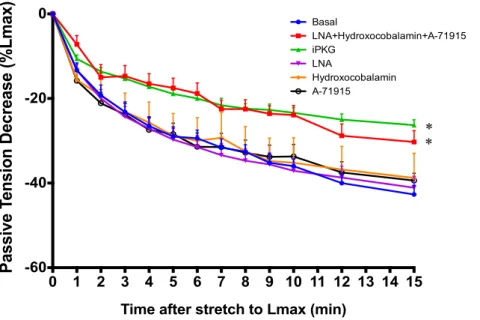
in turn increases the concentration of cGMP in the subsarcolemmal compartment
347
(Francis, 2010) (figure 3). However, in opposition to NO-derived cGMP, whose
348
concentration is kept low through a PDE5 mediated negative feedback mechanism,
CNH-349
derived cGMP triggers a feed-forward mechanism that increases cGMP concentration
350
even more (Castro et al., 2010). While this important difference is not reflected in the
351
relaxation properties of the myocardium, as similarly to NO, CNH exert a positive
352
lusitropic effect through phosphorylation of PLB and TnI, high levels of cGMP stimulate
353
PDE2, reducing cAMP levels, which explains the absence of a positive inotropic effect
354
following CNH release (Potter et al., 2006).
355 356
Both NO and CNH can exert their effects through cGMP; however their physiological
357
role may be quite different. The production/degradation of NO is finely tuned and occurs
358
at a very high pace, meaning that NO must probably be related to adaptations to acute
359
stretch on a beat-to-beat basis (e.g. in inspiration, changing from orthostatic to lying
360
position). Contrastingly, and taking into account their diuretic effects in the kidney and
361
longer half-life than NO, CNH could be more important in hypervolemic, hypertensive
362
states, where stretch is prolonged in time, which explains the importance in relaxing the
363
myocardium without increasing contractility, which would be deleterious.
364 365
Effects of ischemia on the myocardial response to acute stretch 366
367
Myocardial ischemia is a paradigmatic situation of acute hemodynamic overload in which
368
abnormal myocardial loading activates a variety of cellular responses. During an ischemic
369
event, several neurohumoral agents are released, which contribute to the overall
370
physiologic process of cardiac adaptation to hemodynamic overload. Though significant
371
advances in knowledge have been made in the last three decades regarding the processes
372
involved in the pathophysiological consequences of ischemia per se, the cardiac response
373
to stretch under those conditions is just taking its first steps.
374 375
As previously stated, the myocardial response to stretch is a two-step adaptive response.
376
The contractile response following acute stretch under ischemic conditions has been less
377
studied. In a recent study carried out by our group, it was clearly demonstrated that this
378
response is profoundly affected, as shown not only by a blunted FSM but also by the
abolishment of the SFR (Neves et al., 2013), thus supporting some previous indirect
380
observations (Goto et al., 1988;Owen et al., 1993). In the human heart, after an occlusion
381
of a coronary artery, pH falls within 15 seconds (Poole-Wilson, 1989), possibly
382
contributing to the loss of contractility (Steenbergen et al., 1977;Patangay et al.,
383
2009;Decker et al., 2012) due to a reduced sensitivity of the myofilaments to calcium
384
(Kihara et al., 1989;Salas et al., 2006), consequently leading to a diminished Ca2+
385
availability to bind to troponin C. Metabolic abnormalities should not be excluded, as
386
they lead to insufficient energy supply to support cardiac work due to MgADP
387
accumulation, which decreases the sliding velocity in the vicinity ATPase, slowing down
388
the contraction cycle (Sata et al., 1996) and impairing the FSM (Robinson et al., 2002).
389
These mechanisms are probably responsible for the observed ablation of the SFR in these
390
conditions. A possible explanation for this was given recently by our group,
391
demonstrating that this effect was AT1R-dependent and partially AT2R-dependent:
392
blocking AT1R, under ischemic conditions, leads to a soft decline of contraction capacity
393
in response to stretch (Neves et al., 2013). This effect may also be mediated by AT2R
394
activation through bradykinin, PKC and cGMP (Jugdutt and Balghith, 2001).
395 396
Little is known about the role of the PKC under ischemic conditions. As previously stated,
397
PKC activity is more significant during the late phase of the SFR, preventing the
398
development of a slow force decline. Also, under ischemic conditions, PKC was not
399
capable of modifying contractile performance (Neves et al., 2013). This is consistent with
400
its inability to alter the systolic adaptation during early phase of the response to stretch
401
and does not exclude that its downstream mediators may be compromised in such
402
conditions. However, PKC has several isoforms, some of them present within the same
403
cell and activated by the same stimuli but showing different and, at times, opposite effects.
404
For example, in ischemic heart disease, PKCδ induced decreased ATP generation
405
(Inagaki et al., 2003), and therefore, insufficient energy supply may play a role. As such,
406
it would be interesting to study how each one modulates the contractile response to stretch
407
under ischemic conditions.
408 409
Usually, in the setting of myocardial infarction, the PKG signaling pathway through NO
410
and CNH is modulated pharmacologically by nitrates as NO donors. Also, administration
411
of PDE5 inhibitors may be promising therapeutic targets (Kukreja, 2013). However, only
412
recently the specific role of the PKG signaling pathway in ischemic conditions was
413
shown. Intriguingly, activation of PKG pathway did not alter the SFR in ischemic
414
conditions, while PDE5 inhibition significantly mitigated the contractile decline after
415
stretch in ischemia (Castro-Ferreira et al., 2014). This can be explained by
NO/cGMP-416
independent cardioprotective effects (Elrod et al., 2007). However, the mechanisms
417
responsible for the protective action of the sildenafil remain to be fully elucidated.
418 419
Conclusion 420
421
The myocardial response to acute stretch represents a fundamental adaptive capacity of
422
the heart. Although this functional response to hemodynamic overload has been known
423
for long time, the structures responsible for mechanosensing, as well as the pathways
424
involved in the response, are still only partially clarified. The possibility of concomitant
425
diastolic adaptation, although theoretically appropriate, has never been directly assessed.
426
Despite the relevant knowledge accumulated on the cardiac response to acute
427
hemodynamic overload, there is much that we still do not know. The quest for further
428
understanding of the mechanosensing involved in this mechanism, the pathways
responsible for the response and the characterization of the diastolic adaptation may
430
provide new insights, not only in its physiological importance, but also in the prevention
431
and treatment of cardiovascular diseases, such as heart failure with reduced ejection
432
fraction, heart failure with preserved ejection fraction, hypertensive crises or ischemic
433
heart disease.
434 435
PART TWO: A NEW DIASTOLIC PARADIGM
436 437
Introduction 438
Acute hemodynamic load variation represents a permanent challenge to the heart,
439
warranting continuous adaptation to face such changes in cardiac demands. When preload
440
increases, the resulting stretch of chamber walls triggers a classic biphasic response
441
consisting of an immediate (FSM) and sustained (SFR) increase in contractility that is
442
central to cardiac function (Patterson and Starling, 1914; Parmley and Chuck, 1973;
443
Kockskamper et al., 2008).
444 445
As stated in the previous section, this response is stimulated by factors produced or
446
released at the onset of acute cardiac stretch, namely NO (Casadei and Sears, 2003) and
447
B-type natriuretic peptide (BNP), both binding to receptors with guanylate cyclase
448
domains and increasing sarcoplasmic cGMP levels (Potter et al., 2006; Francis, 2010).
449
The systolic potentiation this second messenger produces is attributed, at these
450
concentrations, to pathways including PKG-mediated phosphorylation of several key
451
proteins regulating contraction, including PLB and TNI (Layland et al., 2005; Zhang et
452
al., 2008; Castro-Ferreira et al., 2014), and also of adenosine diphosphate ribosylcyclase,
453
increasing levels of cyclic ADP-ribose, which prolongs opening of sarcoplasmic
454
reticulum Ca2+ channels (Willmott et al., 1996).
455 456
Keeping up with this, the availability and viability of the pathways through which these
457
molecules act appear dysregulated in heart failure (HF), of which decreased preload
458
reserve is a hallmark and a central mechanism of symptomatic congestion (van Heerebeek
459
et al., 2012). The associated oxidative stress scavenges NO into peroxynitrite species,
460
which further impair cardiac function, and the plasmatic concentration of the active
461
fragment of BNP is decreased, blunting natriuretic signalling despite increased levels of
462
its inactive precursor, a commonly used biomarker of this disease (Zois et al., 2014).
463 464
Interestingly, some of these factors acutely modulate diastolic function as well (Paulus et
465
al., 1994; Shah and MacCarthy, 2000). An important target of PKG is titin, the giant
466
sarcomeric protein accounting for most PT within the physiological range of sarcomere
467
length (SL). By phosphorylating its N2B spring element, PKG shifts titin’s resting length
468
downward, making it more distensible and reducing cardiomyocyte stiffness for the same
469
SL (Kruger et al., 2009; Bishu et al., 2011).
470 471
Therefore, there is reason to assume that similar mechanisms could play a role in a
472
diastolic component in the adaptation to acute stretch, which remains unexplored. This
473
hypothesis is further enlightened by evidence that low bioavailability of NO and cGMP,
474
PKG hypoactivity (van Heerebeek et al., 2012; Franssen et al., 2015) and titin
475
hypophosphorylation (Hamdani et al., 2013a) all importantly contribute to the
476
pathophysiology of diastolic dysfunction observed in patients presenting with HF with
preserved ejection fraction (HFpEF). HFpEF represents more than 50% of HF cases, is
478
associated with a poor prognosis and lacks therapeutic agents effective in reducing
479
mortality and morbidity associated with it (Owan et al., 2006; Borlaug and Paulus, 2011).
480 481
Given the grand implications of such a mechanism in cardiovascular physiology and
482
potential impact in understanding the pathophysiology of HF, we aimed to describe a
483
novel evolutionarily conserved mechanism of acute diastolic adaptation to hemodynamic
484
overload in healthy and failing hearts and assess the contribution of PKG-related
485
pathways and titin phosphorylation across several models, including the human heart.
486 487
Material and Methods 488
489
Isolated heart preparation: Wistar rat hearts (n=10) were perfused according to the
490
Langendorff technique at constant pressure (Radnoti LLC, ADInstruments) and
491
electrically paced. Pressure was monitored using a pressure tip catheter (Millar, Inc.)
492
inside a fluid-filled balloon inserted into the left ventricle (LV) and volume was
493
determined and controlled using a syringe pump (New Era Pump Systems). Balloon
494
volume was set to a preload yielding a mean diastolic pressure of 3 mmHg and, after
495
stabilization, inflated to a diastolic pressure of 10 mmHg for 15 minutes. After this period,
496
the left ventricular free wall was rapidly dissected and frozen in liquid nitrogen for
497
posterior analysis.
498 499
Isolated muscle strip preparation: Force measurements were performed in papillary
500
muscles dissected from the right ventricle of male New Zealand white rabbits and human
501
atrial trabeculae (n=14) and human ventricular strips (n=10) strips from patients subject
502
to cardiac surgery. The muscles were vertically mounted in an organ bath containing a
503
modified Krebs-Ringer solution and the upper tendinous end was attached to an
504
electromagnetic length–tension transducer (University of Antwerp, Belgium). The
505
muscle was electrically stimulated to contract isometrically and the length at which active
506
force development was maximal (Lmax) was determined. After stabilization at 92% Lmax,
507
the rabbit papillary muscles were quickly stretched to 100% of Lmax with different groups
508
of muscles being either control (n=11) or incubated with Rp-8-Br-PET-cGMPS [inhibitor
509
of PKG, 10-6M, n=7], L-nitro-arginine [LNA, inhibitor of NO synthase, 10-5M, n=8],
A-510
71915 [natriuretic peptide receptor-A antagonist, 10-6M, n=7], hydroxocobalamin (HCB,
511
NO scavenger, 10-3M, n=8) and LNA, HCB and A-71915 and combined (n=8). At the
512
end of the experiment, the active portion of the muscle was quickly frozen in liquid
513
nitrogen for posterior analysis. Additionally, in a second set of experiments, after 15
514
minutes of stretch, muscles were again set to 92% Lmax over 15 minutes and subjected to
515
another 15 minutes period of stretch to 100% of Lmax plus 92% Lmax over 15 minutes.
516 517
Isolated skinned cardiomyocyte preparation: Force measurements were performed in
518
single and mechanically isolated cardiomyocytes as previously described (Borbély et al.,
519
2005; Falcao-Pires et al., 2011) from rat hearts subjected to the perfusion protocol
520
described above. PT was measured at SL ranging between 1.8µm to 2.3µm (0.1 µm step
521
increases) at 15°C. After PT stabilized at 2.2µm, cardiomyocytes were incubated for 40
522
minutes at 20°C in relaxing solution containing either purified PKG1α, cGMP and
523
dithiothreitol (n=25 and n=21 from stretch and control, respectively); or with protein
524
phosphatase-1, and protein phosphatase lambda (n=25 and n=26 from stretch and control,
525
respectively). After that, the same set of experiments was performed to measure PT at the
526
same SL ranges.
Rat Transverse Aortic Constriction (TAC) banding model: A 3–0 non-absorbable silk
528
suture was placed around the aortic arch of Wistar-Han rats (n=16) with 7 weeks of age,
529
constricting it to the diameter of a 25G needle. Sham-treated animals (n=14), which
530
underwent surgery with placement of a loose non-constrictive suture, were used as
531
controls. After 21 weeks, echocardiographic images (VIVID 7 dimension system;
532
General Electric-Vingmed Ultrasound) were obtained according to standards of
533
American Society of Echocardiography (Lang et al., 2015) under anesthesia with
534
ketamine and xylazine. Afterwards, a 1.4Fr microtip pressure-volume catheter (SPR-838,
535
Millar Instruments) was inserted through an apical puncture wound into the LV cavity.
536
The animal preparation was allowed to stabilize for 15 minutes, after which ventilation
537
was suspended and a basal measurement performed at end-expiration. An acute volume
538
load was the performed by intravenous administration of 15% of total blood volume of
539
hydroxyethyl starch (Voluven®) during 5 minutes. New recordings were made after
540
loading with suspension of the ventilation each 5 minutes until 15 minutes. At the end,
541
the animal was euthanized by exsanguination, and samples were collected.
542 543
Cardiac surgery patients: Hemodynamic evaluation was performed intraoperatively on
544
patients with aortic stenosis and no evidence of LV hypertrophy on transthoracic
545
echocardiogram (n=9) subjected to aortic valve replacement. After weaning
546
cardiopulmonary bypass and in a hemodynamically stable period, pressure and volume
547
were continuously measured by a 5-Fr combined catheter with 1 cm electrode spacing
548
(SPC-551, Millar Instruments, Houston, TX) inserted through the ascending aorta. Once
549
a basal acquisition was obtained, a volume overload was imposed through intravenous
550
bolus of hydroxyethyl starch (Voluven®) and Trendelenburg positioning and values were
551
registered at that moment and 15 minutes after. Four patients were male and five were
552
female, ranging from 49 to 85 years old, and the surgery was performed by the same
553
surgeon.
554 555
Protein kinase activity: The phosphorylation of VASP, a myocardial PKG target, was
556
measured using immunoblots; densitometry (over GAPDH expression) was performed
557
and presented as previously described (van Heerebeek et al., 2012).
558 559
Titin phosphorylation: After 1.8% SDS-PAGE, gels were stained with Pro-Q Diamond
560
(phosphoprotein) for 1 hour and subsequently with Sypro Ruby (total protein) overnight.
561
Phosphorylation signals on Pro-Q Diamond-stained gels were indexed to Sypro Ruby–
562
stained titin signals as previously described (Hamdani et al., 2013a).
563 564
Statistical methods: Values are means ± SEM. Within the same group, the immediate
565
effects of muscle stretch on the various experimental parameters were analysed with a
566
paired Student’s t-test. Comparison of groups at discrete points (length, volume, time)
567
was tested with unpaired Student’s t-test, being the time-dependent effects analysed by
568
repeated measures two-way ANOVA. A p<0.05 was considered statistically significant.
569 570
Results 571
572
Effects of acute cardiac stretch in myocardial stiffness
573 574
Plotting tension of acutely stretched papillary muscles from 92% to 100% Lmax over time
575
clearly illustrates three phenomena (Fig. 4): (1) the Frank-Starling mechanism, with
576
active tension increasing immediately upon stretch; (2) the slow force response, with
active tension further increasing sustainably over time; (3) the diastolic response,
578
characterized by an immediate increase in PT, followed by a progressive and
time-579
dependent fall. Regarding this Stretch-Induced Compliance (SIC), acute stretch resulted
580
in an immediate increase of PT from 1.5±0.3 to 6.3±1.2 mN.mm-2 in rabbit papillary
581
muscles, from 3.0±0.5 to 8.1±1.2 mN.mm-2 in human atrial trabeculae, from 2.4±0.2 para
582
8.2±0.6 mN.mm-2 in human ventricular strips. Subsequently, over the course of 15
583
minutes, PT of rabbit papillary muscles decreased by 43±2% (Fig. 5B), human atrial
584
trabeculae (n=14) by 27±8% and human ventricular strips (n=10) by 27±6% (Fig. 5C).
585
Similarly, left ventricular diastolic pressure of isolated perfused rat hearts fell 43±2%
586
after acute inflation of the LV balloon by 38±2 µL to increase diastolic pressure from 3
587
to 10 mmHg (Fig. 5A).
588 589
At the end of this time period, rabbit papillary muscles were returned to their original
590
conditions by reducing length back to 92% Lmax. At this point, PT was significantly lower
591
than that observed before stretch (0.8±0.2 mN.mm-2) but rose for the following 10
592
minutes (1.1±0.3 mN.mm-2), becoming significantly higher than immediately after stretch
593
but lower than before stretch (Fig. 6). The length-PT relationship in papillary muscles
594
delineated by these “stretch” and “unstretch” interventions clearly illustrates the SIC and
595
its partial persistence over the following 10 minutes (Fig. 7B) and analogous results are
596
obtained in diastolic pressure-volume relationships of isolated rat hearts (Fig. 7A).
597 598
To evaluate whether stress relaxation played a role in SIC, we stretched isolated skinned
599
cardiomyocytes but no significant drop in PT was observed in this setting (Fig. 5D and
600
Fig. 7C). Nonetheless, cardiomyocytes extracted from the LV free wall of five stretched
601
isolated hearts presented with a markedly reduced PT at SL ranging from 1.8 to 2.3 µm
602
compared to those extracted from other five hearts not submitted to the stretch protocol
603
(Fig. 7D), confirming the adaptation occurs only in intact tissue at the myofillamental
604
level.
605 606
Lastly, in cardiac surgery patients evaluated in vivo, volume overload raised end-diastolic
607
volume from 122.5±21.8 to 150.3±21.2 mL. Baseline end-diastolic pressure (EDP) was
608
11.0±2.1 mmHg and, with overload, increased to 17.7±5.5 mmHg. After 15 minutes, EDP
609
measured in cardiac cycles with the same end-diastolic volume of the previous situation,
610
was clearly reduced, at 13.5±1.7 mmHg (Fig. 8).
611 612
TAC banding compromises the decrease in stiffness in response to stretch
613 614
Next, we used a model of cardiac hypertrophy and diastolic dysfunction, rats subjected
615
to transverse aortic constriction (TAC) banding, to test SIC in a pathological context.
616
Compared with sham-operated rats, TAC animals presented a significant increase in
617
cardiac mass (63±16%) and echocardiographic studies revealed significantly higher left
618
ventricular posterior wall dimension in systole and diastole. Furthermore, they presented
619
a significant increase of LV EDP and time constant tau, indicating diastolic dysfunction.
620
LV peak systolic pressure was also significantly higher when compared to Sham, but no
621
significant change was detected in ejection fraction (Table 1).
622 623
In response to acute myocardial stretch induced by volume overload, left ventricular EDP
624
significantly increased in both groups (Sham +60±4%; TAC +79±37%). After 15
625
minutes, EDP significantly decreased in Sham (15±1 vs. 9±1 mmHg), but not in TAC
animals (19±4 vs. 16±3 mmHg), even if end-diastolic LV volume remained unchanged
627
(Figure 9).
628 629
PKG-mediated pathways in the response to acute myocardial stretch
630 631
We hypothesized that PKG-mediated pathways were involved in priming myocardial
632
stiffness to preload elevation. Because both NO and BNP are released upon myocardial
633
stretch and increase cGMP levels, which activate PKG, we confirmed these observations
634
by measuring PKG activity in the LV free wall of rats subjected to Langendorff perfusion.
635
and found that myocardial stretch led to an increase in PKG activity (Fig. 10).
636 637
To assess the individual contributions of these agents to the mechanism, we investigated
638
whether blocking them would interfere with this effect. Incubation with LNA, HCB or
639
A-71915 alone did not significantly alter the myocardial response to stretch and the
640
muscles exhibited a similar behaviour to that observed under basal conditions. However,
641
simultaneous incubation with all three drugs significantly blunted the decrease in PT after
642
acute stretch (30±3 vs. 43±2%), as did the direct antagonism of PKG by
Rp-8-Br-PET-643
cGMPS (26±1 vs. 43±2%) (Figure 11).
644 645
In order to functionally assess the contribution of phosphorylation of myofilamental
646
elements to SIC, we incubated skinned cardiomyocytes extracted from stretched or
647
control isolated rat hearts with protein phosphatases. After 40 minutes, we observed a
648
significant upward dislocation of length-PT relationships in both groups, more prominent
649
in the previously stretched group, approaching control levels (Fig. 12B). On the other
650
hand, incubation of myocytes with PKG for 40 minutes reduced stiffness in the control
651
group, as previously shown (Paulus et al., 1994; van Heerebeek et al., 2012; Hamdani et
652
al., 2013a), but not in the stretched group (Fig. 12A).
653 654
Titin phosphorylation in response to acute myocardial stretch
655 656
Lastly, we compared total titin phosphorylation between groups of muscles subjected to
657
acute stretch for 15 minutes and ones that were kept at constant length throughout the
658
protocol. As illustrated in figure 13, a significant increase in the levels of phosphorylated
659
titin was observed in rabbit papillary muscles (23±3 vs. 13±2%), as well as in human right
660
atrial trabeculae (41±8 vs. 11±1%) and left ventricular strips (71±21 vs. 27±8%),
661
suggesting this as a potential mechanism for the active modulation of myocardial stiffness
662
following stretch. Interestingly, in the hearts from TAC rats not only was titin
663
hypophosphorylated at baseline (73±6% of sham levels), but also the 51±12% increase in
664
titin phosphorylation upon VO observed in sham was blunted to 30±14%.
665 666
Discussion 667
668
With this work, we intended to explore the response to hemodynamic overload in detail,
669
well characterized from a contractile standpoint for decades, but of which a diastolic
670
component had never been described. We observed that the latter, in the form of SIC,
671
allows optimization of filling capacity at lower diastolic pressures and, therefore,
672
potentiation of increased cardiac output triggered by acute myocardial stretch and
673
prevention of pulmonary and systemic congestion. This mechanism augments mechanical
674
distensibility in a potent and reproducible way in various mammalian models, being
675
evolutionarily conserved up to the human species. Additionally, it is evident in
experimental preparations ranging from the isolated cardiomyocyte to in vivo
677
hemodynamic analysis and including in vitro isolated intact hearts and muscle strips from
678
ventricle and atrium.
679 680
Acutely stretching healthy myocardium in vitro is followed by a drop in left ventricular
681
EDP of intact hearts and PT of human and animal isolated muscle strips over time. After
682
this period, EDP-volume and length-PT relationships, respectively, were shifted
683
inferiorly, indicating decreased stiffness acquired after this stimulus. Furthermore, after
684
its establishment, returning to baseline levels of preload reverses the effect incompletely,
685
pointing to a role in both dynamic adaptation on the fly and more durable load
686
conditioning.
687 688
Decreased EDP is also seen after in vivo volume overload both in rats and in patients
689
undergoing aortic valve replacement surgery. Although in this setting central neural
690
reflexes and systemic humoral agents with myocardial effects likely contribute to the
691
effect and cannot be distinguished from an intrinsic myocardial adaptation, its
692
significance in this context still stands.
693 694
Opposite to these situations, stretching isolated skinned cardiomyocytes does not evoke
695
the same response per se. This suggests that the effect requires preserved subcellular
696
compartmentalization to become manifest and, thus, depends on signaling pathways.
697
Different stiffness is found only when comparing cardiomyocytes extracted from
698
stretched and non-stretched hearts, i.e. when the intervention is performed on the intact
699
organ. Taken together, these observations confirm that the effect is, to a grand extent,
700
independent from the passive properties of cardiac extracellular matrix and that the
701
effector players in these pathways must target myofillamentary elements.
702 703
To translate these findings to a pathological context, we used a classic animal model of
704
diastolic dysfunction induced by thoracic aortic constriction. Inducing in vivo acute
705
hemodynamic overload in these animals revealed that, contrarily to sham individuals,
706
EDP, already elevated at baseline, does not decline significantly over time. This
707
difference is relevant, as inability to increase filling capacity in the face of a hemodynamic
708
challenge can lead to congestion. Not only does this underline the importance of the
709
diastolic component of the response to stretch, but also represents the first description
710
that diastolic dysfunction is attributable to both increased myocardial stiffness and
711
compromised modulation after acute load.
712 713
We verified that PKG-related pathways were activated upon myocardial stretch and led
714
to titin phosphorylation, representing the molecular basis explaining SIC and their
715
dysregulation in the hypertrophied heart highlights their pathophysiological relevancy. In
716
our study, the diastolic response to load is highly dependent on the activity of this enzyme,
717
evident by the marked blunting of the effect observed upon its inhibition.
718 719
A similar phenomenon was observed when NO production was inhibited, its intracellular
720
stores were scavenged and the natriuretic peptide receptor A was inhibited, even though
721
these interventions, by themselves, exerted no effect. This suggests a compensatory or
722
redundant interaction between NO and BNP, both activating PKG through increase in
723
intracellular cGMP concentration, although the subcellular compartment whereby this
724
occurs may differ between the two mediators (Castro et al., 2006; Castro et al., 2010), as
725
discussed above in the literature review. In spite of our establishment of the importance
of this pathway in SIC, future study is necessary to ascertain the most relevant NO
727
synthase isoform and cellular cGMP pool in this context.
728 729
At the sarcomeric level, our results corroborate previous studies showing that incubation
730
of isolated cardiomyocytes with PKG increased their distensibility (Kruger et al., 2009;
731
van Heerebeek et al., 2012; Hamdani et al., 2013a). Intriguingly, incubation of myocytes
732
of previously stretched hearts, which already show decreased levels of PT, does not alter
733
their stiffness profile. This points to a degree of saturation of kinase signalling, its target
734
sites probably already phosphorylated by PKG concomitantly to its stretch-induced
735
activation. On the flipside, incubation of cardiomyocytes with protein phosphatases
736
shifted both groups’ length-PT curves upward and brought them very close to each other,
737
thus uncovering the phosphorylation-dependent component of SIC.
738 739
Some known targets of PKG include TNI (Layland et al., 2002), increasing cross bridge
740
cycling, and PLB (Zhang et al., 2005), reducing its affinity for SERCA and boosting Ca2+
741
reuptake during diastole, but none of these actions link acute cardiac loading to decreased
742
myocardial stiffness. We propose that this effect is in fact mediated by titin,
743
phosphorylated by PKG on its N2B elastic element, increasing electric repulsion between
744
its negatively charged amino acids and making it more distensible (Kruger et al., 2009;
745
Kotter et al., 2013).
746 747
Keeping up with this, higher titin phosphorylation levels were measured in stretched
748
myocardial samples from human atria and ventricles and rabbit ventricles. In the latter
749
case, samples analyzed after a period of “unstretch” reveal an incomplete reversal of
750
phosphorylation levels, mirroring the persisting decrease in stiffness observed during the
751
protocol. Furthermore, in vivo hemodynamic overload in rats induced titin
752
phosphorylation in healthy animals but not in the diastolic dysfunction group, which
753
presented baseline hypophosphorylation, confirming the central role of this mechanism
754
in SIC and diastolic function.
755 756
Our line of study may contribute to better understanding of mechanisms implicated in
757
HFpEF-associated diastolic dysfunction. Firstly, it shows that the role of the cGMP/PKG
758
pathway and titin phosphorylation in this context is not limited to modulation of
759
ventricular stiffness but also encompasses the response to hemodynamic stress in
760
different cardiac chambers. Since HFpEF is frequently characterized by exercise
761
intolerance with fatigue, cough and dyspnea and underlying hypoactivity of the
762
previously mentioned molecular mechanisms (Borlaug and Paulus, 2011; van Heerebeek
763
et al., 2012), impaired SIC may be an additional pathophysiological link, leading to
764
inability to optimize diastolic filling pressures after overload. Therefore, its potential
765
diagnostic and therapeutic applications in this condition, which urgently needs new
766
management options to reverse its tendency towards an unfavorable outcome, should be
767
avidly studied.
768 769
As such, the decline in EDP after acute load could be an early disease marker manifesting
770
prior to symptom onset. Noninvasive indices of diastolic dysfunction such as the E/E’
771
ratio on transthoracic echocardiogram before, during and after preload increasing
772
provocative maneuvers (e.g. exercise, limb elevation or Trendelenburg positioning) might
773
be of diagnostic value. Poor adaptation to this stimulus could additionally be used as a
774
disease classification criterion, which is not yet consensual (McMurray et al., 2012;