Evaluation of Cardiovascular Risk Factors in
the Wistar Audiogenic Rat (WAR) Strain
Rubens Fazan, Jr.1, Carlos Alberto A. Silva1, José Antônio Cortes Oliveira1, Helio
Cesar Salgado1, Nicola Montano2, Norberto Garcia-Cairasco1*
1Department of Physiology, School of Medicine of Ribeirão Preto, University of São Paulo, Ribeirão Preto, São Paulo, Brazil,2Department of Clinical Sciences“Luigi Sacco”, University of Milan, Milan, Italy
Abstract
Introduction
Risk factors for life-threatening cardiovascular events were evaluated in an experimental model of epilepsy, the Wistar Audiogenic Rat (WAR) strain.
Methods
We used long-term ECG recordings in conscious, one year old, WAR and Wistar control counterparts to evaluate spontaneous arrhythmias and heart rate variability, a tool to assess autonomic cardiac control. Ventricular function was also evaluated using the pressure-vol-ume conductance system in anesthetized rats.
Results
Basal RR interval (RRi) was similar between WAR and Wistar rats (188±5 vs 199±6 ms).
RRi variability strongly suggests that WAR present an autonomic imbalance with sympa-thetic overactivity, which is an isolated risk factor for cardiovascular events. Anesthetized WAR showed lower arterial pressure (92±3 vs 115±5 mmHg) and exhibited indices of
sys-tolic dysfunction, such as higher ventricle end-diassys-tolic pressure (9.2±0.6 vs 5.6±1 mmHg)
and volume (137±9 vs 68±9μL) as well as lower rate of increase in ventricular pressure
(5266±602 vs 7320±538 mmHg.s-1). Indices of diastolic cardiac function, such as lower
rate of decrease in ventricular pressure (-5014±780 vs -7766±998 mmHg.s-1) and a higher
slope of the linear relationship between end-diastolic pressure and volume (0.078±0.011 vs
0.036±0.011 mmHg.μL), were also found in WAR as compared to Wistar control rats.
More-over, Wistar rats had 3 to 6 ventricular ectopic beats, whereas WAR showed 15 to 30 ectop-ic beats out of the 20,000 beats analyzed in each rat.
Conclusions
The autonomic imbalance observed previously at younger age is also present in aged WAR and, additionally, a cardiac dysfunction was also observed in the rats. These findings make
OPEN ACCESS
Citation:Fazan R, Jr., Silva CAA, Oliveira JAC, Salgado HC, Montano N, Garcia-Cairasco N (2015) Evaluation of Cardiovascular Risk Factors in the Wistar Audiogenic Rat (WAR) Strain. PLoS ONE 10 (6): e0129574. doi:10.1371/journal.pone.0129574
Academic Editor:German E. Gonzalez, University of Buenos Aires, Faculty of Medicine. Cardiovascular Pathophysiology Institute., ARGENTINA
Received:May 18, 2014
Accepted:May 11, 2015
Published:June 1, 2015
Copyright:© 2015 Fazan et al. This is an open access article distributed under the terms of the Creative Commons Attribution License, which permits unrestricted use, distribution, and reproduction in any medium, provided the original author and source are credited.
this experimental model of epilepsy a valuable tool to study risk factors for cardiovascular events in epilepsy.
Introduction
Epidemiological studies have established that the incidence of premature death among epilep-tic patients is markedly higher than in the general population [1,2,3]. There has been growing awareness of sudden unexpected death in epilepsy (SUDEP), which is now acknowledged as a serious problem for epileptic patients [2,3]. Evidence from epidemiological, clinical, and patho-logical studies indicates that in most cases SUDEP occurs after a seizure, although deaths not preceded by seizure have been reported anecdotally [3].
Even though the pathophysiology of SUDEP is unknown in most cases, it is important to consider that sudden death is often attributed to cardiac events. Heart dysfunction and distur-bances in neural control of the cardiovascular system are always associated with morbidity and mortality in epileptic patients [3,4,5,6]. In fact, QT dispersion, reflecting regional heterogeneity of cardiac repolarization and important risk factors for ventricular arrhythmias, have been seen in up to one third of people with epilepsy [7,8]. Moreover, few studies with electrocar-diographic (ECG) recordings during or near to sudden deaths in epileptic patients suggest that cardiac arrhythmias are crucial determinants of some of them [9,10].
Clinical and experimental studies provide evidences that epileptic seizures are accompanied by autonomic cardiovascular imbalance with sympathetic overactivity, resulting in hyperten-sion, tachycardia and electrical instability in the heart [10,11,12]. Although changes in sympa-thetic discharges are known to occur during epileptic seizures, autonomic disturbances, mostly showing enhanced sympathetic cardiovascular tone, were also found in inter-ictal periods in ei-ther epileptic patients [13,14,15,16,17] or experimental models of epilepsy [18].
It has long been recognized that increased sympathetic activity has a profound influence on the electrical and contractile functions of the heart [19]. There is wide evidence that excessive catecholamine release, due to sympathetic activation, produces severe toxic cardiac effects, such as intra-cellular Ca2+overload, high-energy phosphate depletion, life threatening ventric-ular arrhythmias, and sudden cardiac death [19,20,21,22]. Moreover, long-lasting sympathetic overactivity, known to promote myocardial apoptosis [23] and cardiac hypertrophy with chamber remodeling [24,25] is thought to be a major determinant of progressive heart failure.
Remarkable advances in the understanding of autonomic function have been made due to the development of methods to evaluate short-term changes in heart rate (HR) and/or blood pressure (BP). The notion that, in addition to cardiac cycle, other rhythms related to sympa-thetic and parasympasympa-thetic drives to the heart are present in BP and HR series stimulated great interest in using measures of cardiovascular variability as diagnostic tools [26]. Analysis of spontaneous rhythmic fluctuations in HR by spectral analysis offers remarkable insight into the physiological mechanisms of autonomic cardiovascular control [27,28,29,30,31].
Various experimental animal models of epilepsy have been used to examine the pathophysi-ological mechanisms of this syndrome. For example, Wistar audiogenic rats (WAR) [32,33], which are an inbred strain prone to audiogenic epileptic seizures derived from Wistar rats, have been used extensively as an experimental model of epilepsy. A recent study from our labo-ratory [18] showed that young adult WAR are slightly hypertensive, have higher basal HR and a 2-fold higher cardiac sympathetic tone compared with Wistar control rats with no experience of audiogenic epileptic seizures.
Assistência do Hospital das Clínicas (FAEPA/HC) da Faculdade de Medicina de Ribeirão Preto da Universidade de São Paulo (FMRP-USP). RFJ, HCS and NGC hold CNPq Research Fellowships.
It is well established that high BP and chronic cardiac sympathetic overactivity are leading causes of life-threatening cardiovascular events, such as ventricular arrhythmias and are also major determinates of heart failure [23,24,25]. In this context we hypothesize that WAR are at higher cardiovascular risk, especially during aging. Therefore, the aim of this study was to iden-tify spontaneous arrhythmias, as well as to evaluate HR and BP variability as an approach to characterize autonomic modulation of the cardiovascular system in aged, conscious freely-moving WAR. Moreover, cardiac function was also evaluated by analyzing left ventricular pres-sure-volume relationships in anesthetized rats from the WAR strain.
Methods
Ethics Statement
All procedures were performed in accordance with institutional polices and guidelines for ethi-cal use of animals and following the recommendations from the Brazilian Society for Neurosci-ence and Behavior. At the end of the ventricular pressure recordings, with the animals still under the effect of anesthesia (see below), by mean of large laparotomy, they were killed by ample diaphragm opening. Specific protocols for the current experiments were approved by the Commission for Animal Experimentation (CETEA; 117/2011 and 16/2013-1) of the Ribei-rão Preto School of Medicine.
The experiments were conducted on male WAR and their Wistar counterparts (control, n = 9 in each group) at 11 to 12 months of age with no prior induction of audiogenic epileptic seizure (WAR) and sound stimulation (Wistar). The animals were housed individually with free access to food and water and were maintained on a 12/12 h light/dark cycle at 22°C.
Surgery and Recording of Electrocardiogram
The animals were anesthetized with tribromoethanol (250 mg/kg i.p.) and implanted with sub-cutaneous ECG electrodes two days prior to recording. The electrodes tips were suture-fixed subcutaneously and the wires exteriorized at the back of the neck and the rats were allowed to recover for 2 days. The animals were taken to the recording room, where the electrodes were connected to an ECG amplifier (6600 series ECG/Biothac. Amplifier, Gould Instrument Sys-tems In, Valley View, OH, USA) coupled to an A/D interface (DI720 Dataq Instruments Akron, OH, USA) at least 30 min before beginning the recordings. The room was kept at 22 ±1°C, and silence was maintained to minimize environmental stress. After the adaptation peri-od the ECG was digitally recorded (2 kHz) for 60 min.
ECG segments with 20,000 beats were analyzed using the computer software ECG module for LabChart 7.2 (ADInstruments, Mountain View, CA, USA) that automatically detected nor-mal and ventricular premature beats (VPB).
Cardiac Function
end-systolic and end-diastolic volumes (ESV and EDV) as well as cardiac output (CO) were calculated using a cardiac pressure-volume analysis software (PVAN Ultra v3.2, Millar Instru-ments, Houston, TX, USA) [34]. The total peripheral resistance (TPR) was also calculated as the ratio between MBP and CO. A range of left ventricle pressure-volume curves at different pre-load status was assessed by transiently compressing the inferior vena cava. Pre-load inde-pendent indices of heart contractility and stiffness {preload recruitable stroke work (PRSW), +dP/dt vs. end-diastolic volume relationship (+dP/dt-EDV), slopes of end-systolic and end-di-astolic pressure-volume relations [ESPVR and EDPVR]} as well as the efficiency of left ventri-cle work [pressure-volume area] were calculated using PVAN v3.2. Good quality recordings of ventricular pressure and volume were obtained in only 7 WAR and 8 Wistar rats.
Heart Rate Variability
Files with ECG recordings were analyzedoff lineusing the computer software LabChart 7.2 (AD Instruments, Mountain View, CA, USA). R waves were identified every cardiac beat and series of successive RR intervals (RRi) were generated for each animal. Over 5 to 10 stationary RRi series of 500±100 beats were selected by visual inspection of the entire recording.
After calculation of standard deviation of successive normal RRi (SDNN), an index of over-all HRV (time domain) each segment spectral analysis using an autoregressive algorithm with the model order chosen according to Akaike’s criterion to obtain the frequency spectrum of HRV [28,31]. The oscillations of RRi were quantified in 3 frequency bands according to the central frequency of each component modeled by autoregressive algorithm: very low (VLF, below 0.2 Hz), low (LF, 0.2–0.8 Hz) and high frequency (HF, 0.8–2.0 Hz). The LF and HF oscil-latory components were expressed in absolute (ms2) and normalized units as well as a ratio of low to high frequency (LF/HF). Normalization consisted of dividing the power of a given spec-tral component by the total power minus the power below 0.2 Hz and multiplying the ratio by 100 [29,35].
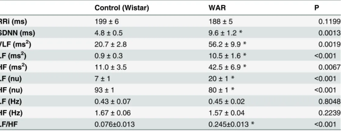
Table 1. Mean values of RR interval (RRi) and RRi variability in time and frequency domain in one-year old conscious freely moving WAR and Wistar control rats.
Control (Wistar) WAR P
RRi (ms) 199±6 188±5 0.1199
SDNN (ms) 4.8±0.5 9.6±1.2* 0.0013
VLF (ms2) 20.7±2.8 56.2±9.9
* 0.0019
LF (ms2) 0.9±0.3 10.5±1.6
* <0.001
HF (ms2) 11.0±3.5 42.5±6.9* 0.0067
LF (nu) 7±1 20±1* <0.001
HF (nu) 93±1 80±1* <0.001
LF (Hz) 0.43±0.07 0.45±0.02 0.8048
HF (Hz) 1.67±0.06 1.57±0.04 0.2239
LF/HF 0.076±0.013 0.245±0.013* <0.001
*P<0.05 compared with Wistar control rats.
RRi: interval between successive R waves, SDNN: standard deviation of normal RRi, VLF, LF and HF: power of oscillatory components of RRi series at very low (<0.2 Hz), low (0.2–0.8 Hz) and high (0.8–3.0 Hz) frequency bands. nu: normalized units. LF and HF (Hz): central frequency of the main oscillatory component modeled at low- and high-frequency bands.
Values are mean±SEM.
Statistical Analysis
Each parameter, i.e. heart rate variability and cardiac function indexes, was firstly tested for normality of the distribution by modified Kolmogorov-Smirnov test (goodness of fit test) Fol-lowing, they were compared between WAR and Wistar control counterparts by 2-tail unpaired Student t test or Mann-Whitney test, accordingly. A value of p<0.05 was considered statisti-cally significant. The data are presented as the mean±SEM.
Results
The basal RR interval (RRi) was similar between WAR (188±5 ms) and Wistar control counter-parts (199±6 ms).
The mean levels, SDNN and power of oscillatory components (autoregressive spectral anal-ysis) of RRi during baseline recordings from both groups of rats are shown inTable 1. While the mean RRi (SDNN) did not differ between the groups, WAR showed higher overall RRi var-iability. Moreover, the normalized LF and the LF/HF ratio were markedly increased in WAR compared to Wistar counterparts.
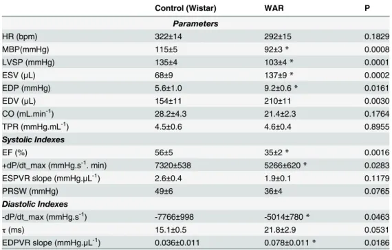
Hemodynamic parameters and indices of cardiac function derived from the left ventricle pressure-volume relationship in pentobarbital-anesthetized rats are shown inTable 2. HR did not differ between the groups while mean BP (MBP) and ventricular systolic pressure (LVSP, mmHg) were lower in WAR than in Wistar control rats. WAR also showed indices of systolic dysfunction, such as higher ventricular end-diastolic pressure (LVEDP, mmHg), lower +dP/ dt_max (mmHg.s-1). Indices of diastolic cardiac function were also altered in WAR compared with Wistar control rats.-dP/dt_max (mmHg.s-1) was smaller in WAR, while time constant of ventricular diastolic pressure decay (tau, ms) and slope of the linear relationship between
end-diastolic pressure and end-end-diastolic volume (EDPVR, mmHg.μL) were both higher in WAR as
compared to Wistar rats.
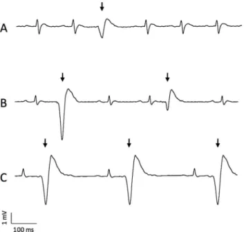
Only 3 out of 9 Wistar control rats exhibited VPB in a range of 3 to 6 premature beats among the 20,000 beats analyzed in each rat (0.1±0.003 ectopic beats/1,000 normal beats). On the other hand, 6 out of 8 WAR showed arrhythmic beats ranging from 15 to 32 VPB out of the 20,000 beats evaluated (0.41±0.2 ectopic beats/1,000 normal beats; P<0.001, compared with Wistar counterparts). All VPB observed for Wistar control rats came from a single ectopic ventricular origin, whereas 4 of 9 WAR studied showed VPB from multiple ventricular origins. Moreover, a short period (approximately 1.5 s) of bigeminy was observed in one WAR, as illus-trated inFig 1.
Discussion
sympathetic imbalance, markedly increases with aging [38,39,40]. In addition, chronic sympa-thetic overactivity is highly recognized as one of the most important determinants in the devel-opment of age-related heart failure, which is an isolated risk factor for cardiovascular events, including sudden death [23,24,25].
Differently to our previous study [18], resting HR was found similar between WAR and aged match Wistar control rats. Two striking differences in the protocols used in both studies can be highlighted and are certainly involved in this discrepancy. First, the age of the rats evalu-ated in both studies. The function of sinoatrial node, the pacemaker of the heart, is known to decline during the ageing process, [41]. Moreover, a loss of autonomic influence to the heart is also associated with ageing [42]. Therefore, the use of aged animals in the present study might explain the similar HR between WAR and Wistar control rats. Second, in our previous study [18] HR was calculated from pulsatile BP recorded from a catheter placed in the femoral artery 24 h prior to the experiment. In the present study, subcutaneous ECG electrodes, placed 48 h before recordings, were used to calculate HR.
WAR exhibited greater overall RRi variability compared with aged-matched control Wistar rats. The spectra of RRi, when calculated in absolute units (ms2), also revealed higher power at both, LF and HF bands in WAR. These findings support the idea of a higher cardiac autonomic
Table 2. Hemodynamic parameters and indices of systolic and diastolic function derived from left ventricle pressure-volume relationship in one-year old pentobarbital anesthetized WAR and Wistar control rats.
Control (Wistar) WAR P
Parameters
HR (bpm) 322±14 292±15 0.1829
MBP(mmHg) 115±5 92±3* 0.0008
LVSP (mmHg) 135±4 103±4* 0.0001
ESV (μL) 68±9 137±9* 0.0002
EDP (mmHg) 5.6±1.0 9.2±0.6* 0.0161
EDV (μL) 154±11 210±11 0.0030
CO (mL.min-1) 28.2±4.3 21.4±2.3 0.1764
TPR (mmHg.mL-1) 4.5±0.6 4.6±0.4 0.8955
Systolic Indexes
EF (%) 56±5 35±2* 0.0016
+dP/dt_max (mmHg.s-1. min) 7320±538 5266±620
* 0.0283
ESPVR slope (mmHg.μL-1) 2.6±0.4 1.9±0.1 0.1179
PRSW (mmHg) 49±6 36±4 0.0765
Diastolic Indexes
-dP/dt_max (mmHg.s-1) -7766±998 -5014±780
* 0.0463
τ(ms) 15.1±0.5 21.8±2.9 0.0531
EDPVR slope (mmHg.μL-1) 0.036±0.011 0.078±0.011* 0.0186
*P<0.05 compared with Wistar control rats.
HR: heart rate, MAP: mean arterial pressure, LVSP: left ventricular systolic pressure, ESV: end-systolic volume, EDP: end-diastolic pressure, EDV: end-diastolic volume, CO: cardiac output, TPR: total peripheral resistance, EF: ejection fraction, dP/dt_max maximal slopes of the systolic pressure increment (+dP/ dt_max) and diastolic pressure decrement (-dP/dt_max), ESPVR and EDPVR: slope of end-systolic and end-diastolic pressure-volume relationships,τ: relaxation time constant, PRSW: preload recruitable
stroke work.
Values are mean±SEM.
drive in WAR, i.e. greater sympathetic and vagal cardiac modulation. Nevertheless, LF power of RRi spectra was remarkably higher in WAR (10-fold) as compared to Wistar counterparts, leading to a higher LF and lower HF power of RRi spectra when expressed in normalized units. Thence, the LF/HF ratio, a well-accepted index of sympatho-vagal balance [28,29,35,43] was notably higher in WAR when compared with control rats. Power of PI spectra at LF band is widely accepted as an index of sympathetic modulation, while HF band power is associated with parasympathetic modulation [28,29,31]. Nevertheless, there is evidence that the LF power of HR spectra also correlates with parasympathetic modulation [43]. Thus, the powers of LF and HF bands more accurately represent autonomic modulation when presented in normal-ized units or as an LF/HF ratio [28,29,35].
Therefore, RRi variability evaluated in the present study in aged WAR strongly suggests an autonomic imbalance with sympathetic overactivity in this strain of aged rats. Substantial clini-cal and experimental evidence links sustained sympathetic activation to increased risk of life-threatening cardiovascular events, especially ventricular arrhythmias [38,44]. Furthermore, there is evidence that the most significant and widely-accepted mechanism of SUDEP involves cardiac arrhythmia induced by seizure discharges acting through the autonomic nervous sys-tem [3,45]. In line with our findings in WAR, there are evidences that epileptic patients also present interictal abnormalities in the autonomic modulation of cardiac activity, with high sympathetic and impaired vagal tone to the heart [14,15,16]. Changes in parasympathetic HR modulation have been noticed in either temporal lobe [13,46] or generalized epilepsies [47,48]. Moreover, anti-epileptic therapy, specifically with carbamezapine may also alter autonomic functions, increasing cardiovascular risk in epileptic patients [13,49].
The balance between sympathetic and vagal influences on the heart is a major regulator of cardiac function and can be a major cause of cardiac dysfunction. Diseases such as myocardial infarction, congestive heart failure and coronary disease are commonly associated with alter-ations in the normal sympatho-vagal balance [50].
Fig 1. ECG recordings illustrating ectopic beats (arrows) in 3 different WAR studied at 1 year of age.
(A) One isolated ectopic beat, (B) two ectopic beats from distinct ventricular origins and (C) a period of bigeminy.
To our knowledge, this is the first study to evaluate cardiac function in an experimental epi-lepsy model using a pressure-volume conductance system, a unique and powerful approach to evaluating systolic and diastolic functionin vivo[34]. Young (70 days) WAR recorded in con-scious freely moving state showed a mild hypertension [18] while aged (1 one year old) WAR, recorded under pentobarbital anesthesia, presented lower levels of basal BP as compared to Wistar control counterparts. The age of the animals, but mainly the effect of anesthesia should explain the difference in BP between these two studies. Aged WAR showed impaired systolic performance accompanied by delayed relaxation and increased diastolic stiffness of the left ventricle.
We found a lower +dP/dt_max and EF in aged WAR. Although +dP/dt_max has historical-ly been used as a cardiac contractile index, this parameter is known to be load-dependent (es-pecially preload-dependent) [51,52,53]. EF is also acknowledged to be influenced by both preload and afterload and cannot be used reliably to assess systolic function in models with changes in preload and/or afterload. The slope of ESPVR was proposed as a relatively load-in-sensitive index of ventricle contractility [51,54]. In the present study, ESPVR was decreased in WAR compared with Wistar rats. TPRI did not differ between the groups.
The data for the end-diastolic and end-systolic pressure volume relationships are presented inTable 2. The ESPVR slope was steeper in Wistar control rats than in WAR, suggesting de-creased systolic performance in WAR. Moreover, EDPVR was inde-creased in WAR compared to controls, indicating increased myocardial stiffness in WAR.
Impaired ventricular relaxation and increased end-diastolic stiffness were also observed in WAR, as reflected in the decreased-dP/dt_max, prolonged Tau, and increased LVEDP and EDPVR seen in these animals. Relaxation is an active process, depending mostly on calcium uptake during the diastole. End-diastolic stiffness is predominantly affected by changes in myocardial structural components [55].
A recent review article from Finsterer and Wahbi [56] regarding neurological diseases af-fecting the heart, describes that epilepsy is rarely directly associated with heart failure. Never-theless, since this disease in either humans or experimental animals is often associated with high blood pressure and autonomic disturbances, the risk of the development of heart failure during aging is expected to be markedly higher in subjects with epilepsy.
Further studies will be extremely important for understanding the mechanisms of autonom-ic imbalance in WAR, for identifying risk factors and triggers of cardiovascular events related to epileptic seizures, and for critically assessing potential strategies to minimize those risks.
Acknowledgments
Special thanks to Leonardo Fidelis Filho and Eduardo Gomes for the care of the animals. We confirm that we have read the Journal’s position on issues involved in ethical publication and affirm that this report is consistent with those guidelines. In addition to that we have no con-flict of interest to disclose.
Author Contributions
Conceived and designed the experiments: RFJr HCS NGC. Performed the experiments: CAAS JACO. Analyzed the data: RFJr HCS NM NGC. Contributed reagents/materials/analysis tools: RFJr NM NGC. Wrote the paper: RFJr NM NGC.
References
2. Tomson T, Nashef L, Ryvlin P (2008) Sudden unexpected death in epilepsy: current knowledge and fu-ture directions. Lancet Neurol 7: 1021–1031. doi:10.1016/S1474-4422(08)70202-3PMID:18805738
3. Devinsky O (2011) Sudden, unexpected death in epilepsy. N Engl J Med 365: 1801–1811. doi:10. 1056/NEJMra1010481PMID:22070477
4. Langan Y, Nashef L, Sander JWAS (2002) Certification of deaths attributable to epilepsy. J. Neurol. Neurosurg Psychiatr 73: 751–752. PMID:12438483
5. Persson H, Kumlien E, Ericson M, Tomson T (2005) Preoperative heart rate variability in relation to sur-gery outcome in refractory epilepsy. Neurology 65: 1021–1025. PMID:16217053
6. Stengl M (2010) Experimental models of spontaneous ventricular arrhythmias and of sudden cardiac death. Physiol Res 59(Suppl1): S25–31.
7. Neufeld G, Lazar JM, Chari G, Kamran H, Akajagbor E, Salciccioli L et al, (2009) Cardiac repolarization indices in epilepsy patients. Cardiology 114: 255–260. doi:10.1159/000233236PMID:19672064
8. Surges R, Thijs RD, Tan HL, Sander JW (2009) Sudden unexpected death in epilepsy: risk factors and potential pathomechanisms. Nat Rev Neurol 5: 492–504. doi:10.1038/nrneurol.2009.118PMID: 19668244
9. Surges R, Sander W (2012) Sudden unexpected death in epilepsy: mechanisms, prevalence and pre-vention. Curr Opin Neurol 25: 201–207. doi:10.1097/WCO.0b013e3283506714PMID:22274774
10. Walton NY. Systemic effects of generalized convulsive status epilepticus (1993) Epilepsia 34 (Suppl1): S54–S58. PMID:8462491
11. Goodman JH, Homan RW, Crawford IL (1999). Kindled seizures activate both branches of the auto-nomic nervous system. Epilepsy Res 34: 169–176. PMID:10210032
12. Hotta H, Koizumi K, Stewart M (2009) Cardiac sympathetic nerve activity during kainic acid-induced limbic cortical seizures in rats. Epilepsia 50: 923–927. doi:10.1111/j.1528-1167.2008.01860.xPMID: 19055488
13. Ansakorpi H, Korpelainen JT, Huikuri HV, Tolonen U, Myllylä VV, Isojärvi JI. (2002) Heart rate dynam-ics in refractory and well controlled temporal lobe epilepsy. J Neurol Neurosurg Psychiatr 72: 26–30. PMID:11784820
14. Frysinger RC, Engel J, Harper RM (1993) Interictal heart rate patterns in partial seizure disorders. Neu-rology 43(10): 2136–9. PMID:8413981
15. Devinsky O, Perrine K, Theodore WH (1994) Interictal autonomic nervous system function in patients with epilepsy. Epilepsia 35(1): 199–204. PMID:8112246
16. Faustmann PM1, Ganz RE (1994) Central cardio-autonomic disorganization in interictal states of epi-lepsy detected by phase space analysis. Int J Neurosci 78(1–2): 43–7. PMID:7883457
17. Mukherjee S, Tripathi M, Chandra PS, Yadav R, Choudhary N, Sagar R et al (2009) Cardiovascular au-tonomic functions in well-controlled and intractable partial epilepsies. Epilepsy Res 85 (2–3): 261–9. doi:10.1016/j.eplepsyres.2009.03.028PMID:19473819
18. Fazan R Jr, de Oliveira M, Oliveira JAC, Salgado HC, Garcia-Cairasco N (2011) Changes in autonomic control of the cardiovascular system in the Wistar audiogenic rat (WAR) strain. Epilepsy Behav 22: 666–670. doi:10.1016/j.yebeh.2011.09.010PMID:22015213
19. Volders PGA. Novel insights into the role of the sympathetic nervous system in cardiac arrhythmogen-esis (2010) Heart Rhythm 7: 1900–1906. doi:10.1016/j.hrthm.2010.06.003PMID:20570754
20. Rona G (1985) Catecholamine cardiotoxicity. J. Mol. Cell. Cardiol. 17: 291–306. PMID:3894676
21. Eisenhofer G, Friberg P, Rundqvist B, Quyyumi AA, Lambert G, Kaye DM, et al. (1996) Cardiac sympa-thetic nerve function in congestive heart failure. Circulation 93: 1667–1676. PMID:8653872
22. Adameova A, Abdellatif Y, Dhalla NS (2009) Role of the excessive amounts of circulating catechol-amines and glucocorticoids in stress-induced heart disease. Can J Physiol Pharmacol 87: 493–514. doi:10.1139/y09-042PMID:19767873
23. Singh K, Xiao L, Remondino A, Sawyer DB, Colucci WS (2001) Adrenergic regulation of cardiac myo-cyte apoptosis. J Cell Physiol 189: 257–265. PMID:11748583
24. Woodiwiss AJ, Tsotetsi OJ, Sprott S, Lancaster EJ, Mela T, Chung ES. (2001) Reduction in myocardial collagen cross-linking parallels left ventricular dilatation in rat models of systolic chamber dysfunction. Circulation 103: 155–160. PMID:11136701
25. Scheuer J (1999) Catecholamines in cardiac hypertrophy. Am J Cardiol 83: 70H–74H. PMID: 10750591
27. Cerutti C, Gustin MP, Paultre CZ, Lo M, Julien C, Vincent M (1991) Autonomic nervous system and car-diovascular variability in rats: a spectral analysis approach. Am J Physiol 261: H1292–1299. PMID: 1833987
28. Malliani A, Pagani M, Lombardi F, Cerutti S (1991) Cardiovascular neural regulation explored in the fre-quency domain. Circulation 84: 482–492. PMID:1860193
29. Task Force of the European Society of Cardiology and the North American Society of Pacing and Electrophysiology (1996) Heart rate variability: standards of measurement, physiological interpretation and clinical use. Circulation 93: 1043–1065. PMID:8598068
30. Su DF, Miao CY (2001) Blood pressure variability and organ damage. Clin Exp Pharmacol Physiol 28: 709–715. PMID:11553028
31. Montano N, Porta A, Cogliati C, Costantino G, Tobaldini E, Casali KR, et al. (2009) Heart rate variability explored in the frequency domain: a tool to investigate the link between heart and behavior. Neurosci Biobehav Rev 33: 71–80. doi:10.1016/j.neubiorev.2008.07.006PMID:18706440
32. Garcia-Cairasco N, Wakamatsu H, Oliveira JA, Gomes EL, Del Bel EA, Mello LE, et al. (1996) Neu-roethological and morphological (Neo-Timm staining) correlates of limbic recruitment during the devel-opment of audiogenic kindling in seizure susceptible Wistar rats. Epilepsy Res 26: 177–192. PMID: 8985699
33. Doretto MC, Fonseca CG, Lôbo RB, Terra VC, Oliveira JAC, Garcia-Cairasco N. (2003) Quantitative study of the response to genetic selection of the Wistar audiogenic rat strain (WAR). Behav Genet 33: 33–42. PMID:12645820
34. Pacher P, Nagayama T, Mukhopadhyay P, Bátkai S, Kass DA (2008) Measurement of cardiac function using pressure-volume conductance catheter technique in mice and rats. Nat Protoc 3: 1422–1434. doi:10.1038/nprot.2008.138PMID:18772869
35. Montano N, Ruscone TG, Porta A, Lombardi F, Pagani M, Malliani A. (1994) Power spectrum analysis of heart rate variability to assess the changes in sympathovagal balance during graded orthostatic tilt. Circulation 90: 1826–1831. PMID:7923668
36. Umeoka EHL, Garcia SB, Antunes-Rodrigues J, Elias LLK, Garcia-Cairasco N (2011) Functional char-acterization of the hypothalamic-pituitary-adrenal axis of the Wistar Audiogenic Rat (WAR) strain. Brain Res 1381: 141–147. doi:10.1016/j.brainres.2011.01.042PMID:21256829
37. Garcia-Cairasco N, Oliveira JA, Wakamatsu H, Bueno ST, Guimarães FS (1998) Reduced exploratory activity of audiogenic seizures susceptible Wistar rats. Physiol Behav 64: 671–674. PMID:9817579
38. Lakatta EG (1993) Cardiovascular regulatory mechanisms in advanced age. Physiol Rev 73: 413–467. PMID:8475195
39. Arshad A, Mandava A, Kamath G, Musat D (2008) Sudden cardiac death and the role of medical thera-py. Prog Cardiovasc Dis 50: 420–438. doi:10.1016/j.pcad.2007.12.003PMID:18474285
40. Dias da Silva VJ, Montano N, Salgado HC, Fazan R Jr (2006) Effects of long-term angiotensin convert-ing enzyme inhibition on cardiovascular variability in agconvert-ing rats. Auton Neurosci 124: 49–55. PMID: 16439186
41. Yann J, Tellez JO, Sutyyagin PV, Boyett MR, Dobrzynski H (2010) Structural remodeling of the sinoatri-al node in obese old rats. J Mol Cell Cardiol 48: 653–662. doi:10.1016/j.yjmcc.2009.08.023PMID: 19729016
42. Moodithaya S, Avadhany ST (2012) Gender differences in age-related changes in cardiac autonomic nervous function. J Aging Res 679345.
43. Appel ML, Berger RD, Saul JP, Smith JM, Cohen RJ (1986) Beat to beat variability in cardiovascular variables: noise or music? J Am Coll Cardiol 14: 1139–1148.
44. Sgoifo A, De Boer SF, Buwalda B, Korte-Bouws G, Tuma J, Bohus B. (1998) Vulnerability to arrhyth-mias during social stress in rats with different sympathovagal balance. Am J Physiol 275: H460–466. PMID:9683433
45. Nei M, Ho RT, Abou-Khalil BW, Drislane FW, Liporace J, Romeo A. (2004) EEG and ECG in sudden unexplained death in epilepsy. Epilepsia 45: 338–345. PMID:15030496
46. Ronkainen E1, Korpelainen JT, Heikkinen E, Myllylä VV, Huikuri HV, Isojärvi JI (2006) Cardiac auto-nomic control in patients with refractory epilepsy before and during vagus nerve stimulation treatment: a one-year follow-up study. Epilepsia 47(3): 556–62. PMID:16529621
47. Harnod T, Yang CC, Hsin YL, Shieh KR, Wang PJ, Kuo TB (2008) Heart rate variability in children with refractory generalized epilepsy. Seizure 17(4): 297–301. PMID:17977751
49. Tomson T, Ericson M, Ihrman C, Lindblad LE (1998) Heart rate variability in patients with epilepsy. Epi-lepsy Res 30(1): 77–83. PMID:9551847
50. Vanoli E, Adamson PB, Hull SS Jr, Foreman RD, Schwartz PJ (2008) Prediction of unexpected sudden death among healthy dogs by a novel marker of autonomic neural activity. Heart Rhythm 5: 300–305. doi:10.1016/j.hrthm.2007.10.021PMID:18242558
51. Kass DA (1995) Clinical ventricular pathophysiology: a pressure-volume view. In: Warltier DC, editor. Ventricular Function. Baltimore, MD: Williams & Wilkins. pp. 131–151.
52. Kass DA, Maughan WL, Guo ZM, Kono A, Sunagawa K, Sagawa K. (1987) Comparative influence of load versus inotropic states on indexes of ventricular contractility: experimental and theoretical analysis based on pressure-volume relationships. Circulation 76: 1422–1436. PMID:3454658
53. Little WC (1985) The left ventricular dP/dtmax-end-diastolic volume relation in closed-chest dogs. Circ Res 56: 808–815. PMID:4006092
54. Nakano K, Sugawara M, Ishihara K, Kanazawa S, Corin WJ, Denslow S. et al. (1990) Myocardial stiff-ness derived from end-systolic wall stress and logarithm of reciprocal of wall thickstiff-ness. Contractility index independent of ventricular size. Circulation 82: 1352–1361. PMID:2401069
55. Pacher P, Mabley JG, Liaudet L, Evgenov OV, Marton A, Haskó G, et al. (2004) Left ventricular pres-sure-volume relationship in a rat model of advanced aging-associated heart failure. Am J Physiol Heart Circ Physiol 287: H2132–2137. PMID:15231502