Development and
application of
bioanalytical
methodologies
based on firefly
luciferase
(Photinus pyralis)
Simone Marin Marques
Doctoral Program in Chemistry
Department of Chemistry and Biochemistry 2014
Supervisor
Joaquim Carlos Gomes Esteves da Silva
Associate Professor with Aggregation, Faculty of Sciences
This work was supported by a Ph.D. scholarship with the reference SFRH/BD/65109/2009,
co-funded by the European Social Fund (Fundo Social Europeu, FSE), through Programa
Operacional Potencial Humano - Quadro de Referência Estratégico Nacional (POPH -
QREN), and by national funds from the Ministry of Education and Science through the
Portuguese Fundação para a Ciência e a Tecnologia, I.P. (FCT, I.P.).
A c k n o w l e d g e m e n t s | VII
Acknowledgements
To my scientific supervisor, Professor Joaquim C.G. Esteves da Silva, without whom
none of this work would exist.
To the Evaluation Panel of the Chemistry scientific area of the Fundação para a Ciência
e a Tecnologia, I.P. (FCT, I.P.) Ph.D. Scholarship Call 2009.
They didn’t know me, I didn’t
know them, yet they changed my life.
To FCT, I.P., for granting the funding for my Ph.D. project.
To my host institution, Chemistry Research Center of the University of Porto (Centro de
Investigação em Química da Universidade do Porto, CIQ-UP), represented by its scientific
coordinator, Professor Manuel A.V. Ribeiro da Silva, for receiving me as a student and
collaborator, and for providing me the conditions to perform my work.
To the Doctoral Program in Chemistry scientific commission, Professor Manuel A.V.
Ribeiro da Silva, Professor Maria João Ramos and Professor Paula Gomes. To
Professor Gomes my special gratitude for always endure my countless e-mails. To my
schoolfellows in the Program. To the teaching staff from the Department of Chemistry
and Biochemistry and the invited lecturers who gave their time and their knowledge to
enrich the curricular part of the Doctoral Program.
To the Scientific Council of Faculty of Sciences, for accepting me as a student and
carefully processing all my bureaucracy over these four years with competence and
attention.
To Doctor Valentin N. Petushkov, for the amazing opportunity of taking part in the
elucidation of Fridericia heliota’s bioluminescent system.
VIII | A c k n o w l e d g e m e n t s
To Zélia Azevedo, for the able assistance in liquid chromatography-mass spectrometry
(LC-MS) analysis.
To Mariana Andrade, from Materials Center of the University of Porto (Centro de Materiais
da Universidade do Porto, CEMUP), for all the help with the nuclear magnetic resonance
(NMR) software. Always available, always so nice.
To the Department of Chemistry and Biochemistry secretariat and the Post-Graduate
students office staff, for all the patience and professionalism to solve all my issues.
To the lab managers Aurora Leal, Maria José Vasconcelos, Maria Arminda Silva, Maria
de Fátima Carvalho and Moisés Xavier, for doing a discrete yet important job.
To my lab buddies Margarida Miranda, Joel Santos, Diana Gomes, Conceição
Mendonça, Helena Gonçalves, Abel
‘MacGyver’ Duarte, Emanuel Ferreira, Ana Reis
and Marcela Oliveira, as well as my adoptive lab buddies Natércia Teixeira, Sónia
Salomé and Isabel Tavares
– always keeping up the fun.
To Sport Lisboa e Benfica, for the supreme joy and fun it gave me in the last few weeks
of the 2012-2013 soccer season.
To all my family. To little misses Carolina and Catarina big kisses and hugs.
To the new generation of Ph.D. students at lab 2.30 of the Chemometric Research of
Chemical, Environmental, Forensic and Biological Systems group, Eliana Simões, Luís
R e s u m o | IX
Resumo
Os recentes progressos da química bioanalítica conduziram a diversas aplicações
de uso generalizado. Neste âmbito destacam-se as técnicas bioluminescentes, com franco
desenvolvimento nos últimos anos, nas quais as luciferases, denominação genérica de
enzimas que, agindo sobre o seu substrato natural, luciferina, promovem uma reacção
bioquímica na qual ocorre libertação de fotões de luz visível, têm um papel dominante.
Este projecto teve como objectivo o desenvolvimento de métodos bioanalíticos visando
determinar espécies de interesse biológico, farmacêutico e ambiental baseando-se na
luciferase do pirilampo norte-americano Photinus pyralis (EC 1.13.12.7) e aplicando
metodologias de desenho experimental estatístico. Os analitos escolhidos foram os
pesticidas organofosforados, sulfato inorgânico, óxido nítrico (•NO) e ácidos gordos livres.
Em paralelo procedeu-se à análise estrutural de compostos semelhantes à luciferina de
Fridericia heliota, uma minhoca siberiana cujas características bioluminescentes foram
recentemente descobertas e que pode constituir um novo sistema bioanalítico de
interesse.
O método bioluminescente acoplado para pesticidas organofosforados apresenta
uma gama linear entre 2,5-15
M, com limite de detecção (LD) de 1,5
M e limite de
quantificação (LQ) de 5,0
M, e foi testado em águas de poços domésticos. O método
bioluminescente para sulfato inorgânico, por sua vez, apresenta uma gama linear entre
14-134 mg·mL
-1, com LD de 10 mg·mL
-1e LQ de 34 mg·mL
-1, e foi testado também em
águas de poços domésticos. Relativamente ao •NO, o método desenvolvido apresenta
uma gama linear entre 10-100 nM, LD de 4 nM e LQ de 15 nM, e foi testado em saliva
humana e meio de cultura de microalgas. Por fim, o método bioluminescente para ácidos
gordos livres apresenta uma gama linear entre 1-20
M, LD de 1,3 e
LQ de 4,5
M, e foi
testado em plasma sanguíneo.
Devido à reduzida quantidade de luciferina de Fridericia heliota que se consegue
extrair, não foi ainda possível estabelecer a sua estrutura química. No entanto, foram
realizados estudos utilizando-se compostos semelhantes à luciferina, que serviram como
modelos. Estes compostos foram informalmente denominados CompostoX (CompX) e
AsLn (acompanhante similar à luciferina). AsLn parece estar intimamente relacionado com
a verdadeira luciferina, como um sub-produto ou um intermediário na sua biossíntese,
enquanto CompX é um fragmento de AsLn.
X | P a l a v r a s - c h a v e
Palavras-chave
Química bioanalítica
Métodos bioluminescentes
Ensaio enzimático
Bioluminescência
Método de vias enzimáticas acopladas com detecção por bioluminescência
Luminometria
Luciferase de pirilampo
Photinus pyralis
Desenho experimental
Pesticidas organofosforados
Sulfato inorgânico (SO
42−)
Óxido nítrico (•NO)
Ácidos gordos livres
Limite de detecção
Limite de quantificação
Gama linear
Curva de calibração
Método das adições de padrão
Enquitreídeo
Fridericia heliota
Luciferina de Fridericia heliota
Compostos relacionados com a luciferina
CompX
AsLn(2)
Determinação da estrutura química
Cromatografia líquida de alto desempenho
Espectrometria de massa
A b s t r a c t | XI
Abstract
Recent advances in bioanalytical chemistry led to several general purpose
applications. In this context the focus is on the bioluminescent techniques, with a fast
development in recent years, in which luciferases, generic name for enzymes that, by acting
on its natural substrate, luciferin, promote a biochemical reaction which release photons of
visible light, have a dominant role. The purpose of this project was the development of
bioanalytical methods to determine species of biological, pharmaceutical and
environmental interest based on luciferase from the North-American firefly Photinus pyralis
(EC 1.13.12.7) and applying statistical experimental design methodologies. The chosen
analytes were organophosphorus pesticides, inorganic sulfate, nitric oxide (•NO) and free
fatty acids. In parallel proceeded the structural analysis of luciferin-related compounds from
Fridericia heliota, a Siberian earthworm whose bioluminescent features were recently
discovered and which may constitute a new interesting bioanalytical system.
The coupled bioluminescent method for organophosphorus pesticides has a linear
range between 2.5-15
M, with a limit of detection (LOD) of 1.5
M and a limit of
quantitation (LOQ) of 5.0
M, and has been tested in water from domestic wells. The
bioluminescent method for inorganic sulfate, in turn, shows a linear range between 14 to
134 mg·mL
-1, LOD 10 mg·mL
-1and LOQ 34 mg·mL
-1and was also tested in domestic wells’
water. Regarding •NO, the developed method has a linear range of 10-100 nM, LOD 4 nM
and LOQ 15 nM and was tested in human saliva and microalgae culture medium. Finally,
the bioluminescent method for free fatty acid has a linear range between 1-20
M, LOD 1.3
M and LOQ 4.5
M and was tested in blood plasma.
Due to the reduced amount of Fridericia heliota luciferin obtained in extracts, it was
not yet possible to establish its chemical structure. However, studies were performed using
compounds similar to the luciferin, which served as models. These compounds were
informally called CompoundX (CompX) and AsLn (accompanying similar to luciferin). AsLn
appears to be closely related to the true luciferin, as either a by-product or an intermediate
in its biosynthesis, whereas CompX is a fragment of AsLn.
XII | K e y w o r d s
Keywords
Bioanalytical chemistry
Bioluminescent methods
Enzymatic assay
Bioluminescence
Coupled bioluminescent assay
Luminometry
Firefly luciferase
Photinus pyralis
Experimental design
Organophosphorus pesticides
Inorganic sulfate (SO
42−)
Nitric oxide (•NO)
Free fatty acids
Limit of detection
Limit of quantitation
Linear range
Calibration curve
Method of standard additions
Enchytraeid
Fridericia heliota
Fridericia heliota luciferin
Luciferin-related compounds
CompX
AsLn(2)
Chemical structure elucidation
High-performance liquid chromatography (HPLC)
Mass spectrometry (MS)
S t r u c t u r e o f t h e T h e s i s | XIII
Structure of the Thesis
This Thesis is divided into four sections, the introduction, the presentation of the
developed methods, the studies made on Fridericia heliota’s luciferin-related compounds,
and a conclusion and future perspectives. Each section, by its turn, is composed of
chapters, corresponding to papers already published or manuscripts intended to be
submitted for publication in specialized peer-reviewed international journals.
The introduction section presents one chapter (Chapter one), in which
photochemical concepts, luciferases’ reaction mechanisms and bioluminescent techniques
principles are described, together with selected examples of applications. This section ends
with a brief description of the main objectives of the project that led to the Thesis.
The second section encompasses four chapters corresponding to the
bioluminescent methods for organophosphorus pesticides (Chapter two), inorganic sulfate
(Chapter three), nitric oxide (Chapter four) and free fatty acids (Chapter five).
The third section has three chapters, dealing with preliminary analyses on AsLn
(Chapter six) and the structural characterization of CompX (Chapter seven) and AsLn(2)
(Chapter eight).
The fourth and last section presents the main conclusions of the performed work,
as well as a book chapter addressing the development of novel bioluminescent methods
using nanomaterials (Chapter nine).
T a b l e o f c o n t e n t s | XV
Table of contents
Acknowledgements ... VII
Resumo ... IX
Palavras-chave ... X
Abstract ... XI
Keywords ... XII
Structure of the Thesis ... XIII
Table of contents ... XV
Section one - Introduction ... 1
Chapter one: There´s plenty of light at the bottom: Bioanalytical and biomedical
applications of luciferases ... 3
Objectives ... 37
Section two – Methods set up and application ... 39
Chapter two: Quantitative analysis of organophosphorus pesticides in freshwater
using an optimized firefly luciferase-based coupled bioluminescent assay ... 41
Chapter three: An optimized bioluminescent assay for inorganic sulfate
quantitation in freshwater ... 55
Chapter four: Nitric oxide quantitative assay by a glyceraldehyde 3-phosphate
dehydrogenase/phosphoglycerate kinase/firefly luciferase optimized coupled
bioluminescent assay ... 71
Chapter five: An optimized firefly luciferase bioluminescent assay for free fatty
acids quantitation in biological samples ... 111
Section three – Study of luciferin-related compounds from Fridericia heliota 125
Chapter six: LC-MS and microscale NMR analysis of luciferin-related compounds
from the bioluminescent earthworm Fridericia heliota ... 127
XVI | T a b l e o f c o n t e n t s
Chapter seven: CompX, a luciferin-related tyrosine derivative from the
bioluminescent earthworm Fridericia heliota. Structure elucidation and total
synthesis ... 143
Chapter eight: AsLn2, a luciferin-related modified tripeptide from the
bioluminescent earthworm Fridericia heliota ... 155
Section four – Future perspectives and conclusions ... 169
Future perspectives ... 171
Chapter nine: New methodologies based on the coupling of luciferase
with nanomaterials ... 173
Section one
Introduction
C h a p t e r o n e | 3
Chapter one
There´s plenty of light at the bottom: Bioanalytical and biomedical
applications of luciferases
Simone M. Marques, Joaquim C.G. Esteves da Silva
Glob. J. Anal. Chem. 2 (2011) 241-271
The text was integrally written and the figures were integrally made by Simone Marques
within the host institution, the Chemometric Research of Chemical, Environmental,
Forensic and Biological Systems group, from the Chemistry Research Center of the
University of Porto (Centro de Investigação em Química da Universidade do Porto,
CIQ-UP), Department of Chemistry and Biochemistry, Faculty of Sciences, University of Porto,
under the supervision of Joaquim Esteves da Silva.
GLOBAL JOURNAL OF ANALYTICAL CHEMISTRY
241
Global Journal of Analytical Chemistry | Volume 2 | Issue 6 | 2011
www.simplex-academic-publishers.com
© 2011 Simplex Academic Publishers. All rights reserved.
There´s plenty of light at the bottom:
Bioanalytical and biomedical applications of luciferases
Simone M. Marques, Joaquim C.G. Esteves da Silva*
Centro de Investigação em Química da Universidade do Porto (CIQ-UP), Department of Chemistry and Biochemistry, Faculty of Sciences, University of Porto, Rua do Campo Alegre, no. 687, 4169-007 Porto, Portugal
*
Author for correspondence: Joaquim C.G. Esteves da Silva, email: [email protected] Received 2 Mar 2011; Accepted 19 Apr 2011; Available Online 18 May 2011
Abstract
Bioanalytical chemistry has been attracting much attention in recent years due to the growing interest in biomolecules in the biochemical, biological, biomedical, pharmaceutical and environmental contexts, allowing detailed and sensitive analysis from the molecular up to the whole-body level. This review focuses on the application of luciferases, a generic name for enzymes whose biochemical reactions lead to a particular phenomenon called bioluminescence, concerning bioanalytical chemistry and biomedicine. Several powerful techniques employing luciferases were developed, among which bioluminescence resonance energy transfer (BRET) assays, split luciferase assays and bioluminescence imaging (BLI) stand out. Luciferase bioluminescent methods were successfully applied to gain insights into drug screening, gene-mediated therapy evaluation, infection progression, cancer therapy, cell tracking, biosensors, basic research and biomedical engineering, which will be illustrated through representative examples.
Keywords: Luciferase; Bioluminescence; Bioanalytical chemistry; Biomedicine; Bioluminescence imaging;
Bioluminescence resonance energy transfer; Split luciferase; Quantum dot
Abbreviations: BLI, bioluminescence imaging; BRET, bioluminescence resonance energy transfer; max, wavelength of maximum emission
1. Introduction
1.1. When analytical chemistry meets life sciences
Bioanalytical chemistry can be regarded, in simple terms, as the result of applying the concepts and techniques of “classical” analytical chemistry towards the study of biomolecules like nucleic acids, proteins, lipids, drugs and antibodies [1]. It is an interdisciplinary field, meeting the interest of chemistry, biochemistry, molecular and cell biology, biomedicine and clinical analysis, environmental toxicology, forensic sciences, food chemistry, industrial and pharmaceutical research, and so forth. Some well-established analytical techniques such as chromatography, mass spectrometry, spectrophotometry and nuclear magnetic resonance are also employed in bioanalysis, taking into account the specificities of biomolecules. Other techniques were developed owing to biomolecule studies, namely the enzyme linked immunosorbent assay (ELISA), the polymerase chain reaction (PCR) and electrophoresis [1].
Bioanalytical methods have to meet essential requirements in order to be suitable for
bioanalysis, namely an enhanced sensitivity, robustness and specificity. Nowadays, with their widespread use in clinical and pharmaceutical analysis, it is also desirable that they can be suitable for high-throughput screening, ease to perform, commercially available and moderate- to low-cost. In this context the enzyme luciferase is an invaluable tool [2], and this review aims to present its most recent application in outstanding research areas.
2. Bioluminescence and luciferases
2.1. Basic photochemical and photophysical concepts
Before focusing attention to the bioluminescence phenomenon itself, it may be convenient to expose some basic concepts on photochemistry and photophysics.
There are two relevant forms of electromagnetic radiation (light) emission, incandescence and luminescence. Originally they were called “hot light” and “cold light” respectively, since incandescence involves the conversion of vibrational energy into radiant energy as a consequence of a raise in the temperature [3], whereas luminescence is about
5
GLOBAL JOURNAL OF ANALYTICAL CHEMISTRY
242 Global Journal of Analytical Chemistry www.simplex-academic-publishers.com | Volume 2 | Issue 6 | 2011 © 2011 Simplex Academic Publishers. All rights reserved.
electronic transitions [4]. But actually, the main differentiation criterion between incandescence and luminescence should not be heat, but instead whether the light emission involves transitions in electronic energy levels among atoms or molecules, in the case of incandescence, or transitions in electronic energy levels within atoms or molecules, in the case of luminescence [5].
Regarding the energy source responsible for the formation of the electronically excited state, luminescence can be classified into different types. For example, photoluminescence is the result of absorption of photons and re-emission of photons of lower energy; electroluminescence occurs due to an electric current; and crystalloluminescence refers to the release of light as a result of the crystallization of a substance [6]. Chemiluminescence designates the process in which a chemical reaction produces the electronically excited substance. When these reactions are encountered in biological systems the effect is then known as bioluminescence [6].
All processes which occur within excited substances can be graphically presented through a Jablonski diagram (Figures 1a and 1b) [4]. In photoluminescence, an electron in the electronic ground state (S0, Figure 1a) is excited (E, Figure 1a) to one of the many vibrational levels in the electronic excited state (S1 and S2, Figure 1a) as a consequence of absorption of photons. Typically, those are excited singlet states (S), so that the electrons remain spin-paired. Once in the excited electronic state, there are many possible ways in which energy can be released to return the electron to its ground state. A molecule in an excited state can lose energy to other molecules through collisions, or distribute its excess energy to its own vibrations and
rotations (VR, Figure 1a) [4]. The electron can also undergo internal conversion (IC, Figure 1a), placing it from a higher vibrational level (S2) to a lower one (S1). It is also possible that the molecule loses its energy in other processes that do not lead to light emission, called non-radiative transfer of energy (NRT, Figure 1a). Finally, an electron in excited singlet states can also return to the ground state by emitting a photon (F, P, Figure 1a). If the emission comes from singlet states it is called fluorescence (F, Figure 1a). The other possibility for the electron is to change its spin in a process called intersystem crossing (ISC, Figure 1a). This results in a triplet excited state. From the triplet state the electron must change its spin again before returning to the ground state. If this is done along with the emission of a photon, the process is called phosphorescence (P, Figure 1a). It is also possible for the triplet state to return to the ground state by non-radiative transfer (NRT, Figure 1a) [4]. Analogous processes occur in bioluminescence (Figure 1b), except that the initial excited state is created by a biochemical reaction. The corresponding emission of photons (Em, Figure 1b) is regarded as bioluminescence emission, although it is indistinguishable from fluorescencent emission.
Another important concept is the quantum yield (Φ). It can be defined as the number of defined events occurring per photon absorbed by the system (1) [6]:
Φ =
(1) For example, in fluorescence processes it can be specifically stated as the ratio between the number of photons emitted to the number of photons absorbed, and in chemiluminescence or
Figure 1. Jablonski diagrams for (a) photoluminescence and (b) bioluminescence. Photoluminescenc (a) is
initiated by the absorption of electromagnetic radiation (wavy blue arrow), whereas in bioluminescence (b) an excited state product is generated in a biochemical reaction; its decay to the ground state releases photons of visible light. See text for more details.
GLOBAL JOURNAL OF ANALYTICAL CHEMISTRY
243
Global Journal of Analytical Chemistry | Volume 2 | Issue 6 | 2011
www.simplex-academic-publishers.com
© 2011 Simplex Academic Publishers. All rights reserved.
bioluminescence it is the amount of photons emitted to the amount of reactant consumed or product formed [6]. In those latter cases, Φ can be divided into three components: (1) the fraction of the reaction that produces the light emitter, Φr; (2) the fraction of emitter that is formed in a excited state, Φex; and (3) the fraction of those excited states that emit light, Φfl (quantum yield for fluorescence) [7, 8]; the overall efficiency resulting is then Φ = Φr Φex Φfl [7, 8]. In this sense, the quantum yield can be viewed as a measure of the efficiency of the system.
2.2. It is only bioluminescence with luciferases As it was seen above, bioluminescence can be regarded as a particular case of chemiluminescence. The main difference is that bioluminescent chemical reactions must be catalyzed by specific enzymes. These enzymes are generally called luciferases, whose name is derived from the Latin words lucem ferre, meaning “light-bearer”. In the same sense, the corresponding substrates are the luciferins. All bioluminescent reactions are oxidations with molecular oxygen, and they may also require co-factors such as adenosine-5’-triphosphate (ATP), magnesium ions or reduced flavin mononucleotides (riboflavin-5’-phosphate, FMNH2). Some reactions involve additional proteins, which will be described later.
Bioluminescence is ubiquitous in nature. Several creatures developed their luciferase-luciferin systems, for example bacteria (Photobacterium, Xenorhabdus, Vibrio),
dinoflagellates (Gonyaulax, Noctiluca, Pyrocystis), coelenterates (Aequorea, Renilla),
annelids (Diplocardia, Fridericia), mollusks (Latia), crustaceans (Cypridina, Metridia, Gaussia), fungi (Panellus, Mycena, Omphalotus)
and insects (Photinus, Photuris, Luciola,
Pyrophorus, Phrixothrix, Pyrearinus, Arachnocampa) [9-12].
However, for practical purposes, the main luciferases in use are those from bacteria, from the marine copepod crustaceans Gaussia
princeps, Cypridina noctiluca and Cypridina hilgendorfii, from the coelenterate Renilla reniformis and from the North American firefly Photinus pyralis, which will be the focus from
this point on.
3. Bacterial luciferase
Bacterial luciferase is classified as alkanal, FMNH2: oxygen oxidoreductase (1-hydroxylating, luminescing), EC 1.14.14.3. All bacterial luciferases isolated so far are heterodimers composed of two non-identical
subunits, an subunit with molecular weights between 40-42 kDa and a subunit between 37-39 kDa, giving a global molecular weight about 76 kDa [10, 12]. The bioluminescent reaction involves the oxidation of FMNH2 and a saturated long-chain aliphatic aldehyde with more than ten carbon atoms, preferably tetradecanal [12]. The aldehyde is considered to be the bacterial luciferin, producing the corresponding carboxylic acid, oxidized flavin (FMN), a molecule of water and light in the blue region of the visible spectrum [wavelength of maximum emission ( max ) = 490 nm] in vitro, with a quantum yield between 0.10 and 0.16 [10, 12].
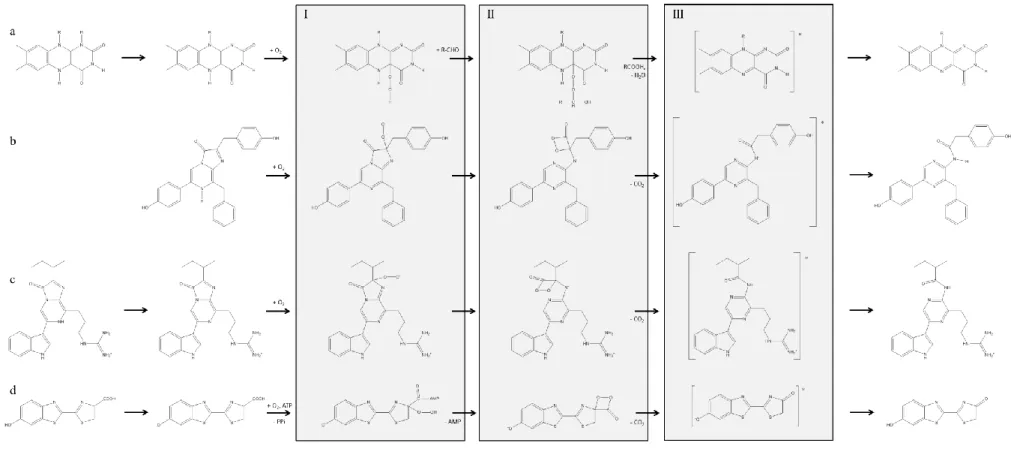
For the bioluminescent reaction to begin, FMNH2 must be produced in bacterial cells from FMN, by reduction with the enzyme flavin reductase using nicotinamide adenine dinucleotide reduced (NADH) as the reducing agent (Figure 2a) [10, 12, 13]. The free form of FMNH2 is extremely unstable in the presence of oxygen, being instantly oxidized. In the presence of bacterial luciferase, however, FMNH2 bounds to it and is deprotonated at a nitrogen atom, forming an FMNH2-luciferase complex that is more stable than free FMNH2 (Figure 2a). The deprotonated flavin in the complex is readily attacked by molecular oxygen, giving a hydroperoxide (intermediate I, Figure 2a). In the presence of a long-chain aldehyde (bacterial luciferin), intermediate I is converted into intermediate II, which contains a peroxyhemiacetal of flavin (Figure 2a). The decomposition of intermediate II, through several steps, yields the excited-state hydroxyflavin-luciferase complex (intermediate III) and the corresponding fatty acid. Although the aldehyde is regarded as the luciferin, the light emitter is considered to be the luciferase-bound hydroxyflavin (intermediate III, Figure 2a). Light is emitted when the excited state intermediate III falls to the ground state, generating water and FMN, which is regenerated by flavin reductase to enter a new reaction cycle [10, 12]. In the same way, the produced carboxylic acid can be reduced again to the corresponding aldehyde. With purified bacterial luciferases, the maximum emission is similar for different species and occurs at 490 nm, but a completely different pattern is seen in vivo [10, 12]. In fact, in vivo emission maxima range from 472 to 545 nm, depending on the bacterium species [12]. This phenomenon is explained by the presence of accessory proteins, namely bacterial blue fluorescent lumazine protein (LumP) and bacterial yellow fluorescent protein (YFP), which absorb the light energy produced by bacterial luciferases and re-emits in longer wavelengths [10, 12]. This process,
7
GLOBAL JOURNAL OF ANALYTICAL CHEMISTRY
244 Global Journal of Analytical Chemistry www.simplex-academic-publishers.com | Volume 2 | Issue 6 | 2011 © 2011 Simplex Academic Publishers. All rights reserved.
Figure 2. Schematic biochemical reactions catalyzed by (a) bacterial luciferase, (b) Renilla and Gaussia luciferases, (c) Cypridina luciferase and (d) firefly luciferase. In general terms,
those reactions involve specific substrate, luciferins, which react with molecular oxygen to generate intermediates I; afterwards, those intermediates originate the energy-rich intermediates II, either by the reaction with long-chain aldehydes (a) or by the formation of four-membered ring dioxetatones (b-d); finally, the excited state light emitter intermediates III are produced, which release photons of light and leads to the oxidized ground-state final products.
GLOBAL JOURNAL OF ANALYTICAL CHEMISTRY
245
Global Journal of Analytical Chemistry | Volume 2 | Issue 6 | 2011
www.simplex-academic-publishers.com
© 2011 Simplex Academic Publishers. All rights reserved.
bioluminescence resonance energy transfer (BRET), will be explained in section 8.6.
Bacterial luciferase genes (lux genes) are arranged in an operon, luxCDABE [14]. The
luxA and luxB genes code for the subunits of
bacterial luciferase, whereas luxC, D and E code for an enzyme, fatty acid reductase, responsible for the biosynthesis of the aldehyde. Other lux genes may be found in certain species, like the regulatory genes luxI and luxR, or luxH involved in riboflavin synthesis [14]. This genetic organization of the bioluminescent system genes in bacteria opens up the interesting possibility of constructing self-illuminating cells and organisms through the introduction of the whole
luxCDABE operon, as it codes for both bacterial
luciferase itself and the enzymes to synthesize bacterial luciferin [15].
4. Renilla reniformis (Renilla) luciferase
The luciferase from the sea pansy
Renilla reniformis is one of the best characterized luciferase-luciferin system from a marine organism. Its systematic name is Renilla-luciferin: oxygen 2-oxidoreductase (decarboxylating), EC 1.13.12.5. Purified Renilla luciferase has a molecular weight of about 35 kDa [10, 12]. In vitro, the bioluminescent reaction presents a pHoptimal at 7.4, at 32 ºC and in the presence of a salt like NaCl or KCl. It generates photons in the blue range ( max = 480 nm) with a quantum yield of about 0.06-0.07 [10, 12]. Renilla luciferin is coelenterazine, an imidazopyrazinone common to other bioluminescent and non-bioluminescent marine organisms [12, 16].
Regarding the in vivo bioluminescent reaction, coelenterazine is stored in the form of coelenterazine enol-sulfate [12]. Part of the coelenterazine sulfate stock is subjected to sulfate removal by a luciferin sulfokinase, allowing coelenterazine to bind to coelenterazine-binding protein, a protein akin to calmodulin in its capacity to bind calcium ions. When calcium concentration is raised, by nerve stimulation, three calcium ions bind to coelenterazine-binding protein, triggering the release of coelenterazine [12]. From this point coelenterazine binds to Renilla luciferase, where it will react with oxygen producing a peroxide intermediate (intermediate I, Figure 2b) [12]. This intermediate undergoes a cyclization to form an energy-rich dioxetanone intermediate (intermediate II, Figure 2b), whose breakage leads to carbon dioxide (CO2) and the oxidized form of coelenterazine, coelenteramide, in the excited state (intermediate III, Figure 2b) [10, 12]. Like in the bacterial bioluminescent system,
in Renilla’s system there is also an accessory protein, Renilla green fluorescent protein (Not to confuse with the better known green fluorescent protein from the jellyfish Aequorea victoria, whose discovery and development granted the Nobel Prize in Chemistry in 2008) [10, 12]. In the presence of Renilla green fluorescent protein, the energy of the excited state coelenteramide is transferred, by BRET, to the fluorescent protein, resulting in the emission of green light ( max = 509 nm); in the absence of the fluorescent protein or in in vitro conditions, however, blue light (λmax = 480 nm) is emitted [10, 12].
5. Gaussia princeps (Gaussia) luciferase
Gaussia luciferase, from the homonymous marine crustacean species Gaussia
princeps, is one of the latest luciferases being
used in bioanalytical applications. It is the smallest luciferase found to date, with a molecular weight about 19.9 kDa [12, 17-19]. It is naturally secreted, in vivo and in vitro, due to a secretory sequence peptide. Moreover, Gaussia luciferase is a coelenterazine-dependent luciferase, and its bioluminescent reaction mechanism is believed to equal that of Renilla luciferase, namely the oxidative decarboxilation of coelenterazine to yield excited-state coelenteramide and photons of blue light ( max = 470 nm) (Figure 2b) [12, 17-19]. Like other coelenterazine-dependent luciferases, this luciferase has a tendency to self-aggregate into inactive forms [12]. In vitro, the bioluminescent reaction presents a pHoptimal at 7.7, and its activity is highly dependent upon the concentration of monovalent cations. Nonetheless, Gaussia luciferase is very resistant to high temperatures (that is, it is a thermostable enzyme) and to extreme acidic and basic conditions, which is important for technological applications [12, 17-19].
Contrary to early discovered luciferases, which were subjected to decades of studies before cloning, genetic engineering improvements and assignement to molecular biology and bioanalytical applications, Gaussia was readily recruited to practical applications. Indeed, Gaussia luciferase is currently commercially available as plasmids (Figure 3). Furthermore, it was already subjected to genetic enhancements. One such important feature includes a recombinant biotinylated version of Gaussia luciferase [20]. Biotin is a small vitamin which is used in laboratorial studies as a biological “hook” to separate proteins [21]. This compound is chemically linked to a protein of
interest. After that, the sample with the biotinylated protein is incubated with
GLOBAL JOURNAL OF ANALYTICAL CHEMISTRY
246 Global Journal of Analytical Chemistry www.simplex-academic-publishers.com | Volume 2 | Issue 6 | 2011 © 2011 Simplex Academic Publishers. All rights reserved.
straptavidin or avidin immobilized into a support. Both of them bind strongly and specifically to biotin, allowing the separation of the biotinylated protein from solution [21]. A biotinylated Gaussia luciferase facilitate its purification, reduces its inactivation due to conjugation with other molecules and facilitates its use as a bioprobe [20]. Another improved Gaussia luciferase is a “humanized”, codon-optimized version which produces 200- (in vivo) to 1000-fold (in vitro) higher bioluminescence signals in mammalian cells compared to Renilla and firefly luciferases [22].
6. Cypridina luciferase
Cypridina luciferase is obtained from marine crustaceans (sea fireflies) of the species
Cypridina noctiluca [23] and Cypridina hilgendorfii (also known as Vargula hilgendorfii) [12, 24]. Its classification is Cypridina-luciferin: oxygen 2-oxidoreductase
(decarboxylating), EC 1.13.12.6. The bioluminescent substrate is Cypridina luciferin, sometimes called vargulin, an imidazopyrazinone chemical compound distinct from the other luciferins described so far [12]. The bioluminescent reaction also requires molecular oxygen, yielding CO2 and oxidized vargulin [12]. Cypridina luciferase is a 61-62-
kDa enzyme, emitting blue light ( max = 452 nm) in vivo, although the color can vary in vitro
according to the buffer used. The pHoptimal is 7.7 and the optimum temperature is about 30 ºC [12]. Salts like NaCl or CaCl2 are important to enhance the reaction output. In vivo, it is secreted to the medium [12], as well as in in vitro assays [25].
The Cypridina bioluminescenct reaction proceeds according to the scheme shown in Figure 2c [12]; the imidazopyrazinone part of Cypridina luciferin is negatively charged when the luciferin is bound to luciferase, making it easily oxygenated by molecular oxygen, and leading to a peroxide anion (intermediate I, Figure 2c). Analogously to Renilla biolumescent reaction, the peroxide cyclizes, forming a dioxetanone ring (intermediate II, Figure 2c), which instantly decomposes by a concerted splitting of the 4-membered ring into CO2 plus an amide oxidized Cypridina luciferin in an excited state (intermediate III, Figure 2c). Light is emitted when the excited state falls to its ground state. The quantum yield is about 0.3 [12].
7. Photinus pyralis (firefly) luciferase
Firefly luciferase, or Photinus-luciferin: oxygen 4-oxidoreductase (decarboxylating,
Figure 3. Principle of luciferase reporter gene assay. A plasmid containing the luciferase gene and the genetic
element under study is constructed (1). These plasmids also contain several genetic elements to allow the correct processing of luciferase gene, like selectable markers and promoters. Once this plasmid is processed by the the cell’s DNA replication machinery, luciferase is transcribed (that is, an mRNA is produced) and translated (2) into functional luciferase (3). By adding luciferin (4), light is emitted (5).
GLOBAL JOURNAL OF ANALYTICAL CHEMISTRY
247
Global Journal of Analytical Chemistry | Volume 2 | Issue 6 | 2011
www.simplex-academic-publishers.com
© 2011 Simplex Academic Publishers. All rights reserved.
ATP-hydrolysing), EC 1.13. 12.7, from the North American firefly (Photinus pyralis), is the most popular luciferase so far regarding bioanalytical applications. It is also one of the best studied and characterized luciferase, thanks to the work of William McElroy and colleagues during several decades, although some details regarding its biochemical mechanism are not yet clear [26].
Firefly luciferase is a 62-kDa [27, 28] enzyme bearing a peroxisome targeting peptide in its native form [29]. The firefly luciferin is the D- enantiomer of a benzothiazolyl-thiazole, [(S)-2-(6’- hydroxy-2’-benzothiazolyl)-2-thiazoline-4-carboxylic acid], or simply D-luciferin [30]. The same way coelenterazine is common to several marine organisms, so all the bioluminescent beetles (order Coleoptera;
families Elateridae, for example click beetles;
Phengodidae, like railroad-worms; and
Lampyridae, the fireflies) share this luciferin in
their bioluminescent reaction [10, 12, 26, 30, 31].
Contrary to the other luciferases described so far, firefly luciferase requires ATP, along with a divalent metallic cation, most frequently magnesium ions, to proceed. Unlike the common sense, however, ATP does not have an energetic role, but rather to provide a good leaving group, adenosine-5´-monophosphate (AMP), which facilitates the subsequent steps of the bioluminescent reaction [10]. In vivo, yellow-green light is emitted; in vitro, however, the emission peak can be the same as in vivo ( max = 562 nm), as firefly’s bioluminescent system does not have accessory fluorescent proteins, but only at basic media (pH about 7.5-7.8) [10, 12, 26, 30, 31]. In fact, firefly luciferase is pH-sensitive, and acidic media (pH about 5-6) can shift the emission to the red ( max = 620 nm), along with higher temperatures or heavy metal cations [10, 12, 26, 30, 31]. The pHoptimal is about 7.8 at 23-25 ºC. The formation of inhibitory by-products can hamper the emission from luciferase. The addition of coenzyme A (CoA) leads to the conversion of these inhibitory by-products to a less inhibitory form, thus stabilizing and prolonging the light emission [10, 12, 26, 30-32]. The detailed mechanism of the bioluminescent reaction catalyzed by firefly luciferase is somewhat complex, and its full presentation is not under the remit of this review (interested readers may consult ref. [26, 30, 31]). In short, the reaction emcompasses the activation of D-luciferin by ATP-Mg2+, generating inorganic pyrophosphate (PPi) and a luciferase-bound adenylated intermediate (Figure 2d), an anhydride formed between the carboxyl group of D-luciferin and the phosphate group of AMP [10,
12, 26, 30, 31]. The adenylate is oxidated by oxygen in the air, forming a hydroperoxide intermediate (intermediate I, Figure 2d) which, by its turn, produces the dioxetanone ring-bearing intermediate II by the departure of AMP. As this high-energy ring is very unstable, it quickly breaks down, yielding CO2 and oxyluciferin in an excited state (intermediate III, Figure 2d). The excited state oxyluciferin release its energy as photons, leaving oxyluciferin in the ground state [10, 12, 26, 30, 31]. The quantum yield of this reaction is about 0.41-0.48 [33, 34]. 8. Bioanalytical methodologies based on
luciferases – then and now
This section aims to present the basic principles underlying the diversified luciferase-based methods established for bioanalytical purposes, preceding the presentation of demonstrative examples of their application. 8.1. Luciferase assay
In initial studies about luciferase, as early as 1947, McElroy observed that the duration of the emitted light by firefly luciferase
in vitro was directly proportional to the amount
of ATP added [35]. Although at that time the role of ATP in the bioluminescent reaction was unknown, and only a qualitative relationship between light and ATP could be inferred [35], it was a hint of the potential of luciferase for ATP assay. And indeed this was the case. In the incoming years, the major application of firefly luciferase was in luciferase assays for ATP [36]. In the simplest assays, a reaction mixture is prepared, composed of luciferase, luciferin and magnesium cations (like MgCl2) in a buffer system at pH 7.6-7.8. Sometimes CoA is also added to stabilize the light emission. A calibration curve with ATP is obtained and the sample is tested. The emitted light is recorded on luminometers, a specific device equipped with photomultiplier tubes with variable degrees of sensitivity. With time, a more complete knowledge of firefly luciferase bioluminescent reaction was achieved, and thus more sophisticated methods were developed [2], for example the ATP measurement inside cells with genetically encoded luciferases (see next section), along with the development of assays for other analytes which participate in the reaction, namely CoA [37] and PPi [38]. Likewise, analytes other than ATP could be assessed with bacterial luciferase, as its reaction allows the assay of long-chain aldehydes [39], NADH [40, 41] and FMN [40].
GLOBAL JOURNAL OF ANALYTICAL CHEMISTRY
248 Global Journal of Analytical Chemistry www.simplex-academic-publishers.com | Volume 2 | Issue 6 | 2011 © 2011 Simplex Academic Publishers. All rights reserved.
8.2. Luciferase reporter gene assay
Reporter gene can be defined as “a gene with a readily measurable phenotype that can be distinguished easily over a background of endogenous proteins” [42, 46]. Reporter gene technology is, then, a molecular biology tool in which a reporter gene is introduced into cells, together with a genetic entity of interest and, once it is translated into protein, it can be readily detected, giving information about molecular or cellular events under study (Figure 3) [43]. The reporter gene and the genetic entity under study are placed together in the same DNA construct, usually in the form of a circular DNA molecule called a plasmid (Figure 3), prior to their introduction into cells. The most commonly studied processes are the following [42, 43]: 1) the expression of a gene of interest. In this case the reporter is directly attached to the gene of interest, being the two genes under the control of the same promoter elements and being transcribed into a single messenger RNA (mRNA). The mRNA is then translated into the protein, and the localization of the protein can be traced by assaying the reporter, for example by adding its substrate when the reporter is an enzyme; and 2) the activity of a particular promoter or other regulatory element, like an enhancer. In this case there is no separate "gene of interest"; the reporter gene is simply placed under the control of the target promoter or element, and its transcriptional strength is then estimated quantitatively from the in vitro activity of the reporter gene product, considering that the action of the promoter upon the reporter will be the same as that upon the native gene [42, 43].
The onset of this technology dates back early 1980’s, for both prokaryotic and eukaryotic cells [44, 45]. At this time popular reporter genes were the enzymes chloramphenicol acetyltransferase, -galactosidase and alkaline phosphatase [46, 47]. Firefly luciferase was introduced as a reporter gene a few years after its cloning in 1985 [28], for example in plant cells and transgenic whole plants [48], for the study of the cauliflower mosaic virus 35s RNA promoter [49], for the analysis of the interleukin-2 promoter [50] and for the study of estrogen regulatory elements in a Xenopus (aquatic frog) model [51]. In the same sense, bacterial luciferase was then applied as reporter, for example in the study of genetic recombinatory mechanisms in prokaryotes [52].
8.3. Bioluminescent Enzyme Immunoassay (BLEIA)
An enzyme immunoassay (EIA) is a biochemical technique used mainly in immunology to detect the presence of an
antibody or an antigen in a sample. In simple terms, a specific antibody is added to a sample with an unknown amount of the antigen or
vice-versa. This antibody is linked to an enzyme and,
in the final step, a substrate is added so that the enzyme can convert it into some detectable signal, most commonly a color change [53, 54]. When the enzyme is luciferase, the assay is called bioluminescent enzyme immunoassay (BLEIA) [55].
Regarding the most common procedure, a buffered solution of the antigen to be tested is added to each well of a microtiter plate, where the antigen will adhere. A solution of a non-reacting protein, such as bovine serum albumin (BSA) or casein, is added to block any plastic surface in the well that remains uncoated by the antigen. Next, a primary antibody is added, which binds specifically to the test antigen that is coating the well. This primary antibody could also be in the serum of a donor, in which case it will be tested for reactivity towards the antigen. Afterwards, a secondary antibody is added, which will bind the primary antibody. This secondary antibody often has an enzyme attached to it, which should have a negligible effect on the binding properties of the antibody. A substrate for this enzyme is then added. Often, this substrate changes color upon reaction with the enzyme. The color change shows that secondary antibody has bound to primary antibody, which strongly implies that the donor has had an immune reaction to the test antigen. The higher the concentration of the primary antibody which was presented in the sample, the stronger is the color change [53, 54]. Often a spectrometer is used to give quantitative values for color strength. In other formulations the primary antibodies can be added to the microtiter plate first, followed by the antigen sample and the secondary antibodies. Also, chemically modified antigens can be marked with the enzyme, instead of the antibody, and mixed to an antigen sample. The labeled antigen competes for primary antibody binding sites with the corresponding sample antigen (unlabeled). The more antigens in the sample, the less labeled antigen is retained in the well and weaker is the signal [53, 54].
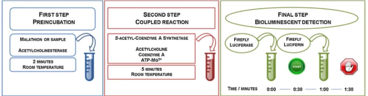
8.4. Coupled Bioluminescent Assay (CBA) A coupled enzymatic assay refers to the determination of a substrate or enzyme activity by coupling one or more enzymatic reaction with another one whose final product is more easily detected. The product of the first reaction is the substrate for the subsequent reaction, and so forth, until the last reaction. In coupled bioluminescent assays, luciferase catalyzes the
12
GLOBAL JOURNAL OF ANALYTICAL CHEMISTRY
249
Global Journal of Analytical Chemistry | Volume 2 | Issue 6 | 2011
www.simplex-academic-publishers.com
© 2011 Simplex Academic Publishers. All rights reserved.
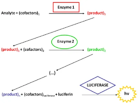
last reaction, and the final output is the measurement of the emitted photons (Figure 4) [55]. This method is often applied when the enzyme-catalyzed reaction or product of interest is difficult to assay directly.
Regarding luciferase, this method is well-established, both in custom-made developed assay, for example for the assay of enolase [56], as well as commercial standard kits [55].
8.5. Bioluminescence Imaging (BLI)
Bioluminescence imaging (BLI) is a technology developed over the past decades which allows the study of the light emitted from living entities to produce an image, where a profile of the emitted light can be visualized and biological information can be inferred [57-60]. This concept was first sketched in early 1990’s for single cell measurements [61] and evolved up to the whole body imaging in small laboratory animals in 1995 [62]. It encloses, therefore, molecular imaging, which is “the visualization, characterization, and measurement of biological processes at the molecular and cellular levels in humans and other living systems” [63], to whole-body imaging in small animals. Today it has a widespread use.
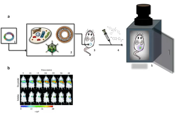
Conceptually, it can be viewed as a fusion between bioluminescent reporter gene and optical imagiology (Figure 5a). First, the gene encoding the luciferase is incorporated into a plasmid (Figure 5a, 1), which is then inserted into cells or other genetic material carriers, like
liposomes or viruses (Figure 5a, 2). Then they are transferred to living small animals like mice (Figure 5a, 3). It is given time for functional luciferase to be produced from the gene and the image is taken [57-60]. The animals are placed in light-tight chambers and, immediately before imaging, they are anesthetized to keep them still. Gas anesthesia with isoflurane or intraperitoneal injection of ketamine and xylazine are safe methods for short-term immobilization, as it is the case of BLI [57-60]. Luciferin, when necessary, is then injected (Figure 5a, 4). Generally, D-luciferin is injected intraperitoneally, whereas coelenterazine is administered via lateral tail vein [57-60]. A normal photography of the animal, in grayscale, is acquired under weak illumination to serve as an anatomic reference. After that, the bioluminescent signal is captured in complete darkness by an ultra-sensitive charge-coupled device (CCD) camera mounted on the top of the chamber (Figure 5a, 5) [57-60]. The duration of the imaging is variable, ranging from a few seconds up to several minutes (maximum of 2 minutes for Renilla luciferase, and 5 minutes for firefly luciferase) [57-60]. The signal intensity is computationally recorded, treated by specific softwares and finally represented as a pseudocolor image superimposed on the greyscale reference photo. The signal intensity is normally quantified as photons per second per cm2 per steradian with a color scale from blue (lowest intensity) to red (highest intensity) [57-60].
Figure 4. Principle of coupled bioluminescent assay. In a chemical or biochemical reaction, catalyzed by
enzyme 1, product 1 is formed. Product 1 will enter into a second reaction cycle with enzyme 2, by its turn generating product 2. The last reaction in the sequence is catalyzed by a luciferase, and the emitted light is measured. A correlationbetween the intensity of the emitted light and the concentration of the initial reagent (analyte) is inferred and quantitative results can be calculated.
GLOBAL JOURNAL OF ANALYTICAL CHEMISTRY
250 Global Journal of Analytical Chemistry www.simplex-academic-publishers.com | Volume 2 | Issue 6 | 2011 © 2011 Simplex Academic Publishers. All rights reserved.
The distinctive component of this technology is the CCD cameras, since they must be capable of detecting very low levels of emitted light [64]. This sensitivity can be enhanced by, for example, cooling the CCD chip to below -100°C, which significantly reduces background dark current signals, and are referred as cooled CCD [57, 64]. Several systems are commercially available; moreover, some models were specifically designed to measure light emission from small animals, for example the IVIS™ imaging system (Xenogen Corporation) with improved functionalities, like the imaging of groups of several animals simultaneously or the incorporation of nose cones to deliver gaseous anesthetics to animals during the course of imaging [57, 58].
8.6. Bioluminescence Resonance Energy Transfer (BRET)
8.6.1. The BRET phenomemon
In sections 3 and 4, it was stated that there are marked differences among the various bacterial species and strains concerning the in
vivo bioluminescence spectra versus the in vitro
one. The emission maxima are spread mostly in a range from 472 to 545 nm in vivo, whereas the
in vitro bioluminescence spectra measured with
purified luciferases obtained from various bacterial species and strains are all similar ( max about 490 nm) [12]. Likewise, the Renilla’s bioluminescent system emits blue light ( max = 480 nm) in vitro, a value that is shifted to green light ( max = 509 nm) in vivo [12]. These spectral shifts of light emission could indicate the occurrence of some energy transfer process involving the luciferase and a chromophore. Today it is known that such process is resonance energy transfer (RET).
RET was observed in the laboratorial context as well as biological systems, like the carotenoid-to-chlorophyll RET in marine diatoms [65]. A theory of RET was proposed by the German scientist Theodor Förster in the 1940’s [66], and hence the common acronym Förster resonance energy transfer (FRET) by which this phenomenon is also known. According to the theory, RET is a photophysical process involving two chromophores, a “donor” and an “acceptor”. The “donor”, initially in an electronically excited state, can transfer excitation energy to an “acceptor” molecule.
Figure 5. Bioluminescence imaging (BLI). (a) Principle of BLI. A plasmid (1) containing a luciferase gene is
introduced into vectors, such as cells, viruses or liposomes (2). These vectors are injected into a living test animal (3). Prior to the analysis, luciferin is injected (4) and the animal is transferred to a chamber mounted with a camera (5). The emitted photons are registered by the device’s software and displayed as colored spots upon the animal’s picture. (b) An example of a BLI analysis. In this study, mice were treated with a powder formulated for pulmonary gene delivery, containing luciferase as a reporter gene. Both its dry and soluble form were tested and, through a BLI analysis, it was verified that the dry powder is more efficient in delivering the gene to the lungs. Figure 5b adapted from ref. [103] and reproduced with permission from Elsevier.
GLOBAL JOURNAL OF ANALYTICAL CHEMISTRY
251
Global Journal of Analytical Chemistry | Volume 2 | Issue 6 | 2011
www.simplex-academic-publishers.com
© 2011 Simplex Academic Publishers. All rights reserved.
This intermolecular energy transfer occurs by dipole-dipole coupling, and not by electron transfer, whereby it is non-radiative [65-67] (Figure 6a).
Bioluminescence resonance energy transfer (BRET) is a particular case of RET in which the “donor” molecule is a luciferase [67] (Figure 6a). In vivo, the “acceptor” is a fluorescent protein, like the bacterial blue fluorescent proteins (LumPs), containing lumazine as their chromophores, and bacterial yellow fluorescent proteins (YFPs), containing a chromophore of FMN or riboflavin [12], or the green fluorescent protein from Renilla reniformis [12].
In order to RET occur, at least two conditions must be fulfilled: firstly, the emission spectrum of the “donor” must overlap with the excitation spectrum of the “acceptor” (Figure 6b); secondly, the two molecules must be in close proximity, from 1 to 10 nm at maximum [65, 67]. This last requirement was readily recognized as a possible application to evaluate protein-protein interactions.
8.6.2. The BRET technology
As an analytical method, the first RET methods involved a fluorescent protein coupled to another fluorescent protein capable of emitting at a different wavelength [68]. In this case, the method is called fluorescent resonance energy transfer (FRET). This denomination refers, hence, to the nature of the “donor”-“acceptor” pairs, and not to the mechanism of light transfer. In both FRET and BRET, to investigate protein-protein interactions, one protein-protein of the pair is genetically fused to the “donor”, and the second
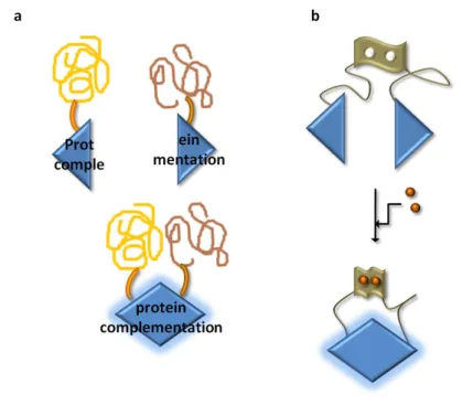
protein to the “acceptor” (Figure 7a). The “donor” protein is excited and, if the two proteins do not interact, only one light signal, corresponding to the “donor” emission, is registered (Figure 7a, 1); however, when the two proteins interact, RET can occur and an additional light signal, corresponding to the “acceptor” emission, is detected (Figure 7a, 2) [69]. The “donor” and “acceptor” reporters can be fused to a single protein, and in this case an intramolecular signal can be monitored, which is useful to study conformational changes upon the binding of a ligand, for example (Figure 7b) [70]. The RET signal can be displayed in several formats, depending on the purpose of the study (Figure 7c).
Although the occurrence of BRET in nature is established a long time ago [71], technological applications of BRET in bioanalysis was not done until 1999, when it was used to demonstrate that the circadian clock protein KaiB, from a cyanobacterium, forms homodimers, by genetically fusing it to Renilla luciferase and a modified green fluorescent protein [72]. Since then, a “boom” of applications based on BRET were developed. For example, FRET- and BRET-based method were extensively applied to study the interactions of cellular receptors with their corresponding ligands, for example the interaction between G protein-coupled receptors (GPCR) and trimeric G protein upon addition of the receptor’s agonist, norepinephrine [73], or to verify receptor’s dimerization [74, 75].
A variant of the BRET methodology recently described is the conjugation of luciferases and quantum dots [76]. Quantum dots
Figure 6. The bioluminescence resonance energy transfer (BRET) principles. (a) Jablonski diagram showing
the “donor” and “acceptor” coupling and resonance energy transfer. (b) Spectral overlap requirement for the occurrence of BRET. A “donor” molecule is excited at its specific wavelength and light emission will occur. If the “donor” emission matches the energy required for a nearby molecule to be excited, energy transfer by BRET will occur and the “acceptor” will posteriorly re-emit this energy as photons.
GLOBAL JOURNAL OF ANALYTICAL CHEMISTRY
252 Global Journal of Analytical Chemistry www.simplex-academic-publishers.com | Volume 2 | Issue 6 | 2011 © 2011 Simplex Academic Publishers. All rights reserved.
are tiny fluorescent and semiconductor crystals of inorganic elements, like cadmium and tellurium, with dimensions in the nanoscale, typically from 3 to 100 nm [77-79]. At this scale, quantum dots present properties differing from the bulk material they came from. In fact, quantum dots have unique optical properties like high quantum yields, large molar extinction coefficients, large excitation spectra, narrow emission spectra, size-dependent tunable emission and high photostability, which make them appealing fluorescent probes for imaging [77-79].
The principle of luciferase-quantum dots BRET is the same as already described (Figure 8a). The first reported protocol was
published in 2006, using a genetically modified Renilla luciferase with improved stability and light output [80]. To obtain such luciferase, eight mutations were performed, and so this mutant was called Renilla luciferase8 [81, 82].
The major advantage is that quantum dots can be produced within a wide emission wavelength, from blue to near-infrared [77-79], which is the desired range for BLI in small living animals. In fact, the interaction of light with tissues involves several kinds of processes (Figure 9). When light is emitted in a bioluminescent reaction inside a living animal, photons passing through the tissues can be scattered by cell and organelles membranes or absorbed by intrinsic cellular chromophores like
Figure 7. The BRET technology. (a) To assess protein-protein interactions, each protein of the presumably
interacting pair is genetically fused to a luciferase and to an “acceptor”, which can be a fluorescent protein, a quantum dot or an organic dye (in this scheme, a fluorescent protein is represented). If the two proteins do not interact, or if they are far apart from each other, BRET will not occur and a single signal, corresponding to the luciferase, will be registered (1); on the other hand, when the two proteins interact, BRET will occur between the luciferase and the “acceptor” and two signals will be recorded, one from the luciferase and a novel peak corresponding to the emission of the “acceptor” (2). (b) Schematic representation of intramolecular BRET. Two BRET pairs are genetically fused to the same protein. By adding its agonist (orange circles), for example, the protein will undergo structuralchanges, bringing the two BRET pairs into close proximity and allowing the occurrence of BRET. (c) BRET data representation. According to the purpose of the analysis, the BRET results can be displayed under several formats.