Development of
molecular tools for
the early warning of
potentially toxic
cyanobacteria
Ana Isabel Bastos de Matos
Mestrado em Biologia Celular e Molecular
Departamento de Biologia2014
Orientador
Agostinho Antunes Pereira, Professor Auxiliar Convidado, Faculdade de Ciências da Universidade do Porto
Co-orientador
Vítor Manuel de Oliveira e Vasconcelos, Professor Catedrático, Faculdade de Ciências da Universidade do Porto
Todas as correções determinadas pelo júri, e só essas, foram efetuadas. O Presidente do Júri,
Acknowledgments
The present work would have not been possible without the help of many people. I want to express my gratefulness addressing them the first words.
First of all, I want to thank to Professor Doctor Agostinho Antunes his availability to be my supervisor throughout this year and for the confidence deposited in me during the development of this thesis. To my co-supervisor, Professor Doctor Vitor Vasconcelos I would like to thank the opportunity of have allowing me to work in his investigation group.
To Fundação para a Ciência e Tecnologia (FCT), through PesT-C/MAR/LA0015/2013, PTDC/AAC-AMB/104983/2008 (FCOMP-01-0124-FEDER-008610) and PTDC/AAC-CLI/116122/2009 (FCOMP-01-0124-FEDER-014029) projects, for financing this project.
To all LEGE elements, I want to thank the good working environment and all the assistance provided. In special, I want to express my gratitude to Doctor Cristiana Moreira who always supported me and shared with me her precious advices and knowledge which allowed me to achieve success on this work.
To Doctor Catarina Churro, from the Instituto Nacional de Saúde Dr. Ricardo Jorge, I would like to thank for the development of the Real-Time PCR standards and to LECEMA, research CIIMAR group, the supply of Real-Time PCR equipment.
To all the master students in particular Catarina C., Joana M., Rita, Susana, Tiago Af. and Tiago Az. for all the help and the always present good humour. In special to Catarina S. that revealed herself as a good friend - sometimes a coffee and a laugh solved many problems.
To all my friends that helped and encouraged me since I began my academic studies I would like to thank them however, I have to highlight Joana R., Leandra, Patricia and Paula as well as Joana F., Margarida, Marta and Tânia for all their friendship and support.
To Ana, Eunice, Mariana, Pedro e Tiago thank you for your support words, good mood and most of all your true friendship that I know that it will be for life.
Finally, I want to express my gratitude to all my family. In special to my parents, my sister Beatriz, my brother José, my sister in law Vanda and my nephews Rita and Tiago for all the support in the most complicated situations and in all the decisions that I take, without them nothing would have been possible.
Abstract
Cyanobacteria are photosynthetic microorganisms that inhabit a wide range of ecological niches. These microorganisms have the capacity to produce secondary metabolites with many biological activities, among them cyanotoxins that represent a human health concern due to their association with cases of human sickness and death, as well as various incidents of animal mortality. Thus, it is necessary to develop new effective tools, such as the molecular methods, for the early warning of water systems for the presence of potentially toxic cyanobacteria species, instead of using solely the traditional microscopy observation and identification. Microcystis aeruginosa is known to be widely dispersed in Portuguese freshwater systems, however, the presence of Cylindrospermopsis raciborskii and Planktothrix agardhii has only been recently described in these water systems. These species can constitute a public health concern due to their potential to form not only blooms but also to produce toxic compounds (cyanotoxins). This study was focused in these three potentially toxic cyanobacteria through the application of molecular techniques for the detection of these species in seven Portuguese freshwater systems located in the North and Centre regions of Portugal, during two consecutive years. In this study, it was also developed a Real-Time PCR protocol for the absolute quantification of M.aeruginosa cells in water systems and also specific primers for Aphanizomenon genus rpoC1 gene since to date they are still unavailable.
The applied techniques allowed the first detection of C.raciborskii in North regions of Portugal, emphasizing its invasive behavior in our national territory, as well as the first report of P.agardhii in North and Centre regions of Portugal. M.aeruginosa was the most dominant species in all the studied water systems. The developed Real-Time PCR protocol for M.aeruginosa permitted a detection limit of 55 copies of gyrB gene per µL of PCR reaction. Similarly, the preliminary assays with the developed primers for Aphanizomenon genus revealed their high specificity and similarity with other sequences of this gene belonging to species of this genus.
In conclusion, this study reinforces not only the need of a continuous monitoring of these three cyanobacteria species in these studied regions but also the need to include other regions of the country. The molecular tools here developed constitute a first step to better characterize the cyanobacteria community in aquatic systems as well as to contribute to the early warning of cyanobacterial blooms.
Keywords: cyanobacteria, molecular tools, freshwater systems, Microcystis
Resumo
As cianobactérias são microorganismos fotossintéticos que habitam uma vasta diversidade de ecossistemas. Um dos grupos de metabolitos secundários mais estudados, produzidos por estes microorganismos, são as cianotoxinas. Estas constituem um perigo ao nível da saúde publica uma vez que estão associadas a casos de morte de animais e intoxicação humana devido ao seu potencial tóxico. Devido a estes factos, é necessário desenvolver novas ferramentas eficazes, tais como os métodos moleculares, para a rápida deteção da presença de espécies de cianobactérias potencialmente tóxicas nos vários ecossistemas de água doce em vez de se usar somente as tradicionais técnicas de microscopia. Microcystis aeruginosa é conhecida por ser uma espécie comummente presente nos vários ecossistemas de água doce portugueses contudo Cylindrospermopsis raciborskii e Planktothrix agardhii só recentemente foram detectadas em Portugal.
Este estudo centrou-se, assim, na monitorização destas três espécies de cianobactérias usando ferramentas moleculares para a sua deteção em sete ecossistemas de água doce localizados em regiões Norte e Centro de Portugal, durante um período de dois anos de amostragem. Neste estudo também foi desenvolvido um protocolo de PCR em tempo real para a quantificação absoluta de células de M.aeruginosa em amostras de água bem como o desenho de novos primers específicos para o gene rpoC1 para o género Aphanizomenon, que até ao momento estão indisponíveis.
As técnicas aplicadas permitiram a primeira deteção de C.raciborskii nas regiões Norte de Portugal, enfatizando uma vez mais o seu comportamento invasor em território nacional, bem como constitui o primeiro relato de P.agardhii em regiões do Norte e Centro de Portugal. M.aeruginosa foi a espécie mais dominante nos ecossistemas de água doce estudados. O protocolo de PCR em tempo real desenvolvido para M.aeruginosa permite a detecção de até 55 cópias de gyrB por µL de reação de PCR. Paralelamente, os testes preliminares para os primers desenvolvidos revelaram elevada especificidade e similaridade para o gene rpoC1 para espécies do género Aphanizomenon. Os resultados obtidos representam não só a necessidade de continuar a monitorização destas espécies de cianobactérias nestas regiões estudadas mas também a necessidade de incluir outras regiões do país.
As ferramentas moleculares desenvolvidas neste estudo constituem o primeiro passo para uma melhor compreensão da comunidade de cianobactérias nos vários ecossistemas aquáticos e contribuem para o alerta precoce de florescências de cianobactérias.
Palavras-Chave: cianobactérias, ferramentas moleculares, sistemas de água doce,
Microcystis aeruginosa, Cylindrospermopsis raciborskii, Planktothrix agardhii, Aphanizomenon
Table of Contents
Figures Index ... vii
Tables Index ... ix
List of Abbreviations ... x
1. Introduction ... 1
1.1 Cyanobacteria ... 1
1.2 Portuguese Freshwater Cyanobacteria Community ... 4
1.3 Detection Methodologies ... 6
1.4 Studied Cyanobacteria Species ... 9
1.4.1 Microcystis aeruginosa... 10 1.4.2 Planktothrix agardhii ... 10 1.4.3 Cylindrospermopsis raciborskii ... 11 1.4.4 Aphanizomenon genus ... 11 1.5 Sampling Sites ... 12 1.6 Objectives ... 12
2. Materials and Methods ... 13
2.1 Sampling ... 13
2.1.1 Description of Sampling Sites ... 13
2.1.2 Sampling Procedure ... 16
2.1.3 Sample Treatment ... 17
2.2 Isolation and Culturing ... 17
2.3 DNA Extraction ... 17
2.4 Agarose gel electrophoresis ... 18
2.5 DNA Quantification ... 18
2.6 Molecular Methods ... 18
2.6.1 Primers development ... 18
2.6.2 Polymerase Chain Reaction ... 19
2.6.3 Real-Time Polymerase Chain Reaction ... 21
2.7 Statistical Analyses ... 22
3. Results ... 23
3.1 Physical and Chemical Parameters ... 23
3.1.1 pH ... 23
3.2 Molecular Results ... 27
3.2.1 Detection of M.aeruginosa ... 27
3.2.2 Detection of C.raciborskii ... 28
3.2.3 Detection of P.agardhii ... 29
3.2.4 Global Analysis - M.aeruginosa, C.raciborskii and P.agardhii... 30
3.3 Microscopy Results ... 32 3.4 Real-Time PCR ... 32 3.4.1 Specificity assay ... 33 3.4.2 Sensitivity assay ... 33 3.5 Primers Development ... 34 4. Discussion ... 35
5. Conclusion and Future Perspectives ... 40
Figures Index
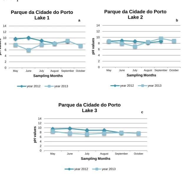
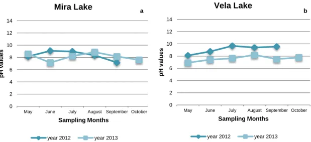
Figure 1 – Representative scheme of the mentioned methods possible to be applied for the detection of cyanobacteria species either in environmental samples or in isolated organisms from laboratory cultures. ... 9 Figure 2 - Sampling site at Torrão Reservoir. ... 14 Figure 3 - Sampling site at Tâmega River (Marco de Canaveses). ... 14 Figure 4 - Sampling sites at Parque da Cidade do Porto. In the largest picture it is possible to observe a satellite view of the lakes (1, 2, 3 and 4) and its distribution in the Park. The smallest pictures are photographs of the Lake 1 (1), Lake 2 (2) and Lake 3 (3) taken at the sampling collections. ... 15 Figure 5 - Sampling Site at Vela Lake. ... 16 Figure 6 - Sampling Local at Mira Lake. ... 16 Figure 7 - pH values registered during the sampling dates in Lake 1 (a), Lake 2 (b) and Lake 3 (c) from Parque da Cidade do Porto in the two sampling years (2012 and 2013). ... 23 Figure 8 - pH values registered at the sampling dates in Torrão Reservoir (a) and Tâmega River (Marco de Canaveses) (b) in the two sampling years, 2012 and 2013. 24 Figure 9 - pH values registered at the sampling dates in Mira Lake (a) and Vela Lake (b) in the two sampling years, 2012 and 2013. ... 25 Figure 10 – Maximum and minimum temperature registered in the sampling dates in Porto/Pedras Rubras weather station (a) and Luzim weather station (b) (North regions) in the two sampling years, 2012 and 2013. ... 26 Figure 11 - Maximum and minimum temperature registered in the sampling dates in Dunas de Mira weather station (a) and Figueira da Foz weather station (b) (Centre regions) in the two sampling years, 2012 and 2013. ... 26 Figure 12 - Frequency of detection, per sampling month, of M.aeruginosa, C.raciborskii and P.agardhii in the first year of sampling (2012), in all of the sampling points, through PCR technique. ... 31 Figure 13 - Frequency of detection, per sampling month, of M.aeruginosa, C.raciborskii and P.agardhii in the second year of sampling (2013), in all of the sampling points, through PCR technique. ... 31 Figure 14 - M. aeruginosa, isolated from Tâmega River (Marco de Canaveses). ... 32 Figure 15 - C.raciborskii isolated from Lake 2 of Parque da Cidade do Porto. ... 32 Figure 16 - Result of the PCR performed to confirm the identification of the
C.raciborskii isolated from Lake 2 of Parque da Cidade do Porto (1); positive control
Figure 17 - Result of the PCR performed to confirm the identification of the
M.aeruginosa isolated from Tâmega River (Marco de Canaveses) (1); positive control
(2). ... 32 Figure 18 - Melt curve chart of the M.aeruginosa strains applied in the Real-Time PCR specificity assay. ... 33 Figure 19 - Standard curve obtained with the application of the tenfold serial dilution of
Tables Index
Table 1 – Main cyanobacteria genera producers of cyanotoxins. Adapted table from
Carmichael (2001). ... 3
Table 2 - Primers applied in conventional PCR and their characteristics. ... 20
Table 3 - PCR programs applied to detect the species in study ... 20
Table 4 - Microcystis aeruginosa strains applied in Real-Time PCR ... 21
Table 5 - Detection of M.aeruginosa in the freshwater systems analysed in the two monitoring programs. The mark (+) indicates the detection of M.aeruginosa and the mark (-) indicates the non-detection of this species, through PCR amplification. The diagonal line (/) means that the sampling was not performed in the marked site or month. ... 27
Table 6 - Detection of C.raciborskii in the freshwater systems analysed in the two monitoring programs. The mark (+) indicates the detection of C.raciborskii and the mark (-) indicates the non-detection of this species, through PCR amplification. The diagonal line (/) means that the sampling was not performed in the marked site or month ... 28
Table 7 - Detection of P.agardhii in the freshwater systems analysed in the two monitoring programs. The mark (+) indicates the detection of P.agardhii and the mark (-) indicates the non-detection of this species, through PCR amplification. The diagonal line (/) means that the sampling was not performed in the marked site or month. ... 29
Table 8 - Overall analysis of the detection of the three cyanobacteria species in the seven freshwater systems analysed. (+) represents the detection of the species in at least one sampling month and (-) represents the non-detection of the species in all the sampling months. ... 30
List of Abbreviations
BLAST - Basic Local Alignment Search Tool BMAA - B-methylamino-L-alanine
BSA - Bovine Serum Albumin DNA - Deoxyribonucleic Acid
dNTP’s - Deoxynucleotide Triphosphates ELISA - Enzyme-Linked Immunosorbent Assay
ERIC-PCR - Enterobacterial Repetitive Intergenic Consensus - Polymerase Chain Reaction
FISH - Fluorescence In Situ Hybridization
gyrA - DNA gyrase subunit A gyrB - DNA gyrase subunit B
HPLC - High-Pressure Liquid Chromatography IPMA - Instituto Português do Mar e da Atmosfera MC-LR - Microcystin-LR
MC-RR - Microcystin-RR MC-YR - Microcystin-YR
NCBI - National Center for Biotechnology Information PCR - Polymerase Chain Reaction
PCR-DGGE - Polymerase Chain Reaction - Denaturing Gradient Gel Electrophoresis PCR-RFLP - Polymerase Chain Reaction - Restriction Fragment Length Polymorphisms
PCR-TGGE - Polymerase Chain Reaction -Temperature Gradient Gel Electrophoresis PP1 - Protein Phosphatase 1
PP2A - Protein Phosphatase 2A PSP - Paralytic Shellfish Poisons
1. Introduction
1.1 Cyanobacteria
Cyanobacteria, one of the major phyla of Bacteria (Madigan et al. 2010), are probably the most ancient organisms living in the Planet earth since it is believed that its appearance occurred between 2.45 and 2.32 billion years ago, contributing to the Great Oxidation Event and to the increasing levels of oxygen molecules in the atmosphere (Bekker et al. 2004; Schirrmeister et al. 2013). These organisms, classified as Gram-negative bacteria, were the first oxygenic phototrophic organisms (Madigan et al. 2010) and the raise in oxygen molecules was responsible for the evolution and emergence of new species.
Besides oxygen, there is also the production of chlorophyll a, carotenoids and phycobilins by these microorganisms. Most of them have a blue-green colour due to the production of phycocyanin, one class of phycobilins, and clorophyll a, however some cyanobacteria species may present red or brown colour due to the production of phycoerythrin, a red phycobilin (Madigan et al. 2010).
It is also well known the production of secondary metabolites by cyanobacteria with many biological activities such as anticancer activity, toxic activity and antiprotozoal properties (Tan, 2013). Through extraction and purification methods these compounds have been isolated, characterized and tested to be applied in pharmaceutical and food industries (Liu et al., 2014). However, the secondary metabolites most studied due to their hepatotoxic, neurotoxic, cytotoxic and dermatotoxic potential are the toxic compounds, also known as cyanotoxins.
Cyanotoxins can be organized, by their mode of action, in neurotoxins (anatoxin-a, -as, homoanatoxin-a, B-methylamino-L-alanine and saxitoxins), hepatotoxins (microcystins and nodularins), cytotoxins (cylindrospermopsin), dermatotoxins (lyngbyatoxin and aplysiatoxin) and irritant toxins (lipopolysaccharides) (Carmichael 2001; Cox et al. 2005; Dittmann and Wiegand 2006). The group of hepatotoxins comprises the microcystins and nodularins, being the first frequently detected in the freshwater systems. Several toxic variants of microcystins are known to exist such as MC-LR, MC-RR and MC-YR (Campos and Vasconcelos, 2010). Hepatotoxins have the ability to inhibit serine/threonine-specific protein phosphatases (PP1 and PP2A), which causes phosphorylation of cellular proteins, due to the presence of a rare amino acid (Adda) in their protein composition (Campos and Vasconcelos 2010; Dittmann and Wiegand 2006). The accumulation of these cyanotoxins occurs mainly in liver leading to cellular damages including cytoskeletal disorganization, DNA (deoxyribonucleic acid) fragmentation and intrahepatic bleeding, which can result in the death of organisms, it
is also associated with these cyanotoxins the promotion of tumours (Dittmann and Wiegand 2006). Alternatively, anatoxins and saxitoxins belong to the neurotoxins group. The group of anatoxins includes anatoxin-a, homoanatoxin-a and anatoxin-a(s) which are alkaloid compounds whose toxicological action occurs mainly in the nervous system of the organisms infected. Anatoxin-a is capable of binding to nicotinic acetylcholine receptors of the sodium channel, because it is a nicotinic agonist (Dittmann and Wiegand 2006), altering the electric transmission which may lead to muscle fasciculation, cyanosis and, in severe cases, in death by asphyxiation due to muscular paralysis (Carmichael, 2001). Anatoxin-a(s) may also over stimulate muscles and generate hypersalivation, tremors and ataxia because it is a cholinesterase inhibitor so, when present, acetylcholine is not removed from the nicotinic receptor generating action potentials leading to exhaustion of the nerve cell (Carmichael 2001; Dittmann and Wiegand 2006). Homoanatoxin-a is a homologue of anatoxin-a which mimic the effect of acetylcholine (Carmichael and Li 2006). Moreover, saxitoxins, also known as paralytic shellfish poisons (PSP’s), are able to block sodium channels leading to the blocking of nervous transmission which may result in nervousness, ataxia, muscle paralysis and death (van Apeldoorn et al. 2007; Briand et al. 2003; Carmichael 2001). Furthermore, B-methylamino-L-alanine (BMAA), may affect brain and central nervous system causing neurologic damage (Al-Sammak et al. 2014). Cylindrospermopsin besides blocking protein synthesis may induce DNA strand breaks and chromosome loss being described as genotoxic and cytotoxic. After ingestion of this cyanotoxin, the first symptoms are kidney and liver failure (Carmichael, 2001; Humpage et al. 2000). The dermatotoxins are potent tumour promoters, Aplysiatoxin is a protein kinase C activator and Lyngbyatoxin A, besides being a promoter of skin tumour, has caused oral and gastrointestinal inflammations in humans (Carmichael and Li 2006). Cyanobacterial lipopolysaccharides are the group of cyanotoxins less studied however it is associated with cutaneous symptoms, headache, fever and respiratory diseases (Stewart et al. 2006).
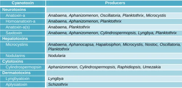
It is worthy to note that each toxin can be produced by more than one cyanobacterium genus or species which means that the production of cyanotoxins is not strain-specific and that the same cyanobacteria genera or species are able to produce more than one type of cyanotoxin (Table 1). There are many hypotheses about the reason why these compounds are produced, some authors suggest the role of these metabolites as iron scavenging molecules (Utkilen and Gjolme 1995), infochemicals (Kaebernick et al. 2000), growth regulators (Sedmak and Kosi 1998) or even as a protective role (Tran et al. 2013). However, there are species which do not express the cyanotoxins coding genes being known as non-toxigenic species, these
are morphologically indistinguishable from the toxin-producing cyanobacteria (Fastner et al. 2001).
Table 1 – Main cyanobacteria genera producers of cyanotoxins. Adapted table from Carmichael (2001).
Cyanotoxin Producers
Neurotoxins
Anatoxin-a Anabaena, Aphanizomenon, Oscillatoria, Planktothrix, Microcystis
Homoanatoxin-a Anabaena, Aphanizomenon, Planktothrix
Anatoxin-a(s) Anabaena, Planktothrix
Saxitoxin Anabaena, Aphanizomenon, Cylindrospermopsis, Lyngbya, Planktothrix
Hepatotoxins
Microcystins Anabaena, Aphanocapsa, Hapalosiphon, Microcystis, Nostoc, Oscillatoria, Planktothrix
Nodularins Nodularia
Cytotoxins
Cylindrospermopsin Aphanizomenon, Cylindrospermopsis, Raphidiopsis, Umezakia
Dermatotoxins
Lyngbyatoxin Lyngbya
Aplysiatoxin Schizothrix
This group of microorganisms is characterized by a huge variety of morphologies, existing unicellular as well as filamentous forms which are widespread across the world, from the tropical to the Polar regions, either in aquatic or in terrestrial habitats (Ionescu et al. 2010; Liu et al. 2014). This diversity of morphologies led to the division of the cyanobacteria into five orders according to the Botanical Code criteria (Henson et al. 2002). The first (Order Chroococcales) comprises colonial or coccoid cyanobacteria whose division is through binary fission. Order Pleurocapsales includes coccoid and pseudofilamentous cyanobacteria which has a combined cell division by multiple and binary fission whereas in the Order Oscillatoriales are grouped filamentous cyanobacteria with absence of heterocytes and akynetes. Filamentous cyanobacteria with heterocytes, akynetes and false branching belong to Order Nostocales whilst Order Stigonematales comprises filamentous cyanobacteria, facultative heterocytes and akynetes and true branching (Komárek 2003).
Furthermore, some cyanobacteria possess cellular characteristics favourable to its dispersion and maintenance in water systems. Many cyanobacteria species have gas vesicles which permit them to regulate its buoyancy and adjust its presence in the water column allowing the establishment in optimum growth conditions. This characteristic is responsible for the occurrence of blooms when cyanobacteria accumulate at water surface (WHO 2003). There is also the presence of heterocytes (specialized cells) which occurs only in filamentous cyanobacteria and they are related with nitrogen fixation under anaerobic conditions and concentration of nutrients.
Akynetes, also another type of specialized cell in filamentous cyanobacteria, enable survival under unfavourable conditions (i.e. dry seasons) because they are capable of long-term sustained dormancy (Komárek 2013).
When the optimum conditions for growth and proliferation of cyanobacteria are present (see section 1.2), it is very likely the occurrence of blooms (increase of cyanobacteria growth rate). These events represent an important public health and biological concern due to the presence of potentially toxic cyanobacteria, whose effects and toxicological mechanisms were described above, alterations which may be induced in the present community as well as in the water quality of the contaminated system (Madigan et al. 2010). However, the increase of nutrients (eutrophication) in the water systems due to anthropogenic activity, discharge of pollutants from diverse industrial sources and from agriculture in the water, lead to the development of blooms which are becoming more frequent (Falconer and Humpage 2005).
The existence of cyanobacteria species until these days as well as its enormous dispersion in the aquatic systems may be explained by the mentioned characteristics of these organisms in this chapter.
1.2 Portuguese Freshwater Cyanobacteria Community
Worldwide, some of the cyanobacteria most frequent in freshwater systems are
Microcystis spp., Cylindrospermopsis raciborskii, Planktothrix rubescens, Planktothrix agardhii, Anabaena spp., Lyngbya spp., Aphanizomenon spp. and Nostoc spp. (WHO
2003). Since the beginning of 20th century that it has been reported the presence of cyanobacteria in Portuguese freshwater systems (Sampaio, 1933; Vasconcelos, 1999). However, the studies have also focused the marine habitats, mainly due to the cyanobacteria species present with a high potential of bioactive secondary metabolites biosynthesis (Leão et al. 2013).
Until now, in the studies performed to investigate the diversity of cyanobacteria present in the Portuguese freshwater systems, Microcystis aeruginosa is frequently detected being in most studies the dominant species. It has been detected from the North to the South regions of Portugal such as in Braça Lake (Amorim 1993; Martins et al. 2009), Vela Lake (Amorim, 1993; de Figueiredo et al., 2006), Mira Lake (Amorim, 1993), Montargil Reservoir (Pereira et al., 2000), Alqueva Reservoir, Alvito Reservoir and Odivelas Reservoir (Galvão et al., 2008), WWTP of Esmoriz (Vasconcelos and Pereira 2001), Guadiana River (Moreno et al. 2003), Tâmega River, Douro River, Aguieira Reservoir, Bastelos Reservoir and Barrinha de Mira Lake (Martins et al. 2009). Other species have been detected in these water systems, such as
Aphanizomenon flos-aquae in Montargil Reservoir and in Vela Lake (de Figueiredo et
al. 2006a; Pereira et al. 2000), Alvito Reservoir (Galvão et al. 2008) and Guadiana River (Moreno et al., 2003), Microcystis wesenbergii in Odivelas reservoir (Galvão et al. 2008), Anabaena circinalis in Alqueva reservoir (Galvão et al. 2008), Anabaena spp. in Guadiana River (Moreno et al. 2003), Planktothrix spp. was detected in Enxoé Reservoir (Galvão et al. 2008), P. rubescens in Beliche reservoir (Paulino et al. 2009),
Planktothrix mougeotii and Pseudoanabaena mucicola in WWTP of Esmoriz
(Vasconcelos and Pereira 2001) and P. agardhii was detected in South regions of Portugal (Churro et al. 2012). More recently, C. raciborskii has been detected essentially in the Centre and South regions of Portugal such as in Vela Lake (Moreira et al. 2011), Odivelas Reservoir, Ardila River, Caia Reservoir, Maranhão Reservoir, Montargil Reservoir, Agolada Reservoir, Bufo Reservoir, Mértola Reservoir and Patudos Reservoir (Saker et al. 2003).
The occurrence of cyanobacteria is being related with conductivity, high temperature, high light intensity, high nutrient concentration (particularly phosphorus) and low ratio between nitrogen and phosphorus concentrations (de Figueiredo et al. 2006; Vasconcelos and Pereira 2001). It is known that the optimum temperatures which lead to cyanobacteria growth and proliferation are around 25ºC, these high temperatures lead to the vertical migration of the phytoplankton due to the decrease of the water resistance and therefore facilitates the bloom’s occurrence by buoyant cyanobacteria (Paerl and Huisman 2009).
These conditions are also reported to be important in the promotion of the cyanobacteria appearance and their dominance in Portuguese freshwater systems (de Figueiredo et al. 2006a; Moreno et al. 2003). However, it is also noteworthy that there is the existence of non-toxic strains which can co-exist with the toxic strains in a bloom event (WHO 2003).
Occurrences of human and animal intoxication by cyanobacteria contact or ingestion have already been reported. In Portugal, it is reported the occurrence of a human intoxication, at Alentejo, related with the consumption of water from Guadiana River which had, at that time, an Ap. flos-aquae bloom occurrence (Oliveira 1991). However, worldwide, other cases have been described, a well-known case of human intoxication due to the ingestion of water contaminated with cyanotoxins, particularly with cylindrospermosin, happened in 1979 in Australia (Palm Island) causing gastroenteritis, hepatomegaly and renal insufficiency either in children and in adults (Humpage et al. 2005). Years later, in 1996, in Brazil (Caruaru) at a dialysis center, after a treatment, the patients suffered from visual disturbances, nausea, vomiting and there were also patients who died of acute liver failure. This incident was due to the
use of water contaminated with microcystins that entered the dialysis systems (Jochimsen et al., 1998). Others cases of human exposure to contaminated water by
Microcystis which led to human deaths, colorectal cancer and primary liver cancer were
described in China between 1977 and 1996, these cases are related with ingestion of contaminated water by microcystins (Bláha et al. 2009). In Sweden in 1994, it was also reported a case of human intoxication which caused gastroenteritis, fevers, abdominal and muscular pain due to the ingestion of water contaminated with microcystins produced by Planktothrix species (Bláha et al. 2009).
It is important to highlight that cyanotoxins have already been detected in food supplements, as reported by Saker and collaborators, since there are cyanobacteria species as Ap. flos-aquae and Nostoc flagelliforme that are cultivated for direct consumption or to be included into dietary supplements (Saker et al. 2005). Cyanobacteria habit desert environments where it was also reported the presence of microcystins and anatoxin-a representing a risk to human health, through soil dust inhalation (Metcalf et al. 2012).
In Portugal, as previously mentioned, it is well known the existence of potentially toxic cyanobacteria in freshwaters systems, in this way the presence and quantification of these species in these water systems is one of the parameters taking into account in the water quality analyses (Decreto-Lei No. 306/ 2007, Diário da República, 1.a série — No. 164). This parameter states that in case of the detection of cyanobacteria potentially microcystin – producing higher than 2000 cells/mL the frequency of sampling should be increased. This fact suggests the need for a constant screening of freshwaters systems.
1.3 Detection Methodologies
Traditionally, the detection and identification of cyanobacteria species in a water system was performed using microscope techniques and morphological characteristics able to distinguish species such as cell size, cell fission type, presence and characteristics of specialized cells. However, this method requires a person with taxonomic skills because despite the taxonomic guides available that can be used to identify an organism, there are species extremely similar in its morphologic characteristics and in the case of small cells they can be undetected or they can be near the limits of resolution of the microscope so it is necessary extreme caution and precision when these analyses are performed. Besides these facts, using microscopic techniques and morphologic characters, it is very difficult, or even impossible, to distinguish in a sample toxic species from non-toxic species. This method is also
time-consuming compared to the molecular methods that exist nowadays which enable the early warning of the cyanobacteria species and its cyanotoxins in the analysed systems. Therefore, the molecular techniques are a useful help in the rapid identification and detection of the cyanobacteria and in the increase of knowledge about their molecular biology, genetic diversity and evolution. Since the development of molecular tools (primers) that conventional Polymerase Chain Reaction (PCR) is the most frequent molecular technique in which these tools, nucleotide sequences which will hybridize to a complementary target sequence, are applied to amplify a specific DNA sequence. In this reaction, occurs the denaturation by heating of the double stranded DNA (template) followed by the annealing step where the primers (forward and reverse) bind to specific DNA sequence to posteriorly be performed the extension step, it is in this step that the addition of dNTP’s (deoxynucleotide triphosphates) by
Taq DNA Polymerase, heat resistant enzyme, happens. This process occurs
repeatedly in a number of cycles previously established so the products of one cycle are templates for the next, therefore the amount of the original target DNA doubles each cycle. At the end of the reaction, the amplified sequence (amplicon) will be accumulated in a high number of copies (Madigan et al. 2010). Particularly, in Portugal since the earlier years of 2000 that molecular techniques have been applied, in the study of bacterial community and species detection in freshwater systems, mainly involving PCR based techniques as PCR - Restriction Fragment Length Polymorphisms (PCR-RFLP) (Saker et al. 2007), a technique that comprises a restriction assay with specific enzymes of the PCR products and the analysis of the results may provide signature profiles specific to the genus, species or even strain (Valério et al. 2009), in this technique, usually, it is obtained several fragments that may difficult the analyses of RFLP profiles. On the other hand, there are studies which prefer the use of conventional PCR (de Figueiredo et al. 2007; Lopes et al. 2012; Martins et al. 2009; Moreira et al. 2011; Paulino et al. 2009; Saker et al. 2005; Saker et al. 2003, 2007). Enterobacterial Repetitive Intergenic Consensus (ERIC-PCR) (Valério et al. 2005), is a technique applied to differentiate cyanobacterial genera based on highly repetitive intergenic sequences (Lyra et al. 2001). Moreover, Real-Time Polymerase Chain Reaction (Real-Time PCR) is becoming a technique highly applied in water monitoring programs (Churro et al. 2012; Moreira et al. 2011; Martins et al. 2011). This method overcomes the limitations of conventional PCR because besides the amplification of the target gene it is possible the quantification of the amplicon in gene copies numbers. This is a more reliable way to monitor the dynamics of cyanobacteria in the water systems and it is more sensitive than the conventional PCR (Moreira et al. 2014).
The principle of this technique is the use of a DNA-binding dye which exhibits fluorescence when it binds to DNA, in this way the fluorescence signal increases at the same time that the target gene is amplified, the fluorescence signal reflects the amount of product formed (Kubista et al. 2006). After the amplification cycles, it is performed a melt curve that is the increase of temperature in small increments and the fluorescent signal is monitored at each cycle until reach the temperature at which 50% of the base pairs of a DNA duplex are separated (melting temperature (TM)) appearing a melting peak and the fluorescent signal decrease because occurs the unbinding of the dye. It can be applied sequence specific probes or non-specific labels as reporters. SYBR® Green, a dsDNA binding dye, is one of the most used. To quantify the number of copies amplified, it is performed a standard curve with standard dilution series of known concentrations through which is calculated the number of amplicons obtained and the efficiency of the reaction (Kubista et al. 2006).
When the main objective of the work is to investigate and study the microbial community and determine the genetic diversity present, it is used PCR-denaturing gradient gel electrophoresis (PCR-DGGE) or PCR-temperature gradient gel electrophoresis (PCR-TGGE) (de Figueiredo et al. 2007; Martins et al. 2010), this method consists in the amplification of a universal genetic marker for the group of organisms in study (i.e. 16S rRNA) obtaining an allelic mixture of fragments of the same length but heterogeneous sequence. The PCR products obtained are visualized by a denaturing gradient gel electrophoresis (PCR-DGGE) or temperature gradient gel electrophoresis (PCR-TGGE) based in the principle that differences in base sequence cause differences in the denaturing profile (Madigan et al., 2010; Muyzer, 1999). Multiplex PCR is also applied (Saker et al. 2007; Valério et al. 2010), it is a technique where two or more genes are amplified in the same reaction, for this reason it is necessary the use of more than one primer pair (Henegariu et al. 1997).
The methods described above are PCR-based techniques however methods such as FISH (Fluorescence In Situ Hybridization), based in the application of fluorescent probes complementary to target organism sequence, and DNA microarrays may also be applied in cyanobacteria detection and in the study of genes expression (Li et al. 2004).
The sequencing reaction is a useful complementary method since it is possible the corroboration of the results previously obtained in the molecular methods performing a nucleotide comparison, BLAST (Basic Local Alignment Search Tool), between the sequences obtained in the sequencing reaction with the sequences present in a public database, NCBI (National Center for Biotechnology Information).
It is worthy to note that these methods do not inform about the toxicity present in a sample, through the application of molecular techniques it is only possible the detection of potentially toxic cyanobacteria. To assess the toxicity present in a sample it is necessary to perform chemical methods such as the High-Pressure Liquid Chromatography (HPLC) and/or biochemical methods such as the Enzyme-Linked Immunosorbent Assay (ELISA). In figure 1, it is represented a scheme of the mentioned methods.
1.4 Studied Cyanobacteria Species
This study focused in three potentially toxic cyanobacteria species, M.aeruginosa,
P.agardhii and C.raciborskii, and in one cyanobacterium genus, Aphanizomenon. Due
to the potential production of cyanotoxins, the occurrence of blooms related with the presence of these mentioned species as well as the recent detection of C.raciborskii and P.agardhii species in Portuguese freshwater systems characterized by their invasive behaviour increase the necessity of monitoring the occurrence and dispersion of this species in Portugal through the application of molecular techniques, mainly in water systems in which the water is used to recreational and human consumption. In this way, in this study, it was applied molecular tools (primers) already developed to detect M.aeruginosa, C.raciborskii and P.agardhii in environmental samples from North and Centre regions of Portugal (see section 2.1.1). For M.aeruginosa, the primers developed by Tanabe et al. (2007) are specific for DNA gyrase subunit B (gyrB) sequence of this species. gyrB and gyrA (DNA gyrase subunit A) are the two subunits
E nviron m e nt a l S a m pl e Is ol a te d Orga ni s m Microscope tecnhiques Molecular Methods Nucleic Acid Extraction Non-PCR-based tecnhiques FISH DNA microarrays PCR-based tecnhiques Conventional PCR PCR-RFLP ERIC-PCR
PCR-DGGE Sequencing Reaction PCR-TGGE Multiplex PCR RT-PCR Biochemical Methods ELISA Chemical Methods HPLC
Figure 1 – Representative scheme of the mentioned methods possible to be applied for the detection of cyanobacteria species either in environmental samples or in isolated organisms from laboratory cultures.
which constitute the bacterial DNA gyrase (Type II DNA topoisomerases) (Huang, 1996). DNA gyrase introduces negative supercoils into double stranded DNA (dsDNA) in an ATP-dependent reaction (Huang 1996; Stanger et al. 2014). The DNA gyrase subunit B is a single-copy gene in all bacteria (Dauga 2002; Holmes et al. 2004). On the other hand, the primers applied to detect C.raciborskii were described by Wilson et al. (2000) with the objective to examine the level of diversity among Australian isolates of C.raciborskii and are specific for the DNA-dependent RNA polymerase (rpoC1) gene. This gene encodes the γ subunit of RNA polymerase and it is present in the cyanobacterial genome as a single copy (Bergsland and Haselkorn 1991). The primers applied to detect P.agardhii in environmental samples are also specific to rpoC1 gene of this species and were developed by Churro et al. (2012) to be applied in Real-Time PCR quantification.
1.4.1 Microcystis aeruginosa
M. aeruginosa (Kützing) Kützing 1846 (Order Chroococcales) is a unicellular
cyanobacterium which has a tendency to form aggregates or colonies. In laboratory cultures, this species exist as single cells whereas in environmental conditions usually occurs as colonies (Bolch and Blackburn 1996). This fact is suggested to be a phenotypic characteristic of this species to face environmental factors that may inhibit its growth (Yang et al. 2006). M.aeruginosa has been detected in many eutrophic freshwater systems being in various studies reported as the dominant species in a bloom as well as the responsible for its occurrence (Saker et al. 2005). As mentioned in section 1.2, M.aeruginosa is widely dispersed in Portugal representing a water quality problem due to the potential production of toxic blooms. The presence of gas vesicles for buoyancy, in the M.aeruginosa cellular constitution, favour the dominance and the occurrence of blooms by this species in the water systems (Saker et al. 2005). This species is able to synthetize microcystins and other compounds with bioactivity such as aeruginosins (thrombin inhibitor), cyanopeptilins (trypsin inhibitor), anabaenopeptins (carboxypeptidase A inhibitor) and microginin (angiotensin-converting enzyme and leucine amino peptidases inhibitors) (Dittmann et al. 2001; Martins et al. 2009).
1.4.2 Planktothrix agardhii
P. agardhii (Gomont) Anagnostidis and Komárek (1988) is a filamentous
cyanobacterium which belongs to Oscillatoriales Order, occurs as single trichomes and it is usually found in hypertrophic temperate freshwaters (Briand et al. 2008). This species is known for its resilient and shade-tolerant characteristics as well as for the synthesis of microcystins (Keil et al. 2002) and saxitoxin (Bonilla et al. 2012; Pomati et
al. 2000). It is also reported the production of other secondary metabolites by some strains which may affect fish and crustaceans (Ernst et al. 2001; Keil et al. 2002). Until now, in Portugal this species has only been known to occur in the South regions (Churro et al. 2012). It was also recorded that the production of microcystins per dry weight by Planktothrix blooms was higher than the production of these toxins by
Microcystis blooms (Fastner et al. 1999).
1.4.3 Cylindrospermopsis raciborskii
The other species in study is C. raciborskii (Woloszynska) Seenayya and Subba Raju, 1972 (Order Nostocales) which is a filamentous and nitrogen fixing species original from tropical environments. It is a heterocystous cyanobacterium having its heterocytes only terminal, at one or both ends of a trichome with rounded-pointed ends (Komárek 2013). It has a worldwide distribution however in Portugal only recently was reported its first detection by Saker et al. (2003) in the South part of the country. Since then, it has been detected in the South and Centre regions of Portugal (Freitas 2009; Valério et al. 2005). This species is known by its invasive behaviour and by the ability to produce cylindrospermopsin and saxitoxin (Lagos et al. 1999) which affect the water quality and represent a public concern since in the past this cyanobacterium species was associated with a human poisoning in Australia in 1979, as mentioned above, and implicated in death of cattle (Thomas et al. 1998).
1.4.4 Aphanizomenon genus
Species from Aphanizomenon genus (Morren ex Bornet et Flahault 1888) (Order Nostocales) are frequently detected in temperate zones. Aphanizomenon is able to produce anatoxin-a, cylindrospermopsin and saxitoxin and some of its analogues (Falconer and Humpage 2005; Stuken et al. 2009). The molecular studies developed, so far to Aphanizomenon genus, are not capable to distinguish this genus from
Anabaena genus. These two genera are genetically very similar, they include species
that are neither phylogenetically nor morphologically separable (Stuken et al. 2009) and some authors suggest these two genera are intermixed and polyphyletic (Gugger et al. 2002; Lyra et al. 2001). However, it is necessary the development of molecular tools able to differentiate these two genera.
It is essential a reliable detection and identification of the cyanobacteria and its metabolites present in the water systems in order to guarantee the safety of public health.
1.5 Sampling Sites
Seven sampling points located at North and Centre regions of Portugal were chosen to conduct this study. These sites were all chosen for their huge importance in public health domain since they are used as a water source for consumption and for recreational activities of the local population. In that way, the selected sites to represent the North region were Torrão Reservoir, Tâmega River (Marco de Canaveses) and Parque da Cidade do Porto (Lake 1, Lake 2 and Lake 3) and to represent the Centre region of Portugal the locals were Mira Lake and Vela Lake.
1.6 Objectives
The main objectives of this work were:
Apply the molecular methods already described to detect the presence of
M.aeruginosa, C.raciborskii and P.agardhii in water samples obtained from seven
sampling sites, representative of North and Centre regions of Portugal, in a two-year study;
Analyse the seasonal variation and spatial distribution of the cyanobacteria in study;
Evaluate the invasive behaviour of P.agardhii and C.raciborskii in Portuguese freshwater systems located in the North and Centre regions;
Isolate and cultivate the cyanobacteria from the collected environmental samples;
Development of a Real-Time PCR protocol to quantify M.aeruginosa in environmental samples;
2. Materials and Methods
2.1 Sampling
2.1.1 Description of Sampling Sites
As mentioned above, seven sampling sites from North and Centre regions of Portugal were screened for the presence of the cyanobacterial species in study, the sites were: Torrão Reservoir, Tâmega River (Marco de Canaveses), Parque da Cidade do Porto (Lake 1, Lake 2 and Lake 3), Vela Lake and Mira Lake.
In order to detect the seasonal variation of the species in study, the sampling was performed in two consecutive years, from May to September of 2012 and from May to October of 2013. In August and September of 2012 it was not possible to do the sampling in Torrão Reservoir because this location was inaccessible for users. In 2012, the sampling was performed by Doctor Cristiana Moreira. It was chosen the mentioned months because these are the months in which, usually, it is recorded the highest concentration of cyanobacteria species.
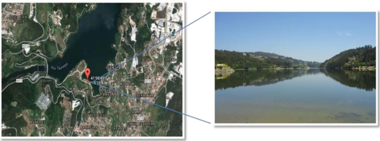
Torrão Reservoir (Figure 2) is situated in the North of Portugal, in Tâmega River, an affluent of the Douro River. The sampling was performed in a leisure park located at this reservoir, Parque de Lazer de Alpendorada e Matos (41°05'45.7"N, 8°15'15.4"W). This reservoir has a total capacity of 77 hm3 and its watershed is about 3280 km2 (Mateus et al. 2014). It is known that the water from this reservoir is used in agriculture, as a source of potable water to the inhabitants of Marco de Canaveses and Amarante or in recreational activities by the local population as well as in energy production due to the existence of a hydroelectric dam in this river (Mateus et al. 2014; Torres et al. 2011). It has been previously reported in this local the presence of cyanobacteria species such as Microcystis spp. (Martins et al. 2011), M.aeruginosa (Vasconcelos et al. 1996) and Ap. flos-aquae (Teles et al. 2008) as well as cyanotoxins such as microcystins (Saker et al. 2007). The second sampling site, located in the Tâmega River, was Tâmega River (Marco de Canaveses) (Figure 3) in Parque Fluvial do Tâmega (41°11'45.9"N, 8°09'38.2"W). It is reported that M.aeruginosa is the dominant species, between July and September (Saker et al. 2005) however it has been already detected the presence of Anabaena sp. and Aphanizomenon sp. (Osswald et al. 2009).
In this river park is situated a children playground, a nautical club and reserved areas for fishing and for sport activities. The distance between these two sampling points, Torrão Reservoir and Tâmega River (Marco de Canaveses), is around 14 km, being this last site situated more upstream of the Tâmega River.
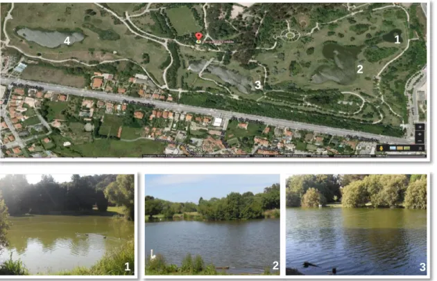
The other three sampling sites located in the North region of Portugal were situated at Parque da Cidade do Porto (Figure 4) which consisted of three consecutive lakes: Lake 1 (41°10'07.1"N, 8°40'20.5"W), Lake 2 (41°10'04.5"N, 8°40'25.6"W) and Lake 3 (41°10'01.5"N, 8°40'39.8"W), (these names were attributed according to its northernmost entrance of the park ). Parque da Cidade do Porto is a recreational park located in the Centre of Porto City and it is composed by four artificial lakes and 83 ha of green area. The water circulation is in succession from the Lake 1 to the Lake 4 and these lakes supply the irrigation system of this park. Lake 4 it was not included in this study because it is the less studied. There is the presence of numerous bird species, fishes and its flora is also very extensive, containing numerous species of arboreal and shrub species (Morais 2009; Moreira 1998). Cyanobacteria species that have been detected in these lakes are Microcystis sp., M.aeruginosa and Planktothrix sp. (Morais 2009; Moreira 1998).
Figure 2 - Sampling site at Torrão Reservoir.
Figure 4 - Sampling sites at Parque da Cidade do Porto. In the largest picture it is possible to observe a satellite view of the lakes (1, 2, 3 and 4) and its distribution in the Park. The smallest pictures are photographs of the Lake 1 (1), Lake 2 (2) and Lake 3 (3) taken at the sampling collections.
Vela Lake and Mira Lake were the chosen sampling sites to represent the Centre region of Portugal. Vela Lake (40°16'23.9"N, 8°47'35.1"W) (Figure 5) is an eutrophic shallow freshwater body which belongs to a system of interconnected reservoirs, being this the largest one, located at Figueira da Foz (Antunes et al. 2002) and it is embedded in a protected area, Ecological European Net - Rede Natura 2000. It has around 70 ha and it is frequently observed the occurrence of blooms and some cyanobacteria species associated with it, such as M.aeruginosa, C.raciborskii, Ap.
flos-aquae and Anabaena flos-flos-aquae (de Figueiredo et al. 2006; Saker et al. 2003). This
lake is surrounded by agricultural fields, in which its water is applied, and by a pine forest. It is common the practice of recreational fishing in this Lake (Abrantes et al 2006).
Mira Lake (40°26'29.8"N, 8°45'07.5"W) (Figure 6), the second sampling site in the Centre region, has around 40 ha of area and the distance between these two sampling sites is around 10 km (Gonçalves et al. 1996). At this Lake, it was already detected the presence of cyanobacteria blooms co-dominant with Actinobacteria (de Figueiredo et al. 2007). This lake is also located in agricultural areas being its water used for the irrigation of crops. In the opposite border to this site it is situated a touristic house.
1 2 3 4 2 1 3
2.1.2 Sampling Procedure
The sampling consisted in collecting water samples from the surface water column, to avoid the collection of sediments, in plastic bottles previously sterilized, with 2L of maximum volume. Besides the water samples collection, it was also performed a drag, using a plankton net with a 55-µm-mesh, to collect the phytoplankton present in the water systems to posteriorly be isolated at the laboratory.
It was also measured, with a pH meter (WTW MultilineP3 – WTW, Germany) the pH of the water systems sampled. However, the water temperature measurement was not possible due to a technical problem. It was necessary to request the atmospheric temperature registered in those sampling sites to IPMA (Instituto Português do Mar e da Atmosfera).
All the samples were maintained in refrigerated conditions until its processing at the laboratory which was performed within the 24 hours after the collection of samples.
Figure 5 - Sampling Site at Vela Lake.
2.1.3 Sample Treatment
In the laboratory, the water samples were filtered with a vacuum filtration system using a Glass-Microfiber filters (grade MG C, 47 mm diameter, 1.2 µm porosity) (Munktell®, Sweden). The obtained biomass was maintained at -20ºC until further DNA extraction.
2.2 Isolation and Culturing
In the laboratory, the phytoplankton samples collected from each sampling site were visualized with an optic microscope (Leica DMLB) and isolated, using the micromanipulation technique, with a Pasteur pipette. It was isolated either colonial or filamentous cyanobacteria. To guarantee that it was only a single cyanobacterium species that was being isolated it was performed washes in distilled water until obtain the isolated organism and transferred it into culture tubes, supplemented with 5 mL of Z8 culture medium (Kotai, 1972) to promote the cyanobacteria growth. The isolated microorganisms were maintained in a culture room under 25ºC temperature with a photoperiod of 14h:10h light-dark and 25 µEm2s of light intensity without aeration.
After the incubation period, the cultures were visualized with an optic microscope (Olympus BX 41) and from the cultures, which did not present any contaminating organisms, it was transferred around 1 mL of culture to 50 mL culture flasks with filter caps (Orange Scientific, EU) supplemented with 25 mL of Z8 medium, in order to maintain the aseptic conditions of the cultures, this procedure was performed under a flame inside a laminar flow chamber (Cruma, Spain). The cultures were afterwards maintained in the same growth conditions as mentioned above. When these cultures reached its exponential phase of growth, they were visualized at the Olympus BX41 optic microscope coupled with a Olympus DP72 photograph machine and using Olympus Cell^B software for image acquisition of the isolated cyanobacteria cultures. It was also collected 1 mL of the culture for DNA extraction to confirm through PCR the organism present in the culture.
2.3 DNA Extraction
DNA was extracted from the environmental samples and from the isolated microorganisms. In the case of the isolated cyanobacterial cultures when the sample was collected from the culture flask, it was centrifuged for 5 minutes at room temperature at a maximum velocity of 17000g (VWR MicroStar 17R, UK). The supernatant was discarded and the pellet was maintained at -20ºC for 48 hours to enhance the cells to break. To extract genomic DNA from the environmental samples it was necessary, to scrape with a sterile blade, the biomass present in the filters, that
resulted from the water filtration. Genomic DNA, from the environmental samples and from the isolated cyanobacterial cultures, was extracted using the PureLink™ Genomic DNA Kit (Invitrogen, Carlsbad, CA, USA), following the protocol for Gram-negative bacteria. DNA was eluted in 50 μL of Elution Buffer.
To confirm the presence of the total genomic DNA, it was performed a 1% (w/v) agarose (UltraPureTM Agarose, Invitrogen Life Technologies, UK) gel electrophoresis (see section 2.4). The DNA was stored at -20ºC until further molecular analysis.
2.4 Agarose gel electrophoresis
The presence of total genomic DNA and amplified DNA was confirmed through a 1% (w/v) agarose (UltraPureTM Agarose, Invitrogen Life Technologies, UK) gel electrophoresis, stained with 3 µL of ethidium bromide (10 mg/mL, Biorad, USA) in 1X TAE buffer (0.4M Tris-acetate, 0.01M EDTA, pH 8.3 ± 0.1, InvitrogenTM, UK), at 100 volts for 30 minutes. In each gel was loaded genomic and amplified DNA with 1 µL of Nucleic Acid Loading Buffer (5x, BioRad – 50 mM Tris-HCl, pH 8, 25% Glicerol, 5mM EDTA, 0.2% Bromophenol Blue and 0.2% Xylene FF) along with 1Kb plus DNA ladder at 1 µg/µL concentration (10 mM Tris-HCl (pH 7.5), 1 mM EDTA, 50 mM NaCl, InvitrogenTM, UK). The gel was photographed, under a UV transilluminator (Cleaver Scientific, Warwickshire, UK), coupled with a Cannon PowerShot G9 photograph machine.
2.5 DNA Quantification
The quantification of total genomic DNA was performed using the Gen5TM Version 2.0 Data Analysis Software using BioTek’s take 3 microplate (Synergy HT, BioTek Instruments, Winooski VT, USA).
2.6 Molecular Methods
2.6.1 Primers development
As mentioned in section 1.6, one of the main aims of this study was to design specific primers, for Aphanizomenon genus, to be applied by conventional PCR. To achieve this aim, it was performed a search in the NCBI database for nucleotide sequences belonging to rpoC1 gene sequences of this genus. The sequences were imported to the Molecular Evolutionary Genetics Analysis 5 (MEGA 5) software (Tamura et al. 2011) and aligned using the ClustalW algorithm. It was performed a BLAST of the alignment consensus sequences comprehending 20-25bp to confirm the primer specificity.
2.6.2 Polymerase Chain Reaction
In all of the PCR reactions, it was applied the GoTaq® protocol from Promega (USA). Each PCR mixture was performed in a volume of 20 µL containing 5-10 ng of genomic DNA, 1 X PCR Green GoTaq® Flexi Buffer, 2.5 mM MgCl2, 250 µM of each
deoxynucleotide triphosphate (dNTP’s), 10 pmol of each primer, 10 mg/mL (w/v) of Bovine Serum Albumin (BSA) and 0.5 U of GoTaq® DNA polymerase (Promega, USA). BSA was used because it is known that the majority of the PCR inhibitors present in the environmental samples are capable to bind to this reagent (Kreader, 1996), it is also reported that BSA is able to stabilize the DNA polymerase used in PCR (Farell and Alexandre, 2012).
Initially, it was performed a PCR in order to detect the presence of cyanobacteria in all the DNA samples, using the 27F and 809R and 740F and 1494R primer pairs for the amplification of the 16S rRNA cyanobacteria sequence. For the amplification of the
rpoC1 gene sequence from C.raciborskii, gyrB gene sequence from M.aeruginosa and rpoC1 gene sequence from P.agardhii, the primers applied were cyl 2 and cyl 4, gyrF
and gyrR, rpoC1_Plank_F271 and rpoC1_P_agardhii_R472, respectively. The sets of primers, used in this study, were already described in previous studies and they were designed to amplify specific sequences of the species in study (Table 2). LEGE Culture Collection strains were used as positive control in the PCR reactions, IZ41 (Microcystis
aeruginosa LEGE 91351) and AQS (Cylindrospermopsis raciborskii LEGE 97047) for M.aeruginosa and C.raciborskii specific PCR, respectively. P.agardhii strain was not
available, in the laboratory, to be applied as positive control in specific PCR for this species.
The PCR conditions are listed in table 3 and all the reactions were carried out in a TProfessional Thermocycler (Biometra, Germany) or in a Bio-Rad MyCyclerTM (Bio-Rad, Hercules, CA, USA). All PCR products were visualized in a 1% (w/v) agarose (UltraPureTM Agarose, Invitrogen Life Technologies, UK) gel electrophoresis (see section 2.4).
For DNA sequencing, it was performed a PCR in a volume of 40 µL and instead of using 1 X PCR Green GoTaq® Flexi Buffer (Promega, USA) it was used 1 X PCR Colourless GoTaq® Flexi Buffer (Promega, USA). The amplified products were purified using the Cut & Spin Gel Extraction Kit (GRiSP, Portugal) in which the DNA band, corresponding to the expected DNA product, was excised from the agarose gel, and it was placed on the top of the column media to be centrifuged at 6200 g for 10 minutes at room temperature in a VWR MicroStar 17R centrifuge. The column was discarded
and the samples were maintained at refrigerated conditions (-20ºC) until be sent for direct sequencing.
The sequencing results were analysed using FinchTV 1.4.0 (Geospiza, Inc.; Seattle, WA, USA; http://www.geospiza.com) and Multalin (Florence 1988). To confirm the amplicons it was performed a BLAST search (Altschup et al. 1990).
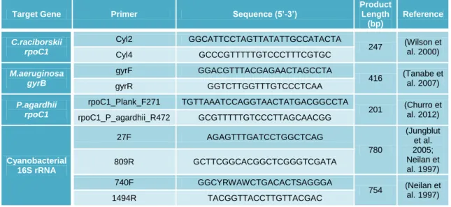
Table 2 - Primers applied in conventional PCR and their characteristics.
Table 3 - PCR programs applied to detect the species in study
Target Gene Primer Sequence (5’-3’)
Product Length (bp) Reference C.raciborskii rpoC1 Cyl2 GGCATTCCTAGTTATATTGCCATACTA 247 (Wilson et al. 2000) Cyl4 GCCCGTTTTTGTCCCTTTCGTGC M.aeruginosa gyrB gyrF GGACGTTTACGAGAACTAGCCTA 416 (Tanabe et al. 2007) gyrR GGTCTTGGTTTGTCCCTCAA P.agardhii rpoC1 rpoC1_Plank_F271 TGTTAAATCCAGGTAACTATGACGGCCTA 201 (Churro et al. 2012) rpoC1_P_agardhii_R472 GCGTTTTTGTCCCTTAGCAACGG Cyanobacterial 16S rRNA 27F AGAGTTTGATCCTGGCTCAG 780 (Jungblut et al. 2005; Neilan et al. 1997) 809R GCTTCGGCACGGCTCGGGTCGATA 740F GGCYRWAWCTGACACTSAGGGA 754 (Neilan et al. 1997) 1494R TACGGTTACCTTGTTACGAC Target Gene
Initial Denaturation Denaturation Annealing Extension Final Extension
Temperature Time Temperature Time Temperature Time Temperature Time Temperature Time
C.raciborskii rpoC1 95 ºC 2 min 95 ºC 90 sec 45 ºC 30 sec 72 ºC 50 sec 72 ºC 7 min 35 cycles M.aeruginosa gyrB 94 ºC 3
min 94 ºC 1 min 60 ºC 1 min 72 ºC
30 sec 72 ºC 5 min 40 cycles P.agardhii rpoC1 94 ºC 3 min 94 ºC 20 sec 58 ºC 20 sec 72 ºC 20 sec 72 ºC 5 min 40 cycles Cyanobacterial 16S rRNA 92 ºC 2 min 92 ºC 20 sec 50 ºC 30 sec 72 ºC 1 min 72 ºC 5 min 35 cycles
2.6.3 Real-Time Polymerase Chain Reaction
One of the main objectives of this work was to develop a Real-Time Polymerase Chain Reaction specific for M.aeruginosa, to be possible its future absolute quantification mainly in water systems. To achieve this goal, it was necessary to perform a specificity assay and a sensitivity assay.
2.6.3.1 Specificity assay
To perform this assay it was applied twelve M.aeruginosa strains representing different geographic origins (Table 4) in order to test the primers specificity as well as the Real-Time PCR protocol developed. In this experiment, the primers applied were the same primers applied in conventional PCR, gyrF and gyrR, at 10 pmol, 1x iTAQTM Universal SYBR Green SuperMix (Bio-Rad, Hercules, CA, USA), 2 µL of DNA to perform the final volume of 10 µL. The Real-Time PCR protocol tested consisted in an initial denaturation of 10 minutes at 95ºC, followed by 35 cycles of 15 seconds at 94ºC, 15 seconds at 60ºC and 50 seconds at 72ºC. It was also tested a Real-Time PCR protocol which consisted in an initial denaturation of 10 minutes at 95ºC, followed by 35 cycles of 15 seconds at 94ºC, 20 seconds at 60ºC and 50 seconds at 72ºC. Data acquisition was monitored after each extension step with temperature increments of 0.5 ºC between 55ºC and 95ºC. Melting temperatures of the products were determined with iQTM5 Optical System Software Version 2.1 (Bio-Rad, USA).
Amplifications were performed in the iQTM5 Multicolor Real-Time PCR Detection Systems (Bio-Rad, USA) and the results analyzed with iQTM5 Optical System Software Version 2.1 (Bio-Rad, USA).
Table 4 - Microcystis aeruginosa strains applied in Real-Time PCR
Strain Continent Country
PCC 786 Europe Netherlands
PCC 7005 America USA
NIVA-CYA 31 America Canada
Ed08 Africa Uganda
Ma Viet Asia Viet Nam
MN Africa Tunisia
27M America Mexico
19M America Mexico
IZ56 Europe Portugal
M6 Europe Portugal
90M America Mexico
2.6.3.2 Sensitivity assay
To perform this assay, it was necessary to obtain standards with known concentrations in terms of gene copy numbers to determine the minimum number able to detect in a given reaction. In this way, it was performed an amplification of a
M.aeruginosa strain (M6 strains; see table 4) through a conventional PCR with a final
volume of 100 µL. The PCR product was sent to Dr. Catarina Churro from the Instituto Nacional de Saúde Dr. Ricardo Jorge (Lisbon, Portugal) for further development of the standards based in a protocol already described by Baxa et al., (2010).
Once the standard DNA arrived at the Laboratory, it was performed a tenfold serial dilutions of the standard in molecular grade water, ranging from 5.5x10⁸ copies/µL to 5.5x100 copies/µL. Posteriorly, they were tested in Real-Time PCR, in triplicate, with the developed program to detect the sensitivity limit. The reagents and its concentrations applied in this reaction were mentioned in the section 2.6.3.1. Amplifications were performed in the iQTM5 Multicolor Real-Time PCR Detection Systems (Bio-Rad, USA) and the results analyzed with iQTM5 Optical System Software Version 2.1 (Bio-Rad, USA).
2.7 Statistical Analyses
Shapiro–Wilk normality test was used to test the normal distribution of maximum and minimum atmospheric temperature data and Levene’s test was used for the analysis of the homogeneity of variance. Statistically significant differences of the maximum and minimum temperatures registered between North and Centre regions of Portugal were calculated using a Student’s t-test with 95% confidence level. The statistical analyses of this work were performed with IBM Statistical Package for the Social Sciences (SPSS Inc.) software version 22 (IBM Corporation, Armonk, NY, USA).