Universidade de Lisboa
Faculdade de Medicina de Lisboa
Treatment of obstructive sleep apneas with a
mandibular advancement device:
A preliminary study about the impact on vigilance and sleep
structure
Miguel Gonçalves Meira e Cruz
Mestrado em Ciências do Sono (3ª Edição)
A impressão desta dissertação foi aprovada pela Comissão Coordenadora do Conselho Cientifico da Faculdade de Medicina de Lisboa em reunião de 28 de Setembro de 2010
Universidade de Lisboa
Faculdade de Medicina de Lisboa
Treatment of obstructive sleep apneas with a
mandibular advancement device:
A preliminary study about the impact on vigilance and sleep
structure
Miguel Gonçalves Meira e Cruz
Mestrado em Ciências do Sono (3ª Edição)
Dissertação orientada pela Prof. Dra. Teresa Paiva
Todas as afirmações efectuadas no presente documento são da exclusiva responsabilidade do seu autor, não cabendo qualquer responsabilidade à Faculdade de Medicina de Lisboa pelos conteúdos nele apresentados.
GENERAL INDEX
Thought …………... 6
Aknowledgments ... 7
Abreviations ... 9
Abstract ... 10
Abstract in Portuguese language ...………. 12
1.Preamble ... 14
2.Review of the Literature ... 16
2.1 Sleep and its related disorders ... 16
2.2 Obstructive Sleep Apnea-Hypopnea Syndrome ... 17
2.3 Daytime sleepiness in OSAHS patients ... 21
2.4 Oral Appliance therapy in OSAHS ... 22
2.5 Clinical protocol ... 32
3.Rational and study goals …... 34
4.1 Patients selection criteria ... 35
4.2 Measurements and protocol ... 36
4.3 Data analysis ……... 40 5.Ethical considerations ………. 41 6.Results Presentation ... 42 7.Discussions ……..………... 74 8.Conclusions …... 81 Appendix ... 82 References ... 95
Close your eyes relax and enter in a darker side of your life Where you can best live your whishes, hopes and fantasies as well as your fears,
Where you can be fully happy even when totally miserable, Where you can touch the untouchable, And where you can be sure that you are alive. I assure you that you will wish to come back every night.
AKNOWLEDGEMENTS
This report is the result of our master thesis project carried out at Lisbon Faculty of Medicine and at Center of Electroencephalography and Clinical Neurophysiology. This is also the last part of our Master of Science degree at Lisbon Medical Faculty.
But this is mainly the result of an effort and an investment for which had to overcome many barriers. I managed to keep the momentum through the indispensable help of many people. To my family, friends, teachers and students, I appreciate the love, the warnings, the teachings, advice and continuous motivation.
Because of the special support, credit, friendship and above all by the dedication and unlimited patience, I want to express special recognition to:
Professor Teresa Paiva, my main supervisor for accepting me as a MSc student, providing excellent work facilities and leading the way into the field of research in Sleep Medicine. She shared with me her knowledge, her enthusiasm and most of all her friendship.
Assistant Professor Edmund Rose, for sharing his experience in the field of oral sleep medicine and teach me the practical foundations of oral appliances therapy.
The late Professor Luis Silva Carvalho for having accepted me in the study group on the physiology of the autonomic nervous system, which was the launching pad for my dedication to the study of neurophysiology, sleep physiology and sleep medicine.
Assistant Professor Marta Drummond, for the friendship and for the willingness to support my training in evaluating and treating patients with sleep related breathing disorders.
Sleep technician Sofia Rebocho, for her kind dedication and for their valuable assistance in staging the sleep of patients who constituted the basis of this thesis.
Dr. João Correia Pinto, for his credit and for having invited me to initiate the sleep consultation in the Stomatology Service of São João Hospital, in Oporto.
Dr. Richard Staats, for his indispensable support to the initial development of my clinical work and research training in Respiratory Sleep Medicine.
ABREVIATIONS
OSAHS Obstructive sleep apnea-hypopnea syndrome OSA Obstructive sleep apnea
OA Oral appliances
MAD Mandibular Advancement Device TRD Tongue Retaining Device
AHI Apnea Hypopnea Index
RDI Respiratory Disturbance Index RERA Respiratory Effort Related Arousal
AI Apnea Index
OEI Obstructive Events Index ODI Oxygen Desaturation Index
PAPs Positive Airway Pressure Systems CPAP Continuous Positive Airway Pressure APAP Automatic Positive Airway Pressure SRBD Sleep Related Breathing Disorders MSLT Multiple Sleep Latency Test
MWT Maintenance of Wakefulness Test UARS Upper Airway Resistance Syndrome FDA Food and Drugs Administration
ABSTRACT
Treatment of Obstructive Sleep Apneas With a Mandibular Advancement Device: A Preliminary Study about the Impact on Vigilance and Sleep Structure Miguel Meira e Cruz, Teresa Paiva
Center of Electroecephalography and Clinical Neurophysiology
Key-words: Obstructive sleep apnea, excessive somnolence, mandibular advancement devices
Introduction: Obstructive sleep apnea-hypopnea syndrome (OSAHS) is a high prevalent and complex sleep disorder with serious consequences to individual and public health mainly because of its association with cardiovascular morbidity and mortality, sleep fragmentation and consequent excessive somnolence. The current American Academy of Sleep Medicine practice parameters recommends oral devices as a plausible option on the treatment of OSAHS. However, there is only a limited data about the efficacy of OSAHS treatment with prefabricated mandibular advancement devices (MAD) regarding to vigilance and sleep structure.
Goals: The aim of this clinical prospective study was to evaluate the efficacy of a prefabricated mandibular advancement device in the treatment of OSAHS and to assess their impact on vigilance and sleep structure.
Methodology: Nine patients (6 males and 3 females) with OSAHS (AHI > 5) recruited from a dental sleep consultation of a sleep clinic were referred to an attended full night polysomnographic study before and after treatment with MAD. Regarding to the therapeutic effect on OSAHS, an AHI < 5 events per hour of sleep after MAD treatment was considered to be a complete therapeutic response or therapeutic success whether an AHI < 50% from the baseline without achieving less than 5 events per hour was considered a partial response. Sleep structure and subjective sleepiness were assessed in order to evaluate the treatment efficacy. Wilcoxon matched pair test was used to test
differences between baseline and follow-up. A p-value of less than 0.05 was considered significant.
Results: Snoring was abolished or became inaudible in all patients as witnessed by their bed partners. There was an improvement on respiratory parameters from the baseline to the therapeutic PSG with 66.7% of patients achieving therapeutic success whereas 11.1% achieved only partial therapeutic response. Slow-wave sleep increased from 14% at the baseline to 21% after therapy with MAD and arousal index was lower after therapy (6/hr) than at baseline (16/hr) even though it was slightly above normal levels. All sleepiness scales scores and sleep complaints as well as sleep quality improved significantly after therapy compared with baseline (p<0.05).
Conclusions: In this short-term study a titratable prefabricated MAD improved PSG respiratory parameters, sleep structure snoring and sleepiness in mild to moderate OSAHS patients thus supporting the use of this kind of option in OSAHS therapeutics. Future studies should focus on the comparison between a titratable prefabricated MAD with a placebo device as well as with a custom made MAD and also evaluate the long-term outcome.
Resumo (Portuguese Language)
Tratamento da Apneia Obstrutiva do Sono com um dispositivo de avanço mandibular: Estudo preliminar sobre o impacto na vigília e na estrutura do sono
Miguel Meira e Cruz, Teresa Paiva
Centro de Electroencefalografia e Neurofisiologia Clínica
Palavras-chave: Apneia obstrutiva do sono, sonolência excessiva, dispositivos de avanço mandibular
Introdução: A síndrome de apneia-hipopneia obstrutiva do sono (OSAHS) é uma doença do sono com elevada prevalência e complexidade que afecta de forma importante a saúde individual e a saúde pública, principalmente pela sua associação com a morbilidade e mortalidade cardiovasculares, fragmentação do sono e consequente sonolência excessiva durante o dia. As recomendações actuais da American Academy of Sleep Medicine incluem os dispositivos orais enquanto opção viável para o tratamento da SAHOS. Contudo, são limitados os dados sobre a eficácia do tratamento da SAHOS com dispositivos de avanço mandibular pré-fabricados (DAM) no que respeita à vigília e no que respeita à estrutura do sono.
Objectivos: O objectivo deste estudo clínico e prospectivo foi avaliar a eficácia de um dispositivo termoplástico de avanço mandibular no tratamento da SAHOS, bem como o impacto deste na vigília e na estrutura do sono.
Metodologia: Nove pacientes (6 homens e 3 mulheres) com SAHOS (IAH > 5) recrutados de uma consulta de medicina dentária numa clínica de sono foram referenciados para um estudo polisonográfico antes e após tratamento com DAM. O sucesso terapêutico foi assumido com um IAH < 5 eventos por hora de sono após tratamento com DAM enquanto uma resposta parcial foi definida por IAH < 50% da linha de base sem que tenha sido atingido um valor menor que 5 eventos por hora. Foram analisados a estrutura do sono e a sonolência de forma a avaliar a eficácia terapêutica. O teste de Wilcoxon foi usado para testar
a diferença entre as condições basal e terapêutica. Um valor de p menor que 0.05 foi considerado como significativo.
Resultados: O ressonar testemunhado pelos parceiros de cama foi abolido ou tornou-se inaudível em todos os pacientes. Existiu uma melhoria nos parâmetros respiratórios na PSG terapêutica comparada com a PSG basal, tendo 66.7% dos doentes atingido sucesso terapêutico enquanto 11.1% atingiram apenas resposta terapêutica. O sono de ondas lentas aumentou de 14% na condição basal para 21% após terapia com DAM e o índice de despertares foi mais baixo após tratamento (6/h) do que na condição basal (16/h) embora tenha persistido ligeiramente superior ao normal. Todas as somas relativas a escalas de sonolência resultaram numa melhoria significativa após terapêutica com DAM. Todas as melhorias foram estatisticamente significativas (p<0.05).
Conclusões: Neste estudo a curto prazo, os dispositivos pré-fabricados tituláveis melhoraram, de forma significativa, os parâmetros respiratórios polissonográficos, a estrutura do sono, a roncopatia e a sonolência nos doentes com SAOS ligeira e moderada apoiando a recomendação deste tipo de dispositivos para a terapêutica da SAHOS. Estudos futuros devem focar-se na comparação de dispositivos intra-orais pré-fabricados tituláveis com placebo e com dispositivos personalizados, e na avaliação dos efeitos destes dispositivos a longo prazo.
1. PREAMBLE
The general aim of this master thesis entitled «Treatment of obstructive sleep apneas with a mandibular advancement device. A preliminary study about the impact on vigilance and sleep structure» was to evaluate the specific role of prefabricated Mandibular Advancement Devices (PF-MAD) in the treatment of obstructive sleep apnea-hypopnea syndrome (OSAHS) with focus on the influence in vigilance and sleep structure.
Although many studies have evaluated the effect of MAD on sleep apneas and oxygenation status during sleep of OSAHS patients, there is little knowledge about their effects on characteristic symptoms among this condition.
A prefabricated MAD was used in the present study because of three reasons: The first one was that although the use of prefabricated devices is not encouraged by official organs, these appliances are probably a frequent option when patients have no financial reimbursement for the treatment, which is the case for many countries today;
Secondly most of the studies of prefabricated appliances used non-titratable devices, thus leading to a difficult interpretation about their isolated strengths and weaknesses;
In third place, the evidence that also in mild cases of the disease there are already some chronic changes that will contribute to a multi-system degeneration turns plausible that patients could benefit from an early control of their condition with easily available treatment as mandibular advancement device, provided when another effective therapeutic option or positive airway pressure systems (PAPs) as continuous or automatic positive airway pressure (CPAP or APAP) are not indicated.
Changes in sleep structure and in vigilance parameters were then analyzed in OSAH patients in order to adequately evaluate the therapeutic efficacy of a
specific mandibular advancement device in obstructive sleep apnea-hypopnea syndrome.
In the introduction, there is a review of the literature, defining the theme, exposing the recent knowledge about general aspects related to OSAH, and positioning oral appliances therapy as an option to the treatment of this condition.
After definition of the specific goals of this thesis, the methodology is described regarding patients selection criteria, protocol and instruments including statistic analysis.
Finally, the results are presented and the discussion of those results is done in order to conclude about the hypotheses that were previously enhanced.
2. Review of the Literature
2.1 Sleep and its related disorders
Sleep is a natural state of relatively suspended sensory and motor activity. This recurrent and vital period of our life time is characterized by total or partial loss of consciousness and mobility, accompanied by specific behavioral pattern as closed eyes and recumbent posture. It really consumes a third part of our lives and, inversely to what was thought until recently, sleep is a highly active rather than inactive process with a crucial role on physical and mental health (1).
Although its primary function still remains to be discovered, the need of sleep is so powerful that we may be not able to avoid it even when doing so could be life-threatening, such as while driving.
Sleep deprivation, either voluntary or involuntary, can be related to absent or decreased sleep and result in excessive sleepiness, changes in the mood, cognition, performance, cerebral function, cardiovascular disorders, endocrine function, metabolism and immune-inflammatory natural response.
Sleep-related disorders, often characterized by sleep disruption and loss of consolidation, can be present in a variety of ways, including insomnia, excessive sleepiness, snoring, sleep apnea, cataplexy, sleep paralysis, sleep hallucinations, nocturnal dyspnea, nocturnal pain, and nocturnal physical phenomena (2).
Sleep disorders cannot be however reduced to a poor night’s sleep problem. The most recent diagnostic classification divides sleep diagnosis into different categories: insomnias, sleep-related breathing disorders, hypersomnias, circadian rhythm sleep disorders, parasomnias, sleep related movement disorders, isolated symptoms, apparently normal variants and unresolved issues, other sleep disorders, sleep disorders associated with conditions classifiable elsewhere and other psychiatry/behavioral disorders frequently encountered in the differential diagnosis of sleep disorders (3).
Sleep disorders, particularly those related to respiratory dysfunction are considered to be one of the most common health problems being estimated that between 82 and 98% of adults with these diseases are undiagnosed (4).
2.2 Obstructive sleep apnea hypopnea syndrome
2.2.1 Definition and prevalence
Obstructive sleep apnea-hypopnea syndrome (OSAHS) is a sleep related breathing disorder (SRBD) characterized by repetitive partial (hypopnea) or complete (apnea) interruptions of respiratory flux (fig.1) at upper airway level occurring during sleep with a minimum duration of ten seconds per event occurring at least five times per hour (5). This is one of the most common conditions observed in the practice of sleep medicine (6) with a high prevalence, occurring in about 4 % of the working male population and 2 % among the women (7).
Fig.1 – Schematic view of airway obstruction during an event on OSAHS patient
OSAHS is associated to a substantial negative impact on quality of life and productivity, due to an increased morbidity and mortality of cardiovascular diseases and sleepiness induced accidents (8).
2.2.2 OSAHS Diagnosis
The most frequent symptoms are diurnal hypersomnia, intense snore and witnessed respiratory pauses during sleep mostly associated to overweight and obesity (5), but neurocognitive, psychiatric, gastrointestinal genitourinary and endocrine or metabolic dysfunction are also common in patients with OSAHS.
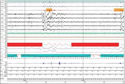
Although clinical features of OSAHS are very characteristic, the final diagnose requires a polysomnographic study (Fig. 2) which should ideally include sleep in all sleep stages and in all sleep positions, and should be at least 6 hours of duration (9). The apnea plus hyponea index (AHI – number of apneas plus hypopneas per hour of sleep) higher than 5, translates the first step of the complex condition commonly subsisting with clinical symptoms. Arousal events without criteria for classification within apneas or hypopneas are classified as respiratory effort related arousals (RERA) and should be taken in account when staging respiratory events. The AHI plus RERAS divided by total sleep time corresponds to the respiratory disturbance index (RDI) and reflects a more complete sleep respiratory status (3,9).
Fig. 2 – Sample of attended PSG focusing two separated obstructive events (obstructive apnea, translated by the loss of signal on the airway flow channel) followed by EEG and EMG (chin and legs) arousal and accompanied by oxygen dessaturation.
2.2.3 Risk Factors
Obesity is an important risk factor for SRBD and namely for OSAHS by means of different mechanisms (10). An obese has 10-14 folds more risk of developing OSAHS than a non-obese has. However, this risk factor alone is not necessary for significant disease and there are other risk factors that can modify its effects. Important risk factors like neck circumference and craniofacial morphology and anthropometry are independently related to OSAHS, even in non-obese patients (10,11).
Gender is a relevant risk factor too, being widely recognized that SRBD affect more men than women, what is related with anatomic as well as functional specific characteristics of each gender (12-14). Several studies found however a higher risk among women than what was though according to previous epidemiologic studies and that risk is clearly influenced by menopausal status (15-18)
.
There are also ethnic differences in the prevalence of OSAHS and nonwhite individuals are more (and more severely) affected and at a younger age than Caucasian individuals (19, 20) although specific causes are not well clarified.
Nasal obstruction was showed to negatively affect PSG parameters of respiratory status like AIH and arousal index in women, suggesting that it plays a role in worsening sleep apnea (21). Furthermore an association between chronic rhinitis with typical signs and symptoms of OSAHS was also found, even after adjustment for age, sex and BMI (22).
Although there is no standardized phenotypic definition of OSAHS, several genetic and familial tendency associations have been emphasized from different perspectives suggesting that, apart from multiple indirect influences affecting obesity, body fat distribution and craniofacial morphology, genetics also play an independent role in OSAHS (23).
Age is other major risk factor for OSAHS, since both snoring and measured respiratory disturbance indices have tended to increase from youth to elderly. SRBD have a peak prevalence of 4.7% in the 50-59-year-old men group. The severity of the disease seem however to decrease in older people (24).
Alcohol (25) and some medications (26) aggravate OSAHS by interfering in physiological control of OSAHS pathogenic associated mechanisms. Smoke also disturbs physiological sleep and particularly affect upper airway predisposing to OSAHS (27).
2.2.4 OSAHS Treatment
Apart from conservative therapeutic measures like weight reduction (28,29), avoidance of alcohol, pharmacological respiratory depressants and smoking (30), as well as sleep hygiene good habits (31) and some adjunctive measures (nasal decongestion and positional therapy) should be undertaken (32-34). At present, OSAHS therapy of choice is continuous or automatic positive airway pressure (CPAP or APAP) devices (35). However, especially in patients with a low AHI, positive pressure therapy is often complicated due to decreased acceptance and compliance by the patient.
It has been shown that OSAHS therapy decreases the cardiovascular risk even in patients with only a moderately elevated RDI (36), providing a valuable argument to treat all the OSAHS patients, including those without indication to PAP therapy. In the last years oral appliances, in particular mandibular advancement devices (MAD) have been increasingly used in patient’s not sufficiently complying CPAP therapy or sometimes as first line therapy in non-obese subjects.
Actual data suggest that treatment with MAD increased the compliance due to a better acceptance, but that treatment success, measured either by AHI and RDI reduction is lower.
Up to today there is only limited information about the impact of MAD treatment on sleep structure and vigilance (37) and although a recent Cochrane review concluded that there is some evidence suggesting oral appliances do improve
subjective sleepiness in patients with sleep related breathing disorders (SRBD), there are recognized limitations which were not only restricted to the small sample size but also to the under-reporting of methods and lack of some important parameters that should be controlled as quality of life evaluation (38).
2.3 Daytime sleepiness in OSAH patients
Excessive Sleepiness causing diurnal impairment is most often the primary complaint of patients presenting with sleep disorders. This can be associated to distinct factors among general individuals and is a common feature either in OSAHS or in cardiovascular disease patients (39).
People are considered excessively sleepy when consistently experience difficulty to maintain wakefulness and alertness to accomplish the tasks of daily living. According to this, degree of sleepiness can be clinically classified in mild, moderate and severe (40).
In OSAHS Patients, excessive sleepiness seems to be related with specific mechanisms affecting sleep consolidation related, although not exclusively, with intermittent hypoxia and consequent sleep fragmentation (41, 42).
In general, individual subjective reports of sleepiness are imprecise, and objective and formal subjective measures of sleep have been devised to quantify the intensity of that symptom (43). Objective assessment of sleepiness severity can be achieved using different laboratorial tests like Multiple Sleep Latency Test (MSLT) and Maintenance of Wakefulness Test (MWT). Formal subjective measures are based on some kinds of sleepiness questionnaires which evaluate either acute (Stanford Sleepiness Scale or Karolinska Sleepiness Scale) or chronic states (Epworth Sleepiness Scale) (44-46).
2.4 Oral appliance therapy in OSAHS
After its development in the early 1980s (47), the continuous positive airway pressure (CPAP) therapy became the gold standard therapy for symptomatic OSAH. It remains the first choice of most sleep medicine practitioners and there is very good evidence that effective CPAP improves the neurobehavioral and cardiovascular consequences of OSAH (48), sleepiness and sleepiness-related motor vehicle accidents (49,50) and quality of life (51). Otherwise, it is clear that effective CPAP therapy involves adherence as a critical target, since not all patients with OSAH accept or tolerate this treatment modality. Many studies show that considering as minimum compliance a regular use of CPAP for at least 4h per night, the success rate is between 46% and 85%, even when considering those sleepy patients with moderate or severe OSAH (52,53).
Those rates tend to decrease when compliance is intended as a regular use of CPAP for more than 4 hours by night. Besides that, in asymptomatic snorers the rate of poor or no compliance to CPAP seems to be higher opening the scope for use of other treatments which could benefit patients. In this context a body of evidence exists towards the use of OA.
It has been accepted by the American Academy of Sleep Medicine (AASM) in 2005, and recently updated in the recommendations (2006) the use of OA as a first line treatment option of snoring and mild to moderate obstructive sleep apnea (54).
OA constitutes a successful, easy and non invasive treatment option (or complement) for many patients with sleep related breathing disorders (55). Because they are simple to use and to manage, they are commonly and easily accepted by the majority of patients. However, due to poor knowledge of both dental professionals and other medical professionals, oral appliances are still an under prescribed option.
The rational that propitiated the enthusiasm for therapeutic oral appliances is remote and its beginning may be reported to 1903, when was described that micrognathic infants could benefit when the tongue is sutured forward to the lower lip (56). Since then, there was a period during which helmets and chinstraps where used by physicians to reposition the mandible forward, and in
1934, the French stomathologist Pierre Robin reported the first use of an oral appliance to reposition the mandible in order to treat a case of glossoptosis due to atresia and hypertrophy of the mandible (57). Nevertheless, it was only almost 50 years later that Cartwright and Samuelson used OA in order to manage snoring and sleep apnea with a specific OA (tongue retaining device) which is still in use (58).
a. b.
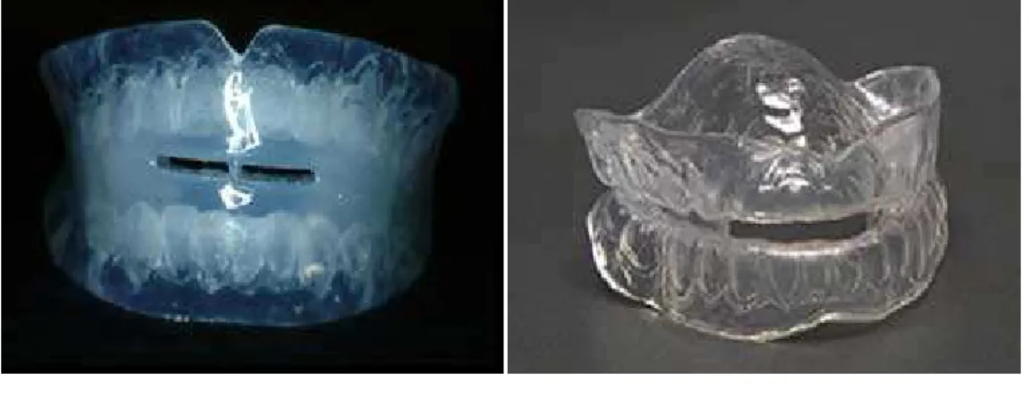
Fig. 3 - Bibloc Type Mandibular Advancement Devices: a. Thermolabyle (Somnofit, Oscimed); b. Custom made (MAS, Somnodent).
a. b. Fig. 4 – Monobloc Type Mandibular Advancement Devices: a. Thermolabyle; b. Custom-made.
Fig. 5 - Tongue Retainer Device (TRD Aveo).
2.4.1 Types and designs of oral appliances
Currently, diverse assortment of designs and materials are being used in OA. Those oral devices can be classified in two main categories: MAD and tongue retaining devices (TRD). The first ones (Fig. 3 and 4) are the most commonly used in clinical practice and the scientific literature supporting their use and effects is far greater than for the other types of oral devices. Besides, if it is true that in the early 80s OA were indicated only in cases of CPAP or surgical failure, many investigations about the effectiveness and successful treatment of OSA with OA brought these devices (mainly, the MAD type) to the medical and scientific arena of today . They are now recognized as an excellent, evidence-based option to a wide range of patients with SRBD, particularly patients with OSAHS.
OA therapy, through the use of MADs is now indicated to several patients, from those non apneics with primary snoring to those with Upper Airway Resistance Syndrome (UARS) or airflow limitation, to those patients with definitive diagnosed OSAHS (59).
2.4.1.1 Mandibular advancement devices (Fig. 3 and 4)
MAD can be constructed with one piece (even called “monobloc”) or two pieces (“bibloc”) designs and may be custom-made or prefabricated (61). A monobloc
MAD rigidly fixes the mandible in an anterior position whereas a bibloc MAD allows for some freedom of mandibular movement, and is liable for individual titration. It is possible that because of this mechanism, this type of MAD is associated also with a lower rate of temporomandibular disorder and discomfort (60)
.
While prefabricated MADs are appliances that only require a simple individual molding of a thermolabile material, custom-made appliances need well defined dental impressions, a bite registration and laboratory involvement.
Although prefabricated MADs are not FDA approved as a definitive therapeutic and are only suggested for temporary use (61), they can be mainly useful in circumstances where patients are renitent to wear an expensive appliance without being sure of its full success or in circumstances in which patients cannot accede to an expensive solution without any reimbursement as happen with the majority of titratable custom made appliances. Furthermore, some evidence which serve the basis for the argument against the use of PF MADs in the continued treatment of SRBD seem to involve some bias since till now, the only study comparing effectiveness of PF MAD with custom made MAD actually compare a bibloc custom made type of appliance with a monobloc PF type of MAD (62), thus conducting to a conflicting interpretation of the results. Moreover, a very recent publication reported that titratable PF MADs were significantly more effective than non-titrable PF MAD either on objective or subjective outcomes regarding OSAHS improvement (63).
A similar argument can be used with respect to mechanical retention. This property is essential for the successful treatment with a MAD, and it can be achieved, in upper and lower arch, by means of clasps, acrylic, or thermoplastic polymer embedded in the appliance, preventing the device pops up during the night due to masticatory muscles relaxation (64). Lack of retention is intended to be greater in PF MADs than in custom made MADs. However, the bias related to different appliance designs (bibloc versus monobloc) may also lead to difficult interpretations about the real independent effect related with the type of OA.
2.4.1.2 Tongue retaining devices (Fig. 5)
TRD are appliances that reposition the tongue in an anterior position by securing it with negative pressure in a soft plastic bulb with or without restraint in the teeth. TRD can be useful as an alternative to MAD in some cases where this appliance cannot be successfully applied (due to insufficient retention related to extensive tooth loss, because an ongoing orthodontic treatment or the inability to get an adequate protrusion). However its full night application may be hampered by the shortness of the nasal passage, discomfort, or loss of negative pressure in the bulb.
2.4.2 Oral appliances mechanisms of action
The primary mechanism of action of a MAD is related with mandibular advancement, indirect anterior reposition of suprahyoid and genioglossal muscles, and the consequently movement of the tongue forward, increasing the anteroposterior dimensions of the oropharynx (65). However, it appears that the cross-sectional area of the velopharynx increases not only in the anteroposterior but also in the latero-lateral dimension, while the oropharynx increases mainly in the lateral dimension (66). Beside anatomical changes, MAD has a significant impact in neuromuscular properties of the upper airway and either both mandible rotation and advancement has been implicated in an increased muscle activity in the upper airway (67-70).
The TRD mechanism of action is somewhat different: Although, usually reserved for patients in whom MAD is not indicated, the forward movement of the tongue out of the mouth tends to be greater and may produce more favorable changes in the retroglossal region. Otherwise, TRD seem to increase cross-sectional area of the upper airways during wakefulness (71) and to increase genioglossal muscle activity in OSAH patients (72), what possibly contributes to a reduction on snoring and apneic events.
a. b.
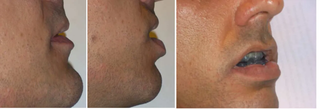
Fig. 6 - Lateral X-Ray from an OSA patient (a) and same patient using a mandibular advancement device (b)
Fig. 7 - Sleep Breathing Disorders Patient a) lateral regular photo; b) and c) using oral appliance
2.4.3 Clinical outcomes of oral appliances therapy
2.4.3.1 Effectiveness
Oral appliance treatment goals involve not only the relief of the clinical signs and symptoms but also the normalization of polysomnographic parameters. Many OSA patients treated with MADs report less somnolence and snoring compared with their basal (not treated) condition. It is however important to be aware of different criteria defining success in the literature and to realize that
many subjective and objective parameters should be evaluated in order to securely recognize that a patient is being treated (not cured). Evidence related to MADs has shown that clinical symptoms such as excessive sleepiness, fatigue, snoring and non restorative or fragmented sleep improve with effective therapeutic measures. That should happen as well with measured polysomnographic associated events, like increased apnea-hypopnea index and respiratory effort related arousals (RERAs), increased oxy-hemoglobin dessaturation levels and increased microarousals (54). MAD therapy also associates to a significant increase of slow-wave and REM sleep (73,74).
It is worth noting that patients commonly report subjective improvement without adequate improvement in PSG parameters (75). Considering however the risk associated to an unperceived increased in AHI and RDI, a follow up sleep study should always be conducted.
Long term success studies of OA therapy show a high success rate after two to five years later (76-79) and demonstrate that a high number (80%) of patients initially successfully treated with OA, also experience a long term control of their OSAHS (80). The success rate tends to decrease with time, what is usually related either to a failure in maintaining mandibular advancement in an optimal therapeutic position or to an increase in body weight (79-81).
2.4.3.2 Oral Appliances therapy versus other treatments
Since CPAP is the gold standard therapy for OSAHS, every study about success should imply comparison with CPAP treatment. In such studies, it is clear that although CPAP results better in reducing AHI particularly in patients with more severe disease (82-86), patients prefer MAD when both treatments are effective (84-88). Comparisons with other treatment options (surgical and non surgical) were made and discussed and, in all of them, effectiveness with OA was never inferior (89), being significantly superior in many important aspects of short (90) and long term success (77).
2.4.3.3 Impact of OA in the modification of health risks
Two major complications of OSAHS (Cardiovascular and neurobehavioural) are related to intermittent hypoxic and sleep fragmentation mechanisms which lead to a sympathetic activation, blood pressure elevation, nitric oxide suppression and increase on oxidative stress, with consequent endothelial dysfunction and eventual impairment of cardiovascular autonomic regulation (91,92). There is also evidence that in OSAHS patients free from cardiovascular disease there are already signs of arteriosclerosis (93).
Although there is a lack of evidence about the effectiveness of OAs in many isolated vascular diseases, recent studies confirm the reduction of arterial pressure with MAD and it was showed that MAD reduces significantly the arterial pressure during the night when compared with CPAP or placebo (94-96) and that MAD therapy is also associated to an improvement of microvascular endothelial function (97).
In a different level, other studies demonstrate that MAD improves neurocognitive function as mood disturbances, deficit of attention and memory (98)
as well as quality of life (99).
2.4.3.4 Oral Appliances as a co-adjuvant therapy
The effectiveness of OA therapy in OSAHS was tested largely as a single treatment when other therapeutic resources failed. Its role as an adjuvant is being debated as well, and it is being proved that OA can be a viable option together with CPAP in some cases where it is pretended to reduce the pressure (100)
or after surgery when outcome is not the expected one (101). On a similar perspective, when problems with PAP adherence persist, OAs can be used concomitantly or intermittently with PAP systems, probably providing better therapeutic outcomes either in mild, moderate or severe OSAHS (102).
2.4.3.5 Therapeutic compliance
One of the major problems of a therapeutic decision is to guarantee a sufficient adherence in order to get an optimized response. Because of the importance of patient compliance knowledge, measures of compliance should be taken periodically. Nevertheless, in contrast to PAP systems, there is not a reliable method of compliance evaluation regarding OA thus treatment compliance must be subjectively measured. This can be usually achieved by means of an OA usage diary. Meanwhile, either objectively or subjectively compliance measurements based studies show that a significant higher compliance is achieved with OA rather than with CPAP.
One blind study (patients were blind to treatment purposes) in which was used a thermo sensitive device in order to get an objectively measurement of 14 days compliance to OA therapy, showed that the average compliance is 6,8 h of OA use per night. The same study has shown that there is an excellent agreement between objective and patient-reported compliance (103). In another study with patients self report of compliance (time and frequency of OA usage), 76% where compliant to a prefabricated monobloc OA whereas only 62% where compliant to CPAP (104). That tendency is being confirmed in some recent studies showing a significant higher compliance when patients are treated with OA (86%) than when those patients are treated with CPAP (67%) (105). Treatment compliance rate seems to have however a decrement over time which is variable. One study had been reported that after a four year period only 32% of patients use appliance as prescribed (106) while other works show higher therapeutic adherence ranging from 48% to 76% after a two to five years (75, 77, 79, 107)
. It is likely, however, that this decrement is related with the appliance specific design as well as the material characteristics, so it probably can be prevented with adequate follow-up, with correction of occurred time-dependent defects.
There is a lack of studies comparing compliance between OA, however MADs are probably better than TRD (108, 109).
Different conditions could affect compliance, and discontinuation of OA therapy is most of the times associated to side effects, treatment complications or the
lack of self perceived benefits (110) but this association is however conflicting because of their common occurrence either in compliant and non compliant patients.
2.4.3.6 Prediction of treatment response
There is a wide variability within clinical and polysomnographic improvement among OA compliant patients: a high success is shown in a large number of studies, but some other studies have shown that about 30% of patients are under-treated (76).
Clinical, polysomnographic and cephalometric features correlate with a better outcome of MAD therapy (60). Computed tomography, magnetic resonance imaging and endoscopy also appear to provide predictive information but their poor accessibility turns limited their use in a routine care basis.
Disease severity (less severe), age (younger), weight (less obese), supine dependent events and reduced airway space are frequently (good) predictive parameters.
2.4.3.7 Side effects
The majority of side effects are usually temporary and related to OA type, usage frequency, degree of protrusion, clinical conditions or patient individual characteristics. They can vary from short-term to long-term respecting to the appearance of signs/symptoms, although the majority of them are considered midterm side effects as excessive salivation, dry mouth, tooth and jaw pain or discomfort and occlusion changes (76,81,108,111), thus requiring regular reevaluations in order to prevent traumatic lesions to the teeth and soft tissues. Longer-term side effects had however been reported in patients compliant to OAs. These are mainly related to dental and skeletal changes which could increase over time (112-114). In this context, overbite and overjet can be reduced and occlusion can open laterally. Patients with normal or mesial occlusion will
probably have unfavorable evolution, while patients with distal occlusion may benefit either from an orthodontic point of view and smaller changes are expected in patients with deeper bites (115,116). These effects are rather common, but usually small. The majority of the patients become accustomed to their slightly changed bite and considers that the treatment benefits largely overweigh the side effects.
2.5 Clinical Protocol
The international societies recommended approach for the initiation of OA therapy involves a prior specialized medical consultation, physical examination, some specific surveys, polysomnography and other complementary exams in order to diagnose the sleep problem and to discard other medical conditions. When a SRBD is diagnosed and OA is indicated, the responsible physician should refer the patient to a specialized sleep dentist.
Sleep dental doctors should than perform a careful anamnesis, physical examination and be responsible for the prescription (or contraindication) and selection of the best oral appliance for the given condition. Apart from its specific role, dentists should also actively advise and monitor the general conditions and measures which can influence the clinical route as adherence to general measures, sleep hygiene, smoke habits and weight control.
After insertion and adequate adjustment of OA, they also have to monitor the efficacy of the OA over time, which can be done either with subjective resources (e.g. scores like Epworth Sleepiness Scale and Visual analogue scale for somnolence and SF36 for the quality of life evaluation) and objective sleep recordings (sleep laboratory or ambulatory home recording).
Particular attention should be given to the initial assessment and documentation of signs, symptoms, cast models and other exams, when necessary, in order to adequately compare and evaluate in the follow up visits.
Short-term follow-up visits (to take place regularly from the date of appliance insertion to the 6th month) should be taken into account for adequately monitoring of compliance and of side effects and to titrate the OA till an optimal
position (often the “possible position”) (54,117). After that period patients should ideally return each 6 months in the first year and then every year afterwards (54). Economical factors may, however, influence this recommended regime. In this context, dentists can also have an important treatment role by systematically monitoring blood pressure, blood glucose and evaluating somnolence regularly as well as all other factors contributing to sleep related breathing disorders.
3. Rationale, Study Goals and Hypothesis
As the introductory text emphasize, oral appliances, particularly mandibular advancement devices seem to play an important role in the treatment of sleep apnea patients with minor or any compliance to PAP systems or even as adjuvant to PAP therapy with lower pressure levels being necessary to achieve therapeutic success. It is however lacking evidence about the influence of oral appliances on reducing excessive somnolence among these patients.
Given the high prevalence of PAP non-compliant patients, many of them with diurnal excessive somnolence which should be treated in order to promote individual and public health (avoiding accidents, labor failings, fatal mistakes), it is of major interest to understand the specific role of mandibular advancement devices in excessive somnolence of patients with sleep apnea syndrome.
The main goal of this thesis is to evaluate if treatment of OSAHS with a specific thermolabile mandibular advancement device changes sleep structure and subjective vigilance parameters.
3.1 Hypotheses of the study
Motivated by the lack of evidence about the impact of oral appliance in daytime sleepiness of mild and moderate OSAHS patients, this study tested some scientific hypotheses regarding this therapeutic outcome. Therefore the null hypothesis (H0) and alternative Hypothesis (H1) are presented:
H0: Treatment of OSAHS with a titratable prefabricated mandibular advancement device doesn’t have impact on vigilance and on sleep structure H1: Treatment of OSAHS with a titratable prefabricated mandibular advancement device has an impact on vigilance and on sleep structure
4. Methodology
4.1 Patients selection criteria
The diagnosis of OSAHS was based on a typical history, including complaints of excessive daytime sleepiness, habitual snoring, nocturnal apneas observed by the bed partner (if applicable), and a sleep study with an apnea-hypopnea index of at least 5 respiratory pauses per hour of sleep, or breathing patterns with signs of sleep-disruptive snoring combined with an elevated arousal index. The patients were treated with a mandibular advancement device because they were not eligible, had refused or did not tolerate nasal continuous positive airway pressure.
Active dental or gingival disease was first treated, if necessary.
Patients had to agree (gave written consent) with the regular use of the MAD and with the medical and follow-up examinations according to the protocol.
Other inclusion criteria
Ages between 35 and 60, Body Mass Index between 19 and 34.9 Kg/m2 and the existence of 8 healthy teeth or more per jaw (superior and inferior) without dentures or mobility and maximal protusion ability of at least 6 mm.
Exclusion criteria
Patients were excluded if they had other sleep disorders but the OSAHS, other severe or uncontrolled diseases, significant nasal obstruction and medicament therapy other than 1 antihypertensive drug or dental status not allowing mandibular advancement device insertion (abnormal dentition, acute dental lesion or periodontal infections or temporomandibular dysfunction according to the Helkimo index). Patients with a MAD compliance rate less than 50% of days usage were also excluded.
4.2 Measurements and Protocol
4.2.1 Evaluation at baseline
A general medical history and a careful dental examination were performed. Snoring and its impact was subjectively assessed trough a Portuguese version of the Thornton snoring scale. A Portuguese translation of the Epworth sleepiness scale, Karolinska Sleepiness Scale, Stanford Sleepiness Scale and Visual Analogue Scale were administered to evaluate the somnolence. Pittsburg Sleep Quality Index was administered to assess self perceived sleep quality.
The diagnosis and severity of OSAHS were confirmed by in lab attended polysomnography with technical supervision. Sleep stages and arousals were scored according to AASM 2007 revised roles (117). An apnea and hypopnea were defined respectively as a decrease in the sum signal of the calibrated respiratory inductive plethysmograph to <= 50% of baseline and total absence of signal for more than 10 seconds with more than 4% of oxygen desaturation. Snore was defined by an excursion in the microphone channel that exceeded baseline noise by more than 50% and that was periodic with respiration.
Lateral cephalometric radiographs and panoramic radiographs according to standard criteria were asked in order to access the anatomic features of the superior airway and to confirm the inexistence of maxillofacial and dental pathological findings, respectively.
The Somnofit (from Oscimed – Switzerland) appliance is a titratable thermoplastic made two piece device joined by a plastic band supplied in different standard lengths, thus allowing corresponding levels of advancement.
In each patient a Somnofit device was fitted at the dental office after softened in warm water according to manufacturer instructions. The protrusion was initially set at 3 mm and, if necessary, adjusted according to the maintenance of symptoms and comfort of the patient.
Patients were asked to register for one month (previous to the therapeutic PSG), the frequency of usage of the device by means of a simple usage diary in which they were asked to point out for each day, if they used the device in the night before. In this standardize diary, other topic respected to the amount of sleep in which patients used the device (all night, more than a half of the night, less than a half of the night). Another topic of this diary respected to self perceived degree of fatigue and/or somnolence comparing the baseline period with the therapeutic period (fatigue and/or somnolence is the same as before, better than before, worse than before). This diary make possible to get a relative compliance rate and a direct (although subjective) impact of the treatment continuity on patient.
4.2.2 Evaluation after therapeutic PSG (with oral appliance)
To evaluate effectiveness of mandibular advancement device therapy, patients were encouraged to use the oral device during 4 weeks before subsequent advancement. If needed (when major symptoms like witnessed snoring, witnessed respiratory pauses and excessive somnolence persists), subsequent advances were made, although limited to the pre-fabricated bands height (of 3, 4.5, 6, 7.5, 9, 10.5 mm) and/or to the physiologic limited range of mandibular protrusion. MAD was considered “titrated” when no additional advance was needed because clinical improvement was achieved (significant reduction or cessation of symptoms) or no more advance was possible because of the anatomic and mechanical limitation. A polysomnography was then performed and all the questionnaires mentioned while the baseline evaluation were repeated. Questions about possible side effects and their contribution to a probable treatment discontinuation were scored by a binominal scale (yes or no).
Treatment success (complete response) was defined by improvement of AHI from the baseline to less than 5 events per hour while partial response was defined by improvement of AHI in at least 50% from the baseline but not below 5 and Epworth Sleepiness Scale score below than 10. All the other conditions were assumed as therapeutic failure.
4.2.3 Instrument for subjective assessment of snoring
Thornton Snoring Scale - TSS
This scale is a self administered questionnaire with 5 questions that characterizes snoring and its social impact. TSS ask people to rate, on a 4 point scale (0-3) how snoring affects their relationship with the bed partner and other persons and how loud is their snoring. The total of TSS is the sum of 5 item-scores and can range from 0 to 15. A score >= 5 highly suggest that snoring may be highly affecting quality of life.
4.2.4 Instruments for sleepiness assessment
Epworth Sleepiness Scale – ESS
This scale is a self administered questionnaire with 8 questions that provides a measure of a person’s general level of daytime sleepiness, or their average sleep propensity in common circumstances during the month before. The ESS ask people to rate, on a 4 point scale (0 – 3), their usual chances of dozing off or falling asleep in 8 different situations or activities that most people engage in as part of their daily lives. The total ESS score is the sum of 8 item-scores and can range between 0 and 24. The higher the score, the higher the patients level of daytime sleepiness. A score >= 10 indicate sleepiness.
Stanford Sleepiness Scale – SSS
Is a seven point Likert-type scale with descriptors ranging from “feeling active, vital alert, or wide awake” (score 1) to “no longer fighting sleep, sleep onset soon and having dream-like thoughts” (score 7). The patients should choose the descriptor which best describes their feeling of sleepiness at the time this scale is administered.
For the purpose of this thesis, the punctuation was the mean of the measurements in three different periods of the same weekday (immediately after wake up, in the middle of the morning at 10 a.m, in the afternoon at 4 p.m).
Karolinska Sleepiness Scale – KSS
Is a standardized ten-level Likert-scale ranging from “very alert” (score 1) to “extremely sleepy” (score 10). The patients should choose the level that better adjusts to their feeling in the moment. The higher the score, the poorer is the alertness.
For the purpose of this thesis, the punctuation was the mean of the measurements in three different periods of the same weekday (immediately after wake up, in the middle of the morning at 10 a.m, in the afternoon at 4 p.m).
Pitsburg Sleep Quality Index – PSQI
Is a self-administered questionnaire that is made up of 19 items in adition to five questions for the bed partner. The 19 items analyze different determinants of sleep quality grouped into seven components: quality, latency, duration, efficiency and sleep alterations, use of sleeping pill and daytime dysfunction whereas the last five questions are used to clinical information assessment but not to contribute for the total score of the index. Each component is scored from 0 to 3 and the total score of PSQI is obtained from the sum of the seven components and ranges from 0 to 21 point (the higher the score, the worse the sleep quality). The usual cutoff value is 5 (>=5 reflect a bad sleep quality).
Visual Analogue Scale for sleepiness assessment – VAS
Is a measurement instrument that tries to measure the amount of somnolence that a patient feels across a continuum of values within two extreme opposite meanings. Operationally is usually applied through the use of an horizontal line with 100 mm in length anchored by words reflecting two extreme and contrary conditions, e.g. Very alert and Very sleepy.
The patient marks on the line the point that represents their perception of current state and the VAS score is determined by measuring in millimeters from the left end of the line to the point that patient marks.
For the purpose of this thesis, VAS punctuation respect to the mean of three applications in different moments (immediately after wake, in the middle of the morning at 10 a.m, in the afternoon at 4 p.m).
4.3 Data Analysis
Statistics
Computations were performed using the Statistical Package for Social Sciences (SPSS 17.0 for windows; SPSS Inc, Chicago). All statistical analyses employed 2-tailed p values.
Comparisons of corresponding mean values at successive time points were performed by Wilcoxon matched pairs test.
Spearman rank correlation was used to determine the association between some variables.
All tests were two-tailed and P values ≤.05 were considered statistically significant.
5. Ethical considerations
The Ethics Committee of Lisbon Faculty of Medicine approved the study on 22 of July 2009.
All participants were given written and oral information about the study. All patients gave their informed consent. Furthermore, they were informed that their participation was voluntary and could be withdrawn by them at any time. The patients who declined to participate were respected without any explanations requested.
The data were handled confidentially. All the results are presented in a way that ensures that it is not possible to identify any of the individuals. All patients were given the same information about the study at hand. We do not consider that participation in any of the studies can have had any negative consequences for the subjects included; rather they received more attention and assessment compared with common clinical handling.
6. Results Presentation 6.1Descriptive Statistics
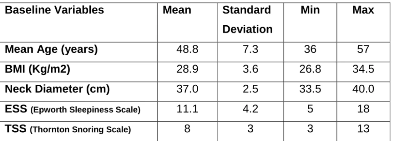
Eleven Caucasian patients who met selection criteria were initially approached to participate and asked to give them informed consent. One patient withdrew from the study because of lack of previously defined minimum compliance as subjectively evaluated by the diary usage (< 50% of the days), and one patient was lost for follow up leaving a total of nine subjects (6 males and 3 females) with a mean age of 49 and ranging from 36 to 57 years old at baseline.
Fig. 8 - Patients inclusion Flow chart
Baseline Variables Mean Standard Deviation
Min Max
Mean Age (years) 48.8 7.3 36 57
BMI (Kg/m2) 28.9 3.6 26.8 34.5
Neck Diameter (cm) 37.0 2.5 33.5 40.0
ESS (Epworth Sleepiness Scale) 11.1 4.2 5 18
TSS (Thornton Snoring Scale) 8 3 3 13
BMI – Body Mass Index; ESS – Epworth Sleepiness Scale; TSS – Thornton Snoring Scale
Table 1.Characteristics of the study group at baseline (n=9).
11 patients selected
9 patients included 2 patients excluded 1 lack of compliance
There were no differences in average (p>0.05), regarding of Age, BMI and Neck diameter between Basal and MAD conditions.
The mean induced mandibular advancement was 4.3 mm, with a median of 4.5 mm (6 patients) and a minimum and maximum of 3 mm and 6 mm, respectively. The titration period (period between the diagnostic PSG and the last mandibular advancement before therapeutic PSG) was 19 weeks in average with a minimum of 0 (when patient was considered titrated with the very first advancement, as assessed by the clinical improvement after that) and a maximum of 48 weeks. The time between the start of treatment and the follow-up sleep recording was 9.5 weeks in average, with a minimum of 4 and a maximum of 13 weeks.
The mean amount of sleep at baseline as subjectively assessed by a sleep questionnaire was 443±11 on week days and 470±10 on weekends (p=0.02). 6.2 Inferential Statistics
Differences between Means
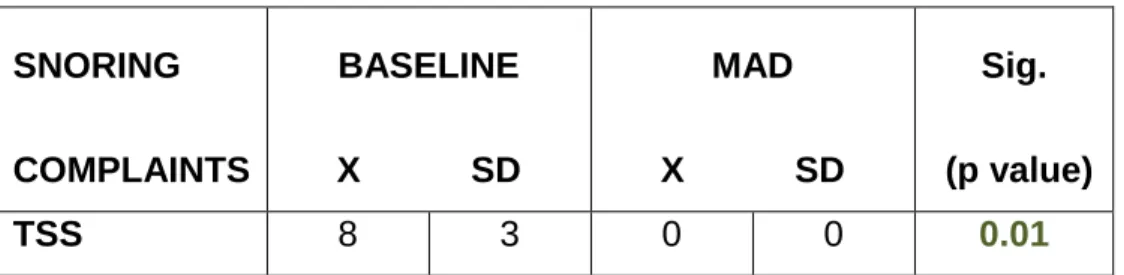
SNORING COMPLAINTS BASELINE X SD MAD X SD Sig. (p value) TSS 8 3 0 0 0.01
TSS – Thornton Snoring Scale score
Table 2.Snoring Score according to the Thornton Snoring Scale before (BASELINE) and after (MAD) treatment with mandibular advancement device (n=9)
Thornton Snoring Scale (TSS)
TSS after treatment (0) was inferior to ESS before treatment (8).
P < 0.05, meaning that existed significant statistical differences between two moments.
Preliminary Conclusions: In this sample, there were differences between two moments regarding to snoring and snoring social negative impact assessed by TSS; snoring and snoring related social problems improved (were reduced) after treatment with MAD compared to basal condition.
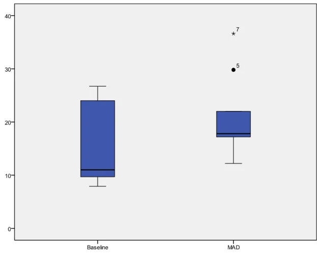
Graph. 1 - Boxplot Chart showing the differences between TSS score at the baseline and after therapy with MAD
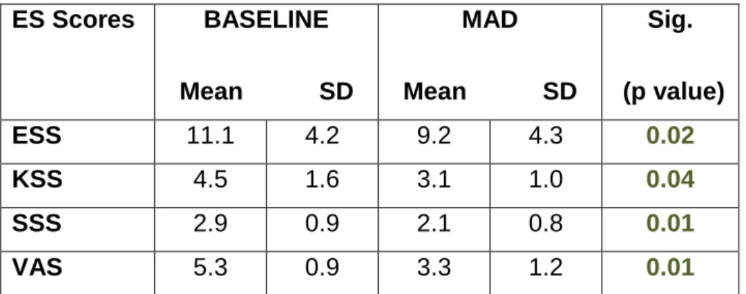
ES Scores BASELINE Mean SD MAD Mean SD Sig. (p value) ESS 11.1 4.2 9.2 4.3 0.02 KSS 4.5 1.6 3.1 1.0 0.04 SSS 2.9 0.9 2.1 0.8 0.01 VAS 5.3 0.9 3.3 1.2 0.01
ESS – Epworth Sleepiness Scale; KSS – Karolinska Sleepiness Scale; SSS – Stanford Sleepiness Scale; VAS – Visual Analogue Scale
Table 6. Excessive somnolence (ES Scores) status before (BASELINE) and after (MAD) treatment with mandibular advancement device (n=9).
SCORE BASELINE Mean SD MAD Mean SD Sig. (p value) PSQI 8.2 1.7 4.4 2.0 0.01
PSQI – Pitsburg Sleep Quality Index
Table 7. Sleep Quality Index Total Score before and after treatment with mandibular advancement device (n=9)
Epworth Sleepiness Score (ESS)
In average, ESS after treatment (9.2) was inferior to ESS before treatment (11.1).
P < 0.05, meaning that existed significant statistical differences between two moments.
Preliminary Conclusions: In this sample, there were differences between two moments regarding chronic excessive daytime sleepiness assessed by ESS; previous month (“chronic”) excessive sleepiness improved (was reduced) 17.0% after treatment with MAD compared to basal condition.
Graph. 17 - Boxplot Chart showing the differences between ESS score at the baseline and after therapy with MAD
Karolinska Sleepiness Scale (KSS)
On average, KSS after treatment (3.1) was inferior to KSS before treatment (4.5).
P < 0.05, meaning that existed significant statistical differences between two moments.
Preliminary Conclusions: In this sample, there were differences between two moments regarding acute excessive daytime sleepiness assessed by KSS; acute daytime sleepiness improved (was reduced) 31.3% after treatment with MAD compared with baseline condition.
Graph. 18 - Boxplot Chart showing the differences between KSS score at the baseline and after therapy with MAD
Stanford Sleepiness Scale (SSS)
On average, SSS after treatment (2.1) was inferior to SSS before treatment (2.9).
P < 0.05, meaning that existed significant statistical differences between the two moments.
Preliminary Conclusions: In this sample, there were differences between two moments regarding acute daytime sleepiness assessed by SSS; acute daytime sleepiness improved (was reduced) 25.8% after treatment with MAD compared with baseline condition.
Graph. 19 - Boxplot Chart showing the differences between SSS score at the baseline and after therapy with MAD
Visual Analogue Scale for Acute Sleepiness Assessment (VAS)
On average, VAS score after treatment (3.3) was inferior to VAS score before treatment (5.3).
P < 0.05, meaning that existed significant statistical differences between the two moments.
Preliminary Conclusion: In this sample, there were differences between two moments regarding acute daytime sleepiness assessed by VAS for acute sleepiness assessment; acute daytime sleepiness improved (reduced) 37.3% after treatment with MAD compared with baseline condition.
Graph. 20 - Boxplot Chart showing the differences between VAS score at the baseline and after therapy with MAD
Pittsburg Sleep Quality Index (PSQI)
On average, PSQI score after treatment (4.4) was inferior to PSQI score before treatment (8.2).
P < 0.05, meaning that significant statistical differences existed between the two moments.
Preliminary Conclusions: In this sample, there were differences between two moments regarding sleep quality assessed by PSQI; On average, sleep quality improved (there was a lower PSQI score) in 46.0% after treatment with MAD, compared with basal condition.
Graph. 21 - Boxplot Chart showing the differences between PSQI score at the baseline and after therapy with MAD
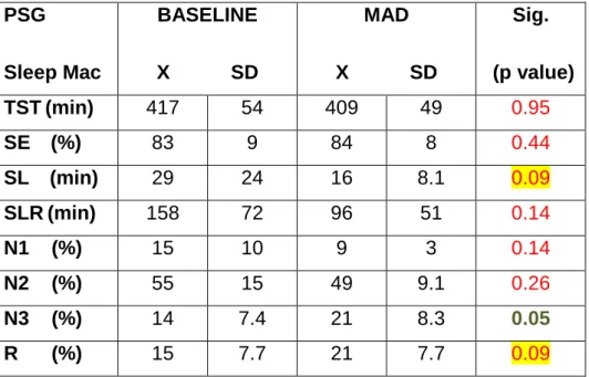
PSG Sleep Mac BASELINE X SD MAD X SD Sig. (p value) TST(min) 417 54 409 49 0.95 SE (%) 83 9 84 8 0.44 SL (min) 29 24 16 8.1 0.09 SLR(min) 158 72 96 51 0.14 N1 (%) 15 10 9 3 0.14 N2 (%) 55 15 49 9.1 0.26 N3 (%) 14 7.4 21 8.3 0.05 R (%) 15 7.7 21 7.7 0.09
TST – Total Sleep Time; SE – Sleep Eficiency; SL – Sleep Latency; SLR – Sleep Latency to REM Sleep Stage; N1 – N1 Sleep Stage; N2 – N2 Sleep Stage; N3 – N3 Sleep Stage
Table 3.PSG Sleep macrostructure (Sleep Mac) components status before (BASELINE) and after (MAD) treatment with mandibular advancement device (n=9)
Total Sleep Time (TST)
TST after treatment (409 min) was inferior to TST before treatment (417 min) P > 0.05, meaning that existed no significant statistical differences between two moments.
Preliminary Conclusion: In this sample, there were no differences between TST before and after treatment with MAD.
Graph. 1 - Boxplot Chart showing the differences between TST at the baseline and after therapy with MAD
Sleep Efficiency (SE)
SE after treatment (84%) was superior to SE before treatment (83%).
P > 0.05, meaning that existed no significant statistical differences between two moments.
Preliminary Conclusion: In this sample, there were no differences between SE before and after treatment with MAD.
Graph. 2 - Boxplot Chart showing the differences between SE at the baseline and after therapy with MAD
Sleep Latency (SL)
SL after treatment (16 min) was inferior to SE before treatment (29 min).
P > 0.05, meaning that existed no significant statistical differences between two moments.
Preliminary Conclusion: In this sample, there were no differences between SL before and after treatment with MAD; a trend for a lower sleep latency was however observed after treatment with MAD (p=0.09).
Graph. 3 - Boxplot Chart showing the differences between SL at the baseline and after therapy with MAD
Sleep Latency to REM sleep stage (SLR)
SLR after treatment (96 min) was inferior to SLR before treatment (158 min). P > 0.05, meaning that existed no significant statistical differences between two moments.
Preliminary Conclusion: In this sample, there were no differences between SLR before and after treatment with MAD although there was a trend.
Graph. 4 - Boxplot Chart showing the differences between SLR at the baseline and after therapy with MAD
N1 sleep stage (N1)
The relative amount N1 sleep stage after treatment (9%) was inferior to the relative amount of N1 sleep stage before treatment (15%), in average.
P > 0.05, meaning that existed no significant statistical differences between two moments.
Preliminary Conclusion: In this sample, there were no differences between the relative amount of N1 sleep stage before and after treatment with MAD.