UNIVERSIDADE DE LISBOA
Faculdade de Medicina
The role of nuclear positioning in muscle function
Mafalda Ramos de Melo PimentelOrientador:
Prof. Doutor Edgar Rodrigues Almeida Gomes
Tese especialmente elaborada para obtenção do grau de Doutor em Ciências Biomédicas especialidade em Biologia Celular e Molecular
UNIVERSIDADE DE LISBOA
Faculdade de Medicina
The role of nuclear positioning in muscle function
Mafalda Ramos de Melo PimentelOrientador: Prof. Doutor Edgar Rodrigues Almeida Gomes
Tese especialmente elaborada para obtenção do grau de Doutor em Ciências Biomédicas especialidade em Biologia Celular e Molecular
Júri: Presidente: Doutor João Eurico Cortez Cabral da Fonseca, Professor Catedrático e Vice-Presidente do Conselho Cientifico da Faculdade de Medicina da Universidade de Lisboa
Vogais: Doctor Antoine Guichet, Group Leader and Principal Investigator, Institut Jacques Monod, Université Paris Diderot;
Doutor Reto Gassmann, Group Leader and Investigador do Instituto de Biologia Molecular e Celular da Universidade do Porto;
Doutor Ramiro Daniel Carvalho de Almeida, Professor Auxiliar do Departamento de Ciências Médicas da Universidade de Aveiro;
Doutora Solveig Thorsteinsdottir, Professora Associada com Agregação da Faculdade de Ciências da Universidade de Lisboa;
Doutora Maria do Carmo Salazar Velez Roque da Fonseca, Professora Catedrática da Faculdade de Medicina da Universidade de Lisboa;
Doutor Edgar Rodrigues Almeida Gomes, Professor Associado Convidado da Faculdade de Medicina da Universidade de Lisboa;
Instituição Financiadora: Fundação para a Ciência e Tecnologia SFRH/BD/52227/2013 2019
A impressão desta tese foi aprovada pelo Conselho Científico da Faculdade de Medicina de Lisboa em reunião de 16 de Outubro de 2018.
As opiniões expressas nesta publicação são da exclusiva respondabilidade do seu autor.
i
Acknowledgments
I would like to express my gratitude to my supervisor Dr. Edgar Gomes, for giving me the opportunity to perform my thesis in his new lab. I am very grateful for his support throughout my PhD and for his sharing of knowledge, experience and out of the box ideas. After this challenging but gratifying project, I know now that science can be “cool”, exciting and curiosity driven.
I would like to thank all former and present members of the Edgar Gomes group, for the productive and friendly working atmosphere and for all the group activities inside and outside the lab. In particular, I am grateful to Bruno Cadot and Valerie Vilmont, for receiving me in Paris, making me feel welcomed and helping me with all the initial technicalities and bureaucracies. I would like to thank Judite Costa for all the support and advice, and for always making me see the better side of things. I am thankful to Sara Ferreira for taking care of our lab and making our lives infinite times easier. Many thanks to the muscle team, for all the hours spent doing primary cells (or trying), for all the troubleshooting and shared frustrations. I am especially thankful to Graciano Leal, who became my mRNP mentor/ encyclopedia, and to Helena Pinheiro, who is my science soulmate and makes me enjoy every little detail in our joint scientific lives. I would like to thank my front neighbor, Francisco Calero, for all the molecular biology teachings no matter how hopeless they were. Most importantly, I am grateful to William Roman for changing my paradigms in science and beyond.
I would like to thank our neighbor lab, especially to Claudio Franco, for all the meaningful input in our meetings and to Pedro Barbacena for being always there for me. I would like to thank the Figueirencios, for being simply the best ex-lab ever and never stopping taking care of me. I acknowledge the IMM community for being so friendly and helpful all the way.
Finally, I would like to express my gratitude to my family and friends for their constant support and immense belief in me. You remind me every day why it is worth it.
iii
Table of contents
List of figures ... v
List of tables ... vi
List of abbreviations ... vii
Summary ... ix
Resumo ... xi
1 Introduction ... 1
1.1 Skeletal muscle biology ... 1
1.1.1 Skeletal muscle structure ... 1
1.1.2 Skeletal myogenesis ... 5
1.1.3 Muscle function ... 6
1.1.4 Muscle disorders ... 9
1.2 Nuclear positioning and nuclear domains ... 11
1.2.1 Nuclear positioning in skeletal muscle ... 11
1.2.2 Nuclear domain theory ... 13
1.2.3 Skeletal muscle research models ... 15
1.3 Subcellular mRNA localization ... 18
1.3.1 Relevance of mRNA localization... 18
1.3.2 Sequence determination and RBPs ... 22
1.3.3 mRNP transport by cytoskeleton motors ... 25
1.3.4 mRNP anchoring and hitchhiking... 29
1.3.5 mRNA localization in muscle ... 30
2 Objectives ... 35
3 Materials and Methods ... 37
3.1 Myoblast isolation and in vitro myofiber differentiation ... 37
3.2 Immortalized human myoblast culture and co-culture ... 38
iv
3.4 Transfection of plasmids and siRNAs ... 39
3.5 RNA extraction and RT-qPCR ... 41
3.6 Drug treatments ... 41
3.7 Immunofluorescence staining and image acquisition ... 42
3.8 Active ribosome labelling (Puromycilation) ... 43
3.9 smFISH and total mRNA FISH ... 43
3.10 SYTO14 live imaging ... 45
3.11 Nuclear movement imaging ... 45
3.12 Light-induced contraction ... 45
3.13 Image analysis and quantification ... 46
3.14 Statistics ... 46
3.15 Protein size and GO term analysis ... 47
4 Results ... 49
4.1 Nuclear positioning in myofibers differentiated in vitro ... 49
4.2 Perinuclear mRNA localization in mature myofibers ... 51
4.3 Giant muscle mRNAs have a particular distribution ... 57
4.4 mRNA localization is cytoskeleton dependent ... 59
4.5 Translation correlates with regular mRNA distribution... 67
4.6 mRNA localization correlates with muscle function ... 70
4.7 Contributions ... 72
5 Discussion ... 73
6 Appendix ... 81
6.1 smFISH probes ... 81
6.2 MATLAB script for spatial analysis of smFISH ... 87
6.3 GO term analysis of top10 biggest CDSs in the genome ... 91
6.4 Publications ... 94
v
List of figures
Figure 1 – Structural organization of skeletal muscle. ... 2
Figure 2 – Sarcomere basic components and organization. ... 3
Figure 3 – Ultrastructure of a skeletal muscle cell. ... 4
Figure 4 – Excitation-contraction coupling. ... 7
Figure 5 – Hematoxylin and eosin staining of healthy and CNM muscle sections 10 Figure 6 – Events of nuclear movement during myogenesis. ... 12
Figure 7 – Myofiber nuclear position and nuclear domains. ... 14
Figure 8 – Differentiation of mouse primary myofibers in vitro ... 17
Figure 9 – Overview of roles and mechanisms for mRNA localization ... 20
Figure 10 – RBP binding to localization elements determines mRNA localization 24 Figure 11 – Regulation of motored mRNA transport. ... 27
Figure 12 – Localization of mRNA in skeletal muscle cells ... 31
Figure 13 – Microtubule regrowth in isolated adult myofibers... 33
Figure 14 – Nuclear movement and positioning are recapitulated in vitro. ... 50
Figure 15 – Total mRNA is enriched perinuclearly. ... 51
Figure 16 – Individual mRNAs detected by smFISH are enriched perinuclearly .. 52
Figure 17 – Acta1 is a highly expressed mRNA clustered around the nucleus .... 53
Figure 18 – Nuclear enrichment and origin of mRNAs in a heterokaryon ... 54
Figure 19 – mRNAs are excluded from cell center to sarcolemma by myofibrils .. 55
Figure 20 – Non-muscle mRNAs also accumulate perinuclearly by default ... 56
Figure 21 – Giant mRNAs are spread and do not accumulate perinuclearly ... 58
Figure 22 – Differential mRNA distribution is also observed in vivo ... 59
Figure 23 – mRNA localization and levels are affected by colchine treatment ... 60
Figure 24 – Kinesin 1 (Kif5b) affects nuclear but not mRNA distribution ... 62
Figure 25 – Inhibition of dynein disperses perinuclear mRNA ... 63
Figure 26 – Dynein is enriched perinuclearly and does not anchor the nucleus ... 64
Figure 27 – Dynactin complex contributes to perinuclear mRNA accumulation ... 66
Figure 28 – Ribosome content is increased in the nuclear proximity ... 68
Figure 29 – Translation is increased at the perinuclear region ... 69
Figure 30 – Protein localization is dependent on nuclear position ... 70
vi
List of tables
Table 1 – Reagents for primary myofiber in vitro differentiation ... 38
Table 2 – Plasmids transfected for overexpression ... 40
Table 3 – Silencer select siRNAs from Ambion ... 40
Table 4 – Primers used for RT-qPCR ... 41
Table 5 - Antibodies used for immunofluorescence ... 42
Table 6 – Characteristics of mRNAs studied by smFISH ... 57
vii
List of abbreviations
A band - anisotropic band AchR - Acetylcholine receptor ADP - Adenosine diphosphate ANOVA - analysis of variance ATP - Adenosine triphosphate bp - Base pairs CDS - coding sequence ChR2 - Channelrhodopsin-2 CNM - centronuclear myopathy DHPR - dihydropyridine receptor E-C
coupling - Excitation contraction coupling ECCE - excitation-coupled Ca2+ entry EDL - extensor digitorum longus ER - endoplasmic reticulum GO term - Gene Ontology Term h - hours
hLamA/C - human Lamin A/C I band - isotropic band
IF - intermediate filaments kb - Kilobases
kD - kiloDalton Kif - Kinesin family LE - Localization element LUT - Look up table
MAP - microtubule associated protein MBP - myelin basic protein
MCI - mRNA clustering index mDa - megaDalton
MHC - Myosin Heavy Chain
MIP - Maximum intensity projection MRF - myogenic regulatory factors mRNA - messenger ribonucleic acid mRNP - messenger ribonucleic particle ms - milliseconds
MT - microtubules
MTJ - Myotendinous junction
MTOC - Microtubule organizing center MW - molecular weight
NMJ - neuromuscular junction PAX - paired box gene
RBP - RNA binding protein RNA - ribonucleic acid RYR - Ryanodine receptor
viii
SD - spinning disk sec - second
SERCA - sarco/endoplasmic reticulum Ca2+-ATPase smFISH - single molecule fluorescence in situ hybridization SOCE - store-operated Ca2+ entry
SR - sarcoplasmic reticulum SUM - image SUM projection TL - Transmitted light um or μm - micrometers
UTR - untranslated regulatory region wt - wild type
ix
Summary
Skeletal muscle is formed by multinucleated myofibers, the biggest cells in the human body. The multiple nuclei in these cells are regularly positioned so that the distance between them is maximized. It was previously found that nuclear positioning is important for skeletal muscle function (Metzger et al., 2012). However, mechanistic insight was missing since no evident structural abnormalities were found as a consequence of nuclear mispositioning. We hypothesized that each nucleus influences the nearby cytoplasm by determining mRNA localization along myofibers. As a consequence, protein translation and regulation would be hampered in situations of nuclear mispositioning, such as in centronuclear myopathies.
Using highly matured mouse myofibers differentiated in vitro, we found that overall mRNA distribution depends on nuclear position. Using smFISH we observed that during myofiber maturation and myofibril organization, mRNAs are pushed towards the sarcolemma. We also validated the nuclear domain theory (Pavlath et al., 1989) by detecting total mRNA clustering around peripheral nuclei. This seems to be the default localization of mRNAs in myofibers since both muscle specific and housekeeping transcripts display the same pattern.
This perinuclear clustering is an active mechanism, dependent on the minus end directed microtubule motor dynein and its activator dynactin. We have also established that the levels of protein translation can depend on nuclear location. Ribosome content is higher in the nuclear region, independently of Dynactin2 expression. Using a heterokaryon system, we show that at least some proteins in the cell remain localized close to their nucleus of origin. Moreover, contractibility of the cells correlates with the position of the nucleus and thus with overall mRNA localization.
Interestingly, a peculiar subset of mRNAs localizes regardless of where the nucleus is placed. A common feature of these transcripts is their extremely big length. We confirmed that this differential distribution is also happening in vivo. We propose that an active mechanism is responsible for this “giant” mRNA localization
x
in order to ensure and facilitate the localization of the encoded proteins. Understanding the mechanisms of mRNA transport and anchoring that govern its subcellular destinations in myofibers may be the key to understand how nuclear positioning impacts muscle activity.
Keywords: skeletal muscle, mRNA localization, microtubules, translation,
xi
Resumo
O músculo esquelético é formado por longas células excitáveis e contrácteis denominadas fibras musculares. Estas são as maiores células no corpo, altamente complexas e especializadas (Marieb and Hoehn, 2007). As fibras musculares têm origem na fusão de dezenas a centenas de células percursoras – os mioblastos – durante a embriogénese. O seu citoplasma está maioritariamente preenchido pelas miofibrilas, compostas pelos filamentos de actina e miosina, efetores da contracção muscular. A fibra muscular é um dos raros sincícios existentes no corpo humano. Os múltiplos núcleos existentes em cada fibra organizam-se durante o desenvolvimento de modo a posicionarem-se à periferia da célula e a que se maximize a distância entre eles (Bruusgaard et al., 2003; Roman and Gomes, 2017). Este posicionamento é altamente conservado evolucionariamente, o que sugere relevância biológica (Liu et al., 2009). Adicionalmente, em certas patologias o posicionamento do núcleo encontra-se afectado, apresentando-se ao centro da célula e muitas vezes em agregados (Biancalana et al., 2012). As consequências desta alteração morfológica na função muscular dos pacientes não são totalmente entendidas (Romero, 2010).
Ainda não é clara a extensão da influência que cada núcleo pode exercer no citoplasma de uma fibra muscular. Foi reportado anteriormente que o posicionamento do núcleo afecta a função muscular, mas até então não se sabia exactamente através de que mecanismo (Metzger et al., 2012). Nós colocámos a hipótese de que cada núcleo é responsável por uma porção do citoplasma envolvente através do controlo da localização do RNA mensageiro (mRNA) que transcreve e exporta. De acordo com esta hipótese, um posicionamento incorrecto dos núcleos levaria a uma distribuição anormal de produtos de expressão génica potencialmente importantes para a contracção e homeostasia do músculo. A localização do mRNA já foi descrita como importante para diversos mecanismos biológicos, nomeadamente a formação e manutenção de sinapses no sistema nervoso (Sutton and Schuman, 2006). A deficiência dos mecanismos moleculares necessários para a correcta localização de certos transcritos também já foi associada a diversas patologias (Brinegar and Cooper, 2016; Wurth and Gebauer, 2015). Embora todos os mecanismos descritos até à data sejam específicos para
xii
cada espécie de mRNA, geralmente é comum a todos a ocorrência de transporte activo através de uma proteína motora do citoesqueleto e proteínas adaptadoras ligadas ao transcrito, muitas vezes através do 3’UTR (Buxbaum et al., 2015).
No músculo esquelético a localização do mRNA tem sido alvo de interesse, mas a sua estrutura e complexidade dificultaram estudos mais aprofundados e com maior especificidade. Adicionalmente, dada a delicadeza fisiológica destas células, são escassos os estudos dinâmicos com relevância similar ao que acontece em músculo completamente formado e funcional. Utilizando um sistema in vitro para o desenvolvimento de fibras musculares altamente diferenciadas nós confirmámos que a distribuição do mRNA depende do posicionamento nuclear. Este sistema permite desenvolver fibras musculares de ratinho de modo a apresentarem as características de fibras musculares in vivo (Pimentel et al., 2017). Permite também a manipulação e observação microscópica com alta resolução de todo o processo de diferenciação. O desenvolvimento inicia-se com mioblastos recolhidos de recém-nascidos que durante 10 dias formam fibras musculares com forma tubular, miofibrilas alinhadas, contracção espontânea, núcleos à periferia e tríades em dupletos a flanquear o disco Z dos sarcómeros (Falcone et al., 2014).
Através de smFISH (hibridação de sondas fluorescentes in situ para marcação de moléculas individuais) observámos que durante a maturação da fibra muscular e dos seus filamentos (mofibrilas) os mRNAs são excluídos para a periferia das células levando à sua acumulação perto da membrana citoplasmática. Confirmámos adicionalmente a teoria dos domínios nucleares de Pavlath que durava há décadas no campo da investigação muscular (Pavlath et al., 1989) ao detectar um enriquecimento significativo de mRNA na zona envolvente dos núcleos à periferia da célula. Esta restrição da distribuição de transcritos já tinha sido observada na junção neuromuscular mas não em núcleos não sinápticos, dada a maior dificuldade em entender a origem dos transcritos no sincício (Merlie and Sanes, 1985). Esta parece ser a localização preferencial dos transcritos em geral dado que tanto transcritos específicos de musculo como transcritos housekeeping partilham desta localização. A disrupção do posicionamento nuclear através da depleção de kif5b leva a regiões cuja densidade de transcritos é diminuída.
xiii A localização perinuclear do mRNA é um mecanismo activo dado que é dependente do motor Dineína, um complexo proteico que transporta cargas para a extremidade positiva dos microtúbulos. O complexo auxiliar Dinactina também é importante para a manutenção de mRNAs em volta do núcleo. Não observámos o envolvimento de nenhuma das Cinesina testadas na localização de mRNA em fibras musculares. No entanto, algumas delas afectaram consideravelmente o desenvolvimento celular sendo possível que estejam implicadas no transporte de mRNA. Adicionalmente, também observámos que os ribossomas estão enriquecidos na zona perinuclear através da marcação do RNA ribossomal 18S e das proteínas P. Utilizando o ensaio de puromicilação, confirmámos que os níveis de tradução são proporcionalmente mais elevados perto do núcleo do que no em zonas longe dos mesmos. Para determinar com precisão a localização de proteínas específicas relativamente ao seu núcleo de origem, optimizamos a formação de heterocários em que um núcleo humano é incorporado numa célula contendo múltiplos núcleos de ratinho. Utilizando anticorpos específicos para proteínas humanas detectamos um enriquecimento das mesmas perto do único núcleo humano na célula. Em células contendo apenas núcleos de ratinho não foi observado um enriquecimento proteico na região perinuclear. Isto deve-se possivelmente ao facto de que o espaçamento nuclear permite que as proteínas se encontrem devidamente distribuídas em fibras musculares saudáveis.
Para tentar compreender a possível implicação desta assimetria na distribuição do mRNA e respectiva tradução, medimos a função muscular através da contracção. Utilizando uma ferramenta optogenética que consiste num canal de catiões activado pela luz (Channelrodopsin2) pudemos concluir que a região nuclear da fibra muscular é mais facilmente induzida a contrair do que regiões afastadas do núcleo. Estes resultados apontam para a importância da distribuição equidistante dos múltiplos núcleos nas células de músculo.
Paralelamente, encontrámos um conjunto de mRNAs que não se acumula na periferia do núcleo. A única característica comum que conseguimos apurar entre eles foi o seu tamanho acima do normal. Um deles é o mRNA para a Titina, a maior proteína codificada no genoma, específica e essencial para o músculo. De facto, várias das maiores proteínas musculares são anormalmente grandes em parte devido à sua função estrutural. Os mRNAs que codificam para estas
xiv
proteínas encontram-se amplamente distribuídos nestas células. Apesar de não termos encontrado nenhuma proteína motora que afecte o transporte dos mesmos (em parte devido à possível toxicidade do seu fenótipo de depleção), observámos que estes transcritos se encontram altamente concentrados nas extremidades celulares. Essa localização sugere uma dependência da orientação positiva dos microtúbulos, embora não tenhamos estabelecido uma conexão com nenhuma das Cinesinas testadas. O transporte diferencial de mRNAs “gigantes” traria benefícios que poderiam ser passiveis de selecção evolucionária. Ao localizar estes mRNAs ao longo de toda a célula, as várias proteínas traduzidas a partir dos mesmos não teriam de percorrer distâncias tão elevadas e exigentes energeticamente. A topologia destas proteínas também pode requerer que estas sejam traduzidas localmente, tendo especialmente em conta a elevada densidade do citoplasma muscular (sarcoplasma).
Em conjunto estes resultados demonstram a relevância do posicionamento nuclear em fibras musculares ao nível da distribuição dos mRNAs em geral. Também implicam que um incorrecto positionamento pode potencialmente original zonas da célula em que a contracção não é tão eficiente. Este estudo revela a localização especial de um conjunto de transcritos, os mRNAs “gigantes” que nunca tinha sido descrita anteriormente. A distribuição particular destes mRNAs constitui um novo exemplo que fundamenta a importância da localização de certos transcritos para a optimização de funções biológicas específicas.
Palavras-chave: músculo esquelético, localização de mRNA, microtúbulos,
1
1 Introduction
1.1 Skeletal muscle biology
Skeletal muscle tissue is by far the most abundant in a mammalian organism, composing up to 40% of the human body (Janssen et al., 2000). It can be divided in two groups – striated and smooth muscle – based on the internal arrangement of contractile filaments. Striated muscle exhibits clear arranged striations under a brightfield microscope in comparison to the smooth counterpart. It can be further subdivided into skeletal and cardiac tissues. Although with a very similar contractile machinery, they are quite distinct not only in function but also in cellular organization. Skeletal muscles attach to bones through tendons and are responsible for all voluntary movements of the body, posture and heat generation. Each muscle is composed of long multinucleated cells that span the entire organ length. On the other hand, cardiac muscle generates involuntary heart beat and is generally composed of mononucleated cells connected by specialized junctions called intercalated disks. Despite their different biogenesis, many proteins and pathways are shared between the two types of striated muscle and so the two fields of research are often connected.
1.1.1 Skeletal muscle structure
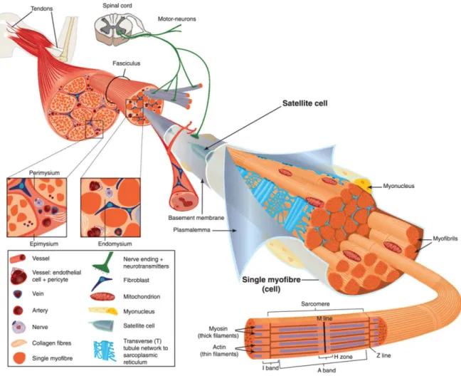
Skeletal muscle is a highly complex and organized organ composed of several types of tissue (Figure 1,Figure 1 – Structural organization of skeletal muscle. Aminoff, 2005). The predominant cell type – skeletal muscle fibers or myofibers – are long multinucleated cells encapsulated by a basement membrane. Several myofibers surrounded by connective tissue (endomysium) bundle into a fascicle. Multiple fascicles are bound by an epimysium and ultimately compose the muscle organ, connected to bone usually through tendons. In addition to the supportive layers of connective tissue, each muscle has an intricate network of small capillaries. These are derived from a central artery and branch along each myofiber in order to serve its high metabolic needs. Furthermore, each muscle is innervated by at least one motor neuron being each myofiber controlled by only one axon branch, at the neuromuscular junction.
2
Figure 1 – Structural organization of skeletal muscle.
Skeletal muscle is highly vascularized and is innervated by axon branches of motor neurons. It is mainly composed of several fascicles which aggregate multiple myofibers (muscle cells), spanning the organ length. Each cylindrical myofiber has numerous myofibrils containing arrays of contractile units, the sarcomeres. The multiple nuclei are positioned at the cell periphery, under the sarcolemma. The organization and function of muscle is also dependent on its several layers of connective tissue. Adapted from Tajbakhsh, 2009.
At the myofiber level, intracellular organization is also highly complex (Marieb and Hoehn, 2007). Each tubular cell has multiple nuclei positioned at the periphery, under the membrane, known as sarcolemma. Inside, the sarcoplasm (muscle cytoplasm) surrounds a dense arrangement of filament bundles termed
myofibrils. These cylindrical myofibrils are sequential repetitions of the
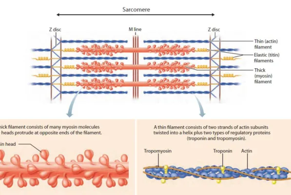
contraction units, the sarcomeres, in which filaments of myosin slide over actin filaments to generate force (Figure 2)Figure 2 – Sarcomere basic components and organization.. Each sarcomere extends from one Z line (or Z disk) to another, a very dense structure containing α-actinin for actin filaments anchorage (Clark et
3 al., 2002). The center of the sarcomere is termed M line, given that myosin tails are fixed in this region. By opposition, the region around the Z lines contains only actin and it is known as the I band. The I band has Isotropic light properties in comparison to the Anisotropic nature of the complementary A band (where myosin polarizes light). The gigantic protein Titin spans all the way from the Z line to the M line (Tskhovrebova and Trinick, 2003). Being the biggest protein encoded in the genome, it is 1 µm in length and 4 MDa in weight. Importantly, the elastic properties of the Titin filament provide resistance to excessive stretching while keeping the Myosin filament in place.
Figure 2 – Sarcomere basic components and organization.
Each sarcomere is bordered by the Z lines (or Z disks) where the actin filaments get anchored to α-actinin. The myosin filaments stem from the center of the structure with their heads towards the actin filaments. During contraction, troponin binds to Ca2+ and changes tropomyosin conformation, making actin accessible to myosin. The myosin binds and slides to the next actin site at the expense of one ATP. Titin is a gigantic protein that spans half of the sarcomere. It keeps the myosin filament in place and its elasticity offers resistance to stretch. Adapted from Marieb and Hoehn, 2007.
4
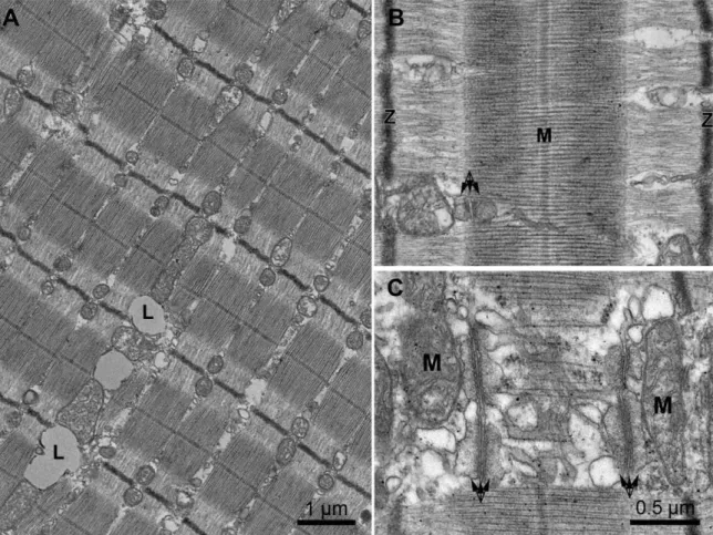
Each sarcomere is laterally aligned with the adjacent sarcomeres in neighboring myofibrils. This ultrastructural myofibril alignment and the different filament density are the reasons behind the striated pattern appearance, typical of striated muscle (Figure 3). This precise patterning enables the crosslinking of all myofibrils through their Z lines, by the intermediate filament Desmin (Capetanaki et al., 2007). It also paves the way for the organization of Triads, membrane structures crucial for muscle contraction (described in section 1.1.3). These also span transversely the whole cell section, residing immediately next to each A band.
Figure 3 – Ultrastructure of a skeletal muscle cell.
A) Low magnification electron micrograph of human vastus lateralis biopsy displaying the typical striated pattern of aligned myofibrils. B) Sarcomere detail with aligned Z line (Z) and M band (M). C) Membrane components positioned at the edge of the A band (triple arrows indicate a triad), next to mitochondria. (Pietrangelo et al., 2013)
5 Importantly, sarcomere and thus myofibril alignment results in efficient muscle force generation. Yet, this contractile machinery has to be anchored to the cell membrane for force transmission to the muscle tissue. Given the magnitude of the contraction force, the subsarcolemmal area has a specialized structure termed
costamere for connection to the extracellular matrix at the Z line (Ervasti, 2003;
Jaka et al., 2015). The dystrophin–glycoprotein complex is a main costamere component, linking the intermediate filaments network of desmin to the extracellular matrix. The costamere is subjected to immense straining, being the origin of a multitude of muscular dystrophies (Cardamone et al., 2008).
1.1.2 Skeletal myogenesis
Myofibers are the biggest human cells, originated from the fusion of numerous muscle precursors – the myocytes (Bentzinger et al., 2012). This happens intensively throughout embryogenesis, as well as sparsely during adulthood in order to maintain tissue homeostasis.
The main intrinsic signaling pathways underlying embryonic progenitor and adult satellite cell fusion are broadly similar and well established. Essentially, a cascade of hierarchical transcription factors is induced to orchestrate the transition of progenitors through specification and commitment into the myoblast stage. The most often referred players are paired-homeobox transcription factors (e.g. Pax3 and Pax7) which regulate early specification, and myogenic regulatory factors (MRFs) which are common markers for committed myoblasts (e.g. Myf5 and MyoD) (Buckingham and Relaix, 2015).
Following proliferation, myoblasts give place to myocytes ready to fuse expressing MyoG and MRF4. The nuclei from these fusing myocytes are placed in the cell center giving rise to a multinucleated myotube (Cadot et al., 2015). The myotube differentiates into a myofiber once the excitation and contraction components are properly expressed and assembled. The process of myofibrillogenesis starts with arrays of sarcomeres being assembled close to the cell membrane (Sparrow and Schöck, 2009). It is believed that integrins anchor premyofibrils, which resemble actin stress fibers containing α-actinin and non-muscle myosin II. While premyofibrils develop, they incorporate titin and muscle myosin II. The correct
6
length of the actin and myosin is regulated by several components (e.g. Titin and Nebulin) as Z disks are formed. The newly formed myofibrils become aligned in a contraction dependent manner. Concomitantly, the nuclei move to the cell
periphery and spread so that the distance between them is maximized
(Bruusgaard et al., 2003; Roman et al., 2017).
Finally, the mature myofiber can undergo hypertrophy (increase in size) in response to exercise. Interestingly, new myoblasts can fuse during hypertrophy suggesting that the number of nuclei is proportional to the cell volume in certain muscles (Bruusgaard et al., 2010; Gundersen, 2016).
Some muscle progenitor cells do not fully engage in the myogenic process and become quiescent after specification (Bentzinger et al., 2012). These will give rise to the adult satellite stem cell pool that upon activation replenishes muscle with myoblasts for hypertrophy or muscle damage repair.
1.1.3 Muscle function
The main function of muscle tissue is the voluntary generation of force. This is why the main switch to induce contraction is an action potential from a somatic motor neuron. In the same muscle, one motor neuron can have multiple axon branches connecting to multiple myofibers. This is known as a motor unit. The smaller the average motor unit size, the more precise and controlled is a muscle.
Once the action potential has reached the axon terminal it has to be passed on to the myofiber. This occurs at the neuromuscular junction, a unique site where both the neuron and myofiber specialized in order to communicate. In there, Acetylcholine (ACh) is released and binds to its receptors at the muscle postsynaptic membrane. As a consequence, the activated receptors open and lead to local membrane depolarization (K+ efflux). Since only one neuromuscular junction exists per myofiber, the excitation signal has to be propagated throughout the entire cell length for contraction to occur. Essentially, this is made possible by the voltage gated Na+ channels spread along the sarcolemma that open sequentially upon the initial depolarization (Na+ influx).
7 After the myofiber has been thoroughly stimulated, the contractile machinery has to be activated. This link between the two events is termed
Excitation-Contraction (E-C) coupling and relies on the specialization and organization of
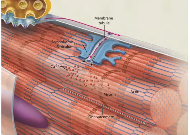
two different membrane structures (Figure 3C and 4). The first has origin in the sarcolemma, which invaginates and forms transversal tubules that go into the cell center while surrounding the myofibrils. These so called T Tubules transmit the action potential from the cell surface to every sarcomere, and flank each Z line at the junction between the A and I bands. The second originates from the
sarcoplasmic reticulum (SR), the endoplasmic reticulum of muscle that governs
calcium levels (Rossi and Dirksen, 2006). The SR has two domains: the longitudinal SR, which is tubular and surrounds myofibrils; and the junctional SR, composed of terminal cisternae which are also at the A-I band junction. Invariably, each T tubule is bordered by two terminal cisternae and this structure is a triad. The triad is where E-C coupling occurs. Briefly, a structural change in the voltage dependent calcium channel DHPR (at the T tubule) leads to Ryanodine receptor (RyR) opening which massively releases Ca2+ from the SR.
8
After local depolarization at the neuromuscular junction, an action potential is generated and travels along the sarcolemma into the T tubules all the way to the cell center. The t tubules are flanked by two terminal cisternae of the SR, making one triad. It is due to the close proximity triad proteins that the membrane depolarization signal is transmitted to the contractile apparatus. DHPR senses the T tubule voltage inducing RyR opening and massive calcium release in the SR. Adapted from Marieb and Hoehn, 2007.
Muscle contraction structurally consists of linking myosin globular heads to accessible actin attachment sites (Figure 2). These cross bridges are formed upon Ca2+ release and binding to troponin, which in turn changes tropomyosin configuration leaving actin exposed. Consecutively, myosin binds actin, releases previously hydrolyzed ATP (ADP + Pi) and moves to the following actin site. A new ATP readily binds to myosin and as a consequence myosin detaches from actin. The unbound but energized myosin head undergoes ATP hydrolysis and is ready for a new cycle of attachment, as long as Ca2+ and ATP are available. This sequential sliding of multiple myosins over actin, will lead to muscle shortening if the combined force produced by all sarcomeres in several myofibers surpasses the resistance offered to the muscle organ. Of note, whereas an action potential lasts 1-2 ms, the consequent contraction lasts at least 10ms and up to hundreds of milliseconds.
Contraction needs to be tightly controlled at all levels for muscle homeostasis: At the neuromuscular junction, Ach is rapidly degraded by acetylcholinesterase after binding to its receptors for neuronal control precision; As a consequence of membrane depolarization by Na+ channels, voltage gated K+ channels are quickly activated (K+ efflux). During this brief period of membrane repolarization (1-2 ms) an action potential cannot be triggered; To compensate this Na+-K+ ionic unbalance, the ATP-dependent Na+-K+ pump works at a relatively slow rate over the course of several contractions until fatigue (contraction inability) eventually occurs (Allen et al., 2008); Calcium stocks are also limited in the SR and so after each contraction they are at least partially restored by the sarco/endoplasmic reticulum Ca2+-ATPase (SERCA).
There are many other levels at which muscle function can be regulated, on the short and long term. The functional interaction of the numerous proteins involved is usually modulated by a third party. For example, Ca2+ is buffered in the SR by
9 Calsequestrin, which regulates Ryr opening through Triadin and Junctin (Beard et al., 2009). Total calcium levels can also be controlled by store-operated Ca2+ entry (SOCE) (Kurebayashi and Ogawa, 2001) or excitation-coupled Ca2+ entry (ECCE) (Cherednichenko et al., 2004). Different myofibers can also have different contraction kinetics, due to the expression of different protein variants and usage of energy sources. Myofibers can be classified in three types: slow oxidative (type 1), fast oxidative (type 2A and 2X) and fast glycolytic (type 2B) (Schiaffino and Reggiani, 2011). In particular, they express different myosin isoforms and use either the aerobic oxidative pathway or the glycolysis for ATP production. Different muscles will have different proportions of these fiber types depending on the kind of contraction they are used for. Altogether, the intrinsic ability for a muscle to contract sustainably depends on the correct expression, at the right place, of numerous proteins with countless possible interactions.
1.1.4 Muscle disorders
Most muscle inherited disorders can be classified as either a Dystrophy or a Myopathy (Cardamone et al., 2008). The pathogenesis of dystrophies is very heterogeneous but often related to structural muscle proteins, mostly at the costamere or its interacting proteins (Mercuri and Muntoni, 2013). Dystrophy symptoms have on average a later onset than Myopathies and there is progressive degeneration over time. Histologically, the dystrophic muscle shows severe necrosis, fibrosis and regeneration signs. The most common and best studied dystrophy is the Duchenne Muscular Dystrophy (DMD). In DMD the Dystrophin gene is mutated so that the protein is absent, affecting the structural integrity of myofibers and possibly mechanotransduction (Cohn and Campbell, 2000). Muscle weakness is one of the first symptoms although patients eventually die of heart or respiratory failure.
Myopathies are rarer than dystrophies, and usually the cause is a mutation affecting the efficiency of contraction. Myopathy biopsies show no signs of necrosis or regeneration. Instead, myofibers have distinct morphological changes such as the centrally located rows of nuclei in Centronuclear myopathies (CNM) (Figure 5, Biancalana et al., 2012). Several genes have been linked with CNM (e.g. DNM2, BIN1, MTM1, RYR1, TTN) although 20% of patients do not have a
10
genetic origin identified so far (Romero, 2010). The etiology of some structural abnormalities has been elucidated over the recent years, being mostly due to mutations in the E-C coupling machinery or in upstream components of membrane trafficking and metabolism (Jungbluth and Gautel, 2014). Myopathies have a much smaller incidence than dystrophies but are mostly congenital and usually present severer symptoms and mortality rates (Cardamone et al., 2008). Unfortunately, there is no cure for any of the disorders but disease specific interventions can sometimes improve the quality of life and longevity of the patients (Manring et al., 2014).
Figure 5 – Hematoxylin and eosin staining of healthy and CNM muscle sections
Healthy muscle biopsies display spaced peripheral nuclei (A,B) whereas CNM patients often have chains of centrally located nuclei (C,D), without showing signs of necrosis or regeneration. A,C transversal cuts; B,D longitudinal cuts. Adapted from Julio, 2013; Park et al., 2014b; Song et al., 2012.
11
1.2 Nuclear positioning and nuclear domains
1.2.1 Nuclear positioning in skeletal muscle
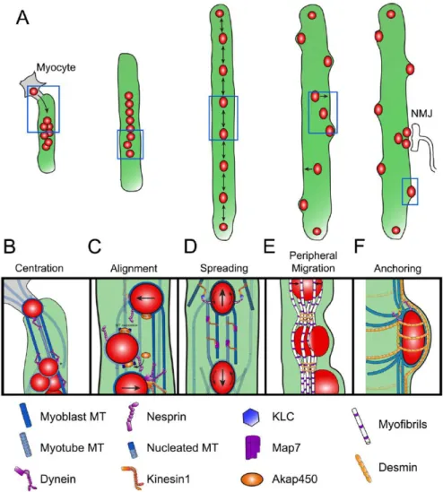
The multiple nuclei in a mature myofiber are positioned at its periphery, under the sarcolemma. In order to reach this location, several intermediary movements occur sequentially during myogenesis: centration, alignment, spreading, peripheralization and anchoring (Figure 6)Figure 6 – Events of nuclear movement during myogenesis. The steps leading to movement to the periphery have been well characterized, being dependent initially on microtubules and later on desmin filaments (Cadot et al., 2012; Falcone et al., 2014; Gimpel et al., 2017; Metzger et al., 2012; Roman and Gomes, 2017; Roman et al., 2017).
Once at the periphery, nuclei eventually get anchored and stop their longitudinal microtubule dependent movements (Bruusgaard et al., 2003; Englander and Rubin, 1987). Importantly, the nuclei are positioned so that the distance between them is maximized, in a non-random manner (Figure 7, Bruusgaard et al., 2003, 2006). The exact trigger for this nuclear caging by microtubules and desmin remains to be elucidated (Roman and Gomes, 2017). The LINC complex components (Nesprin1α2 and Sun1/2) and desmin have been shown to be impact anchorage (Chapman et al., 2014; Lei et al., 2009; Milner et al., 1996; Stroud et al., 2017; Zhang et al., 2007b). It is still unclear whether nuclear spacing and anchorage are interdependent at the periphery, as most phenotypes reported are static observations of nuclear clustering. This is in part due to a lack of appropriate system to dynamically address the question, independently of the preceding nuclear movements.
In a fully matured myofiber, three different areas can be distinguished: the neuromuscular junction (NMJ), at the center of the cell where around 5 subsynaptic nuclei are clustered under the axon terminal (Englander and Rubin, 1987); the myotendinous junction (MTJ), at the tips of the myofiber for attachment to tendons; and the extra-junctional area, where the remaining and majority of nuclei reside. The subsynaptic nuclei in the NMJ express specific genes important for the respective local functions (Fontaine and Changeux, 1989; Nazarian et al., 2005). The tyrosine kinase receptor Musk, when activated by the neuro-secreted
12
agrin, induces the transcription of specific NMJ genes with N-box elements (Hippenmeyer et al., 2007; Shi et al., 2012). Subsynaptic nuclear clustering and maintenance was also shown to be Desmin-Plectin and Nesprin1-Sun1 dependent (Grady et al., 2005; Lei et al., 2009; Mihailovska et al., 2014). Proteins important for membrane integrity, signaling and adhesion also accumulate specifically at the MTJ (Can et al., 2014; Dix and Eisenberg, 1990; Wang et al., 2013). However, nuclear clustering is only occasionally observed at the MTJ, probably as a consequence of regeneration or myocyte fusion (Bruusgaard et al., 2003). Thus, a specific pool of nuclei at the MTJ with a particular expression signature has not been described so far.
Figure 6 – Events of nuclear movement during myogenesis.
After myocyte fusion, dynein clusters nuclei at the center of the cell (B) and are afterwards aligned with the microtubule array in a Nesprin and dependent manner (C). Anti-parallel microtubules later allow the spreading of nuclei via Map7 and Kif5b (D). With myofiber differentiation, nuclei move to the periphery of the cell due to the tension generated by contraction and Desmin crosslinking (E).
13 Throughout differentiation nuclei move longitudinally inside the myofiber, except at highly mature stages where they get anchored by ITs and MTs (F). From Roman and Gomes, 2017.
1.2.2 Nuclear domain theory
The role for nuclear positioning is intuitive in certain circumstances, such as diving cells (Gundersen and Worman, 2013). However, the nucleus can also be asymmetrically positioned in terminally differentiated cells. The developed myofiber represents such cases in which the role for nuclear positioning might not be as evident (Folker and Baylies, 2013).
Bruusgaard and Gundersen contributed immensely to the current knowledge on nuclei number and distribution depending on muscle type and volume. By analyzing specifically myonuclei, they have undoubtedly established that: 1) nuclear distribution is fairly equidistant and not random; 2) the number of nuclei is proportional to cell volume in the slow/oxidative soleus muscle and 3) the number of nuclei is related to the cell surface area in the fast/glycolytic EDL muscle (Bruusgaard et al., 2003). Contradicting studies have sparked controversy, although most did not take into account the cellular heterogeneity of muscle tissue thus giving rise to skewed conclusions (Discussed in Gundersen, 2016; Gundersen and Bruusgaard, 2008). The authors have further confirmed by in vivo imaging that myonuclei number increases as a consequence of hypertrophy through satellite cell fusion (Bruusgaard et al., 2010). Moreover, they have unarguably shown that myonuclei number does not reduce during atrophy, contrarily to muscle size (Bruusgaard and Gundersen, 2008; Bruusgaard et al., 2010). These and other results have suggested the hypothesis of “muscle memory” in which the number of nuclei in a myofiber reflects its maximum size in the past. Accordingly, myofibers with increased myonuclear number but normal size due to testosterone induced hypertrophy and a period of withdrawal, have a much faster regrowth than the control and do not incorporate new myonuclei (Egner et al., 2013). These findings emphasize the importance of myonuclei position and number as they seem to be tightly controlled.
The reason for the particular position of myonuclei and its number regulation is still uncertain although Pavlath et al. provided a possible explanation by stating the
14
nuclear domain theory (Pavlath et al., 1989). Accordingly, each nucleus in a
myotube is surrounded by a region of limited distance where its genetic products can exert their effects (Figure 7E, top). The formation of these nuclear domains by some mRNAs and proteins was shown in a myotube context, by fusing cells of different genetic backgrounds (Ralston and Hall, 1992; Ralston et al., 1997). In fact, this exactly the case for the subsynaptic nuclei clustered at the NMJ. In this functionally specialized region of the muscle cell, the respective mRNAs and proteins accumulate and do not spread (Merlie and Sanes, 1985). It remains to be demonstrated that the majority of myofiber nuclei also have domains of influence where E-C coupling takes place. If Pavlath’s theory applies, mispositioning of nuclei might impede crucial mRNAs and respective proteins to completely reach their cellular targets and exert their functions (Figure 7E).
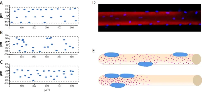
Figure 7 – Myofiber nuclear position and nuclear domains.
(A-C) Bruusgaard et al. compared in vivo nuclear positioning (B) with computational predictions of random distribution (B) and optimal distance between nuclei in 3D (C). All representations are of a myofiber flattened surface. Adapted from Bruusgaard et al., 2003 (D) Example of nuclear distribution in an isolated human myofiber, adapted from Qaisar and Larsson, 2014 (E) Simplified representation of nuclear domains in wild-type (top) and in nuclear mispositioning (bottom) conditions. Nuclei in all panels are depicted in blue.
In accordance to the nuclear domain theory, proper nuclear positioning seems to be important for drosophila skeletal muscle function (Metzger et al., 2012).
15 Moreover, mispositioned nuclei are a not yet understood hallmark of Centronuclear Myopathies (Al-Qusairi and Laporte, 2011), without being a consequence of regeneration. Interestingly, myoblast transplantation into DMD patients led to expression of dystrophin restricted to the new nuclei surroundings (Gussoni et al., 1997). Muscle fiber identity was also shown to decline in elder individuals in distinct nuclear associated domains (Andersen, 2003).
1.2.3 Skeletal muscle research models
There are multiple skeletal muscle models, depending on the biological question. The two most used systems are extremes opposite to one another: the murine in vivo experiments and the in vitro C2C12 culture. In vivo approaches provide by far the most physiologically complete results, with the drawbacks of being mostly static and excessively complex (Meng et al., 2014). They integrate the neurological and systemic response of matured myofibers modulated by the surrounding cells, to the experimental manipulation (e.g. induced damage, gene knock-out or contraction stimulation/inhibition). The second one, although more malleable, is highly limited by the differentiation level that can be reached and by the absence of a neuronal component. C212 cells were isolated from an adult CH3 mouse tight after injury (Yaffe and Saxel, 1977) and immortalized by serial passaging and subcloning (Blau et al., 1983). They constitute an excellent early developmental model, being accountable for most of the knowledge we have on myotube formation and development.
Another frequently used model is the isolation of adult mouse myofibers, either mechanically or enzymatically (Cheng and Westerblad, 2017; Pasut et al., 2013). This delicate ex vivo approach is particularly useful for studying satellite cell activation and fusion, as well as myofiber structure and contraction. It provides a slightly more dynamic insight into adult muscle biology, although limited technically and in time by the biophysical and physiological properties of these cells.
An approach that is being increasingly adopted is the use of in vitro systems with a degree of differentiation significantly higher than classic immortalized cultures. Early work with neonatal rat myoblasts unraveled not only the possibility for in vitro culture improvement but also underlined the different species inherent
16
differentiation potential (Flucher, 1992; Flucher et al., 1991). In fact, human myofiber in vitro differentiation is still limited despite all the investment driven by therapeutic interest (Guo et al., 2013).
With the recent progress of biotechnology, 3D systems were able to greatly enhance greatly in vitro myofiber development (Hinds et al., 2011; Madden et al., 2015). All 3D setups point out the importance of the extracellular matrix structure and composition for proper differentiation. In particular, the technique by Falcone and Roman differentiates primary neonatal mouse myoblasts into highly mature myofibers with peripheral nuclei, transversal triads and twitching capability (Figure 8Figure 7, Falcone et al., 2014; Pimentel et al., 2017). This method does not require specific hardware or highly-skilled manipulation and it is amenable to genetic manipulation and continuous imaging. Because of the simplicity of the setup, it can be adapted for combination with other techniques (e.g. neuron co-culture (Vilmont et al., 2016a)).
Given that in vitro developed myofibers have a smaller diameter, the utilization of high 3D resolution techniques is facilitated (due to increased sample permeation, higher specimen proximity and reduced auto-fluorescence). Additionally, the live imaging and developmental aspects provide a more integrated understanding of muscle biology compared to mammalian in vivo studies. For instance, nuclear dynamics can provide valuable insight into how muscle is compartmentalized and how other organelles are relatively positioned. As such, this in vitro system constitutes a unique skeletal muscle model of great potential in the field.
17
Figure 8 – Differentiation of mouse primary myofibers in vitro
(A-C) Transmitted light images showing the differentiation of myoblasts into myofibers with at day 2, 3, 6 and 11 respectively. Inset in D shows peripheral nuclei and striations of a highly matured myofiber. Scale bar 50μm. (E) Contraction event visualized through the expression of a cytoplasmic calcium sensor (20 ms/frame). Adapted from Pimentel et al., 2017.
18
1.3 Subcellular mRNA localization
The first in situ observation of polarized mRNA distribution dates to 1983 (Jeffery et al., 1983). The egg of the ascidian Styela has three visually distinct cytoplasmic domains, each giving rise to different cell lineages. William Jeffery observed that contrarily to total mRNA, the non-muscle actin mRNA was enriched at the myoplasm in the egg periphery. The potential functions for mRNA localization and localized protein expression were hypothesized, with translation control for cytoplasmic fate determination being proposed. Remarkably, the authors interrogated the mechanism for this cytoplasmic segregation and speculated on a contribution from the cytoskeleton, membranes and organelles. The discussed theories were proven right later on, being still applicable to countless transcripts and spanning many types of organisms.
1.3.1 Relevance of mRNA localization
The field of mRNA localization flourished with further developmental biology studies showing critical roles for specific mRNAs in oocyte, egg and embryo patterning. A classical functional example is the Xenopus Vg1 mRNA. This maternal transcript localizes to the oocyte vegetal pole being necessary and sufficient for mesoderm induction (Birsoy et al., 2006; Dale et al., 1993; Melton, 1987; Thomsen and Melton, 1993).
Eventually, the most widely used model to study mRNA localization became the Drosophila oocyte. In particular, the localization of the maternal mRNAs gurken, bicoid, oskar and nanos is a textbook example of anteroposterior (AP) and dorsoventral (DV) patterning. The localized translation of Gurken in the posterior pole initiates a signaling cascade that leads to cytoskeleton reorganization, nuclear repositioning and DV axis determination (González-Reyes et al., 1995; Guichet et al., 2001; Neuman-Silberberg and Schüpbach, 1993; Roth et al., 1995).
As a consequence, bicoid and oskar can diverge to the anterior and posterior poles respectively, specifying the AP axis (Berleth et al., 1988; Ephrussi and Lehmann, 1992; St Johnston et al., 1991). The posterior translation of Oskar enables the localization of nanos at the posterior pole, which is crucial for abdominal and germline development in the embryo (Figure 9 A and A’, Ephrussi
19 and Lehmann, 1992; Gavis and Lehmann, 1992; Gavis et al., 2008; Wang and Lehmann, 1991).
Several purposes for mRNA localization are recognized nowadays beyond embryonic determination, from bacteria to mammals (Buxbaum et al., 2015; Holt and Bullock, 2009). In 1986, Lawrence and Singer described the polarized localization of cytoskeletal mRNAs in migrating myoblasts (Lawrence and Singer, 1986). In particular, the localization of β-actin mRNA at the lamellipodia of migrating fibroblasts became one of the most studied examples (Figure 9). Abolishment of β-actin mRNA transport leads to altered cell morphology and decrease in the directionality and persistency of cell movement (Kislauskis et al., 1994, 1997; Shestakova et al., 2001). More precisely, these phenotypes were shown to be due to impairment of local translation of β-actin and consequent reduction of focal adhesion stability (Katz et al., 2012; Rodriguez et al., 2006).
In epithelial cells mRNA localization is also polarized, and this seems to be important for adherens junction assembly and signaling (Gutierrez et al., 2014; Kourtidis et al., 2017; Nagaoka et al., 2012). Recent work on the mouse intestinal epithelium has shown that apical mRNA polarization upon feeding increases translation efficiency, required for nutrient absorption (Moor et al., 2017).
Independent genome wide studies emphasize how common mRNA localization seems to be. In one particularly striking study, over 70% of the observed mRNAs localize to specific subcellular compartments in the drosophila embryo, usually at the same location as the encoded proteins (Lécuyer et al., 2007). In line with this, a significant number of mRNAs was found to be enriched in specific cytoplasmic regions of mammalian cells (Cajigas et al., 2012; Mardakheh et al., 2015; Mili et al., 2008; Poon et al., 2006; Weatheritt et al., 2014). Many of these global studies were performed in neurons, with the localization of several individual mRNA species nowadays confirmed and well described (Doyle and Kiebler, 2011; Jung et al., 2012; Spaulding and Burgess, 2017).
20
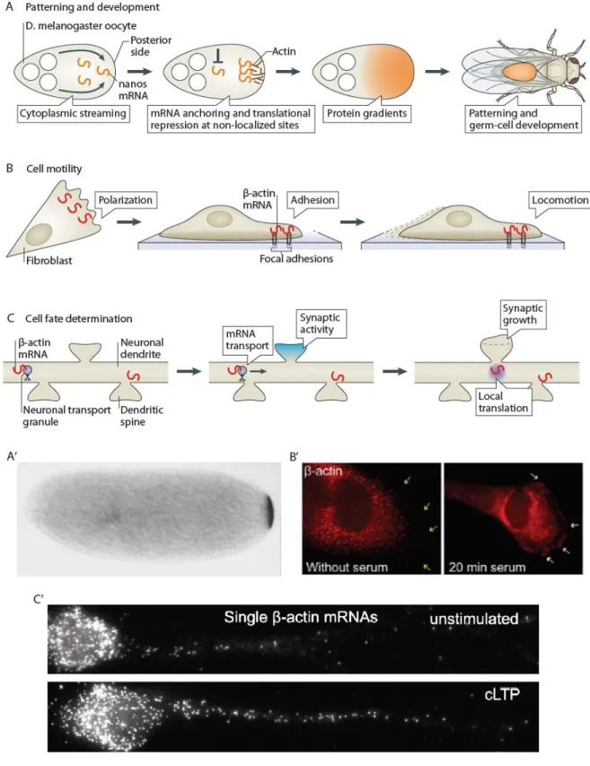
Figure 9 – Overview of roles and mechanisms for mRNA localization
A and A’) mRNA localization in oocytes determines developmental fates (e.g. nanos mRNA at the posterior pole determines abdomen and germ cell lineage) (Wang and Lehmann, 1991). B and B’) Localization of cytoskeletal mRNAs (e.g. β-actin) at the cell edge determines the efficiency of cell migration (Ben-Ari et al., 2010). C and C’) mRNA localization and local translation in synapses is crucial for their development and plasticity (Buxbaum et al., 2014). Schemes adapted from Buxbaum et al., 2015.
21 Neurons constitute an excellent model for mRNA localization studies, since they are highly polarized and their functionality can be easily evaluated. Given that axon length can reach the meter range, it seems intuitive that transport of mRNA in a repressed form would be a very effective way to rapidly localize proteins upon local stimulation (Figure 9C and C’). In fact, β-actin mRNA localization and local translation is also important in neurons for dendritic morphology, neuronal outgrowth and long-term potentiation (Eom et al., 2003; Hüttelmaier et al., 2005; Ramachandran and Frey, 2009).
Defects in the RNA localization machinery have been associated with neuronal and oncogenic disorders (Brinegar and Cooper, 2016; Wurth and Gebauer, 2015). However, out of a in vivo context the functional consequences of abolishing mRNA targeting may appear only mild (e.g. β-actin in migrating fibroblasts; Katz et al., 2012). This indicates that the proteins from remaining sources can still partially execute their functions under certain experimental conditions. Nevertheless, there are undoubtedly many advantages at the molecular level that can explain the evolutionary conservation of this mechanism: increased cost effectiveness by transporting few mRNAs that can generate many protein copies at the destination; facilitation of protein complex assembly by approximation of functionally related mRNAs; synthesis of proteins with distinct properties such as posttranslational modifications depending on the subcellular environment; possibility for local control of translation by repression alleviation in response to cues and thus finer control of protein localization and activity (Eliscovich et al., 2008; Hüttelmaier et al., 2005; Mingle et al., 2005; Weatheritt et al., 2014).
The ability to locally translate is important regardless of mRNA localization. By keeping mRNAs repressed but poised for translation, the relative efficiency of local protein enrichment is improved and ectopic action of potentially detrimental proteins is prevented. A good example is the myelin basic protein (MBP) mRNA localized and translated solely at the distal oligodendritic processes, avoiding aberrant myelination patterns (Lyons et al., 2009). In fact, it is generally believed that mRNAs need to be kept in a repressed state for processive transport, although simultaneous translation and transport have been reported (Katz et al., 2016; Wu et al., 2016). Through local translation, a decentralized and faster control of protein expression occurs at the cytoplasmic regions that directly
22
perceive extracellular cues. This mechanism is the basis of synaptic plasticity and memory formation, since strengthening and weakening of synapses (long-term potentiation and depression) have to be restricted in space while continuous in time (Sutton and Schuman, 2006). Thus, mRNA localization and local translation are mechanisms that often hold hands and allow for fine-tune post-transcriptional gene expression control.
1.3.2 Sequence determination and RBPs
What determines the destination of an mRNA in the cell? There is no consensus answer, as different mRNA species can exhibit very different localization mechanisms. Nevertheless, the involvement of specific regulatory proteins and the cytoskeleton in the process seems to be ubiquitous. mRNAs are constantly associated with RNA-binding proteins (RBPs) in the form of mRNA–protein complexes (mRNPs). When these complexes reach large sizes they can be loosely termed RNA granules, particularly in neurons. Several different RBPs will bind to a transcript depending on the cis-acting elements in its nucleotide sequence, known as localization elements (LEs) or zipcodes.
LEs are found typically in the 3’UTR but can also be located in 5’UTRs, coding sequence, retained introns, exon-junctions and even promoter regions (Buckley et al., 2011; Ghosh et al., 2012; Macdonald and Struhl, 1988; Saunders and Cohen, 1999; Zid and O’Shea, 2014). The higher frequency of LEs in UTRs may reflect their ability to evolve without constrains of retaining coding information. Importantly, RBPs often recognize secondary structures instead of the nucleotide sequence itself (Ferrandon et al., 1994, 1997). Thus, it is not surprising that LE sequences are not conserved across mRNAs known to be bound to the same RBP. Additionally, each transcript can have multiple LEs, either different or repeated. Redundant LEs can act cooperatively towards increased efficiency whereas diverse LEs can also function as modules dedicated to intermediate steps or different contexts for localization (Chartrand et al., 2002; Macdonald and Kerr, 1997; Macdonald et al., 1993). To add even more complexity, in some cases the transcripts must oligomerize for efficient mRNP assembly and localization (Ferrandon et al., 1997).
23 Translation may also be required to localize some proteins, as it is the case of some secreted and transmembrane proteins that get their nascent signal recognition particle anchored to ER resident proteins (Cui and Palazzo, 2014). Given that multiple RBPs can bind one transcript, it is the combinatorial composition of each mRNP that will dictate its localization in a particular cellular context (Figure 10).
A particularly complex mechanism localizes bicoid in the anterior of the Drosophila oocyte (Figure 10B). The different LEs in the 3’UTR of the transcript form stem loops necessary for its stepwise transport, from nurse cells to the anterior of the oocyte where it is anchored (Ferrandon et al., 1997; Macdonald and Kerr, 1997; Macdonald and Struhl, 1988; Macdonald et al., 1993). Moreover, dimerization of the mRNA is necessary for binding to the RBP Staufen, necessary for bicoid localization in the later steps of oogenesis (Ferrandon et al., 1997; St Johnston et al., 1991; Weil et al., 2006).
The detection of LEs facilitates the discovery of its respective RBPs, especially when different RBPs have redundant effects among their multiple mRNP targets. Once the sequence is known, it can be manipulated and used in reporters for better understanding of the function of its binding partners. This was the case for the β-actin zipcode that led to the identification of zip-code binding (ZBP) proteins (Figure 10A, Kislauskis et al., 1994; Ross et al., 1997). The recognition of the 54-nucleotide motif in the 3’UTR of the transcript by ZBP1 is sufficient and necessary for localization at the leading edge (Oleynikov and Singer, 2003). The hexanucleotide sequence ACACCC in the motif is evolutionarily conserved in the β-actin transcript of other species and the chicken ZBP1 also has orthologues like the mammalian IMP1 and the Xenopus Vg1RBP/Vera. More RBPs are now known to bind the β-actin mRNA, such as ZBP2 that binds co-transcriptionally and mediates the rapid engagement of ZBP1 upon its release (Gu et al., 2002; Pan et al., 2007). ZPB2 illustrates how the journey of each transcript starts being determined early in the nucleus, despite its absence in the cytoplasmic mRNPs.
24
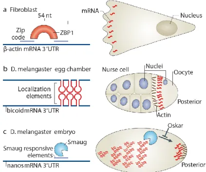
Figure 10 – RBP binding to localization elements determines mRNA localization
A) The zip code sequence of the β-actin 3’UTR recruits zipcode-binding protein1 (ZBP1) that will determine its transport to the leading edge and to synapses. B) Several 50nt stemloops in the bicoid 3’UTR allow its dimerization and binding to Staufen for anchoring at the anterior pole of the Drosophila oocyte. C) Smaug binds to its responsive elements in the 3’UTR of nanos in the absence of Oskar, leading to its degradation in the embryo anterior. Adapted from Buxbaum et al., 2015.
Biochemical approaches have determined that the same RBP can be linked to different mRNAs and vice versa (Fritzsche et al., 2013). Yet, these approaches do not elucidate the functions of these interactions nor specify how diverse each type of granule can be. In fact, the mode of action of most identified RBPs remains undemonstrated in the context of mRNA localization. Three main roles have
been assigned for RBPs: active transport, anchoring and local stabilization/degradation. The most commonly observed is the facilitation of active-transport by interaction with motor proteins. Although evidence for direct binding is scarce, RBPs have been shown to increase the binding affinity of mRNPs to motors, their processivity and run length (Alami et al., 2014; Amrute-Nayak and Bullock, 2012; Fusco et al., 2003; Sladewski et al., 2013).
The two remaining functions for RBPs in mRNA localization are well represented by the localization of nanos in the Drosophila embryo (Figure 10C). During late