0
Oncobiologia
The role of mitochondrial metabolism in
breast cancer aggressive phenotype
induced by hypercholesterolemia
Maria Beatriz Cordeiro Morgado
1
Oncobiologia
The role of mitochondrial metabolism in
breast cancer aggressive phenotype
induced by hypercholesterolemia
Maria Beatriz Cordeiro Morgado
Orientado por:
Professor Doutor Sérgio Jerónimo Rodrigues Dias
2
Abstract
Introduction: Cancer cells are no longer thought to obtain all their energy from aerobic glycolysis (Warburg’s effect) and are able to adopt metabolic programs that better fit their needs. Recently, it was shown that metastatic triple negative breast cancer (TNBC) obtains high levels of ATP through fatty acid oxidation (FAO). In Sérgio Dias lab, it was previously shown that LDL-cholesterol exposure promotes TNBC cells (MDA-MB-231) proliferation, migration and loss of adhesion in vitro. LDL exposed cells have increased mitochondrial number, lower lactate production and higher ATP levels, suggesting a preferential adoption of mitochondrial respiration by more aggressive TNBC cells. Objectives: The aim of this study is 1) to characterize the metabolic alterations induced by hypercholesterolemia and 2) to elucidate their requirement for the acquisition of aggressiveness by LDL exposed MDA-MB-231 cells in vitro. Methods/Results: We confirmed the increase in lipid droplets content in LDL exposed MDA-MD-231 cells by Bodipy staining and the increase in mitochondrial mass by MitoTracker Deep Red staining and analysis of Transmission Electron Microscopy imaged sections. By qPCR analysis, we showed that LDL exposed cells have an increased expression of oxidative phosphorylation chain components (COX5b, ATP5q1 and NDUFB5) and the FAO component CPT1a; a decreased expression of lipid synthesis enzymes (FASN and HMGCOA) and no differences in the expression of mitochondrial biogenesis factors (PGC-1α, PGC-1β, NRF1 and TFAM). Additionally, the CPT1a inhibitor Etomoxir does not affect proliferation, but abolishes the increased migratory capacity of LDL exposed MDA-MB-231 cells. Conclusions: LDL exposed MDA-MB-231 cells have more mitochondria, increased lipid droplets content and rely on FAO for the increase migratory capability.
Key-Words: Metabolism, mitochondria, breast cancer, lipids
3
Resumo
Introdução: Atualmente sabe-se que as células tumorais adotam o metabolismo que melhor satisfaz as suas necessidades energéticas, contrariamente ao previamente estabelecido pelo “efeito Warburg”. As células cancro de mama triplo negativo, em particular, obtêm elevados níveis de ATP através da ß-oxidação. Segundo o trabalho previamente desenvolvido no Sérgio Dias lab, a exposição a LDL favorece a proliferação, migração e perda de adesão de células de cancro de mama triplo negativo (MDA-MD-231) in vitro. Estas células apresentam um aumento da massa mitocondrial, maior produção de ATP e menor produção de lactato, sugerindo a adoção preferencial da respiração mitocondrial. Objetivo: 1) Caracterizar as alterações metabólicas induzidas pela hipercolesterolemia e 2) determinar a sua implicância no fenótipo agressivo das células MDA-MD-231 expostas a LDL-colesterol in vitro. Resultados: As células MDA-MD-231 expostas a LDL apresentam um aumento de gotículas lipídicas (avaliado por marcação com Bodipy) e um incremento da massa mitocondrial (avaliado por Microscopia Eletrónica de Transmissão e marcação com MitoTracker Deep Red). A análise de expressão génica por qPCR revelou um aumento da expressão de genes de complexos da cadeia respiratória (COX5b, ATP5q1 e NDUFB5) e da ß-oxidação (CTP1a), uma diminuição da expressão de genes da síntese lípica (FASN e HMGCOA) e ausência de alterações na expressão de genes da biogénese mitocondrial (PGC-1α, PGC-1β, NRF1 e TFAM). A utilização do inibidor de CPT1a Etomoxir não interfere com a proliferação das células MDA-MB-231 expostas a LDL, mas impacta a sua capacidade migratória. Conclusão: As células MDA-MB-231 expostas ao LDL têm mais mitocôndrias, maior conteúdo de lípidos e a sua alta capacidade migratória encontra-se dependente da ß-oxidação.
Palavras-Chave: Metabolismo, mitocôndria, cancro da mama, lípidos
4
Resumo do Trabalho Final de Mestrado em Língua Portuguesa
Introdução
O metabolismo tem sido reconhecido como um fator decisivo na iniciação e progressão tumorais. Otto Warburg relatou que as células tumorais adotam preferencialmente a glicólise, independentemente da disponibilidade de oxigénio, devido a defeitos mitocondriais. No entanto, sabemos atualmente que os tumores são massas celulares heterogéneas e vários estudos têm demonstrado a importância da respiração mitocondrial, em particular nas células quiescentes - responsáveis na formação de metástases e resistência às terapêuticas anti tumorais. Considerando esta flexibilidade metabólica, é plausível que as células beneficiem de meios ricos em lípidos, cujos ácidos gordos podem servir de substrato à respiração mitocondrial para produção de energia.
O cancro de mama triplo negativo é um dos subtipos de cancro de mama mais agressivos e para o qual não existem terapêuticas dirigidas, o que prediz um mau prognóstico. Torna-se crítico conhecer a sua assinatura metabólica, com o objetivo de encontrar potenciais alvos para terapêuticas dirigidas.
Recentemente, foi demonstrado que o cancro de mama triplo negativo produz altos níveis de ATP através da ß-oxidação associada à respiração mitocondrial e que a inibição desta via suprime o crescimento tumoral. Congruentemente, as células de cancro de mama triplo negativo apresentam um aumento da expressão de genes associados à ß-oxidação e fosforilação oxidativa (como o PGC1α) e uma baixa expressão de genes associados à síntese lipídica. Esta assinatura molecular apresenta uma forte correlação com o cancro de mama triplo negativo comparativamente com os outros subtipos de cancro de mama, nos quais esta regulação metabólica não se verifica. Por outro lado, existem dicotomias a ressalvar face a outros tipos de cancro, como o papel do PGC1α que apresenta efeitos pro-tumorais no cancro de mama (inclusive no subtipo triplo negativo), mas efeitos opostos noutros tipos de cancro (próstata, melanoma, pâncreas ou carcinoma renal). No cancro de mama triplo negativo, o PGC1α fomenta a migração celular e formação de metástases – efeito este que se coaduna com a hipótese de que a ß-oxidação e fosforilação oxidativa se associam a um fenótipo tumoral mais agressivo.
5
A obesidade e dislipidemia têm sido associadas ao aumento do risco de cancro e a ativação do metabolismo lípido tem sido implicada como um evento precoce na carcinogénese. Contudo, a síntese lipídica de novo não é suficiente para suprir as necessidades das células em proliferação; estas apresentam necessidades aumentadas de lípidos exógenos. Neste âmbito, foi demonstrada uma associação positiva entre os níveis de colesterol total sérico e a incidência aumentada de cancro de mama e mortalidade. Tal associação é particularmente forte para cancros ER-. As células de cancro de mama triplo negativo, em particular, apresentam expressão de recetores de LDL mais elevada do que qualquer outro subtipo de cancro de mama. Deste modo, apresentam maior capacidade de internalização de lípidos exógenos, dos quais tiram partido, despendendo menos energia na síntese lipídica de novo – o que lhes confere vantagem em processos altamente dependentes de energia como a migração.
No laboratório do Dr. Sérgio Dias, demonstrou-se que as células de cancro de mama triplo negativo MDA-MB-231 expostas a LDL apresentavam um aumento da proliferação, perda de adesão e migração, resultando num fenótipo mais agressivo comparativamente às células não expostas a LDL. O fenótipo agressivo das MDA-MB-231 expostas a LDL ocorre concomitantemente com um aumento do conteúdo mitocondrial, aumento da produção de ATP e diminuição da produção de lactato, sugerindo que estas células utilizam preferencialmente a respiração mitocondrial.
No presente trabalho, estudou-se a implicação da respiração mitocondrial na aquisição do fenótipo agressivo das células MDA-MB-231 expostas a LDL. Face às evidências supramencionadas, foi colocada a hipótese de que a respiração mitocondrial é responsável pela maior agressividade tumoral das células de cancro de mama triplo negativo expostas a LDL. Para tal, foram estabelecidos os seguintes objetivos: 1) caracterizar as alterações metabólicas induzidas pela hipercolesterolemia e 2) determinar a sua implicância no fenótipo agressivo das células MDA-MD-231 expostas a LDL-colesterol in vitro.
6
Métodos e Resultados:
Foi utilizada a linhagem celular humana de cancro de mama MDA-MB-231. As células cresceram em cultura com DMEM suplementado com FBS 10% durante 24h. No 2º dia, foram transferidas para 1% FBSLF ("Fetal Bovine Serum Lipoprotein-free") DMEM. Ao 3º dia, estabeleceram-se dois grupos: grupo controlo (1% FBSLF DMEM) e grupo exposto a LDL (100 μg/ml LDL) durante 48h. Todos os resultados foram obtidos ao 5º dia de incubação.
Através da marcação com Bodipy, verificou-se que as células MDA-MD-231 expostas a LDL apresentam um aumento de gotículas lipídicas. Utilizando Microscopia Eletrónica de Transmissão e marcação com MitoTracker Deep Red, verificou-se um aumento da massa mitocondrial nas células expostas a LDL. A análise de expressão génica por qPCR revelou um aumento da expressão de genes da cadeia respiratória (COX5b, ATP5q1 e NDUFB5) e da ß-oxidação (CTP1a), uma diminuição da expressão de genes da síntese lípica (FASN e HMGCOA) e ausência de alterações na expressão de genes da biogénese mitocondrial (PGC-1α, PGC-1β, NRF1 e TFAM). Por fim, a utilização de um inibidor de CPT1a (Etomoxir) não condiciona a proliferação das células MDA-MB-231 expostas a LDL, mas impacta a sua capacidade migratória.
Discussão
As células tumorais podem apresentar um estado proliferativo (associado à síntese lipídica de novo) ou estado migratório (com aumento da internalização de ácidos gordos exógenos).
As células MDA-MB-231 expostas a LDL apresentam mais gotículas lipídicas por provável internalização ativa do LDL que apresenta um componente lipídico (além do proteico). Estudos anteriores já haviam demonstrado esta relação entre os lípidos do meio extracelular e as gotículas das MDA-MB-231, tendo verificado a depleção de gotículas lipídicas quando as células crescem em meio “lipid-free”. In vivo, o aumento dos ácidos gordos intracelulares nas células glandulares mamárias sugere um aumento da fosforilação oxidativa.
7
Estudos prévios já haviam sugerido que as células MDA-MB-231 apresentam um aumento do conteúdo mitocondrial, dado aumento do mtDNA. No presente trabalho, esses resultados foram confirmados com a utilização de Microscopia Eletrónica e com o aumento da marcação por Mitotracker, o que sugere aumento da massa e atividade mitocondriais. A massa mitocondrial é considerada um biomarcador metabólico determinante da atividade “mammosphere-forming”, essencial para a propagação de células quiescentes. Estudos anteriores correlacionam o aumento dos marcadores metabólicos mitocondriais ao fenótipo stem cell-like em células de cancro de mama triplo negativo. Deste modo, o aumento da massa mitocondrial poderá estar associado a um fenótipo semelhante ao das stem cells e maior quimiorresistência. Por outro lado, o aumento do conteúdo mitocondrial nas células em migração parece ganhar importância à medida que estas alcançam regiões com maior teor de oxigénio, permitindo uma maior produção de ATP e consequentemente a migração à distância. A dinâmica mitocondrial determina também o equilíbrio entre a produção energética, a reprogramação metabólica e a apoptose. Durante o metabolismo respiratório, as mitocôndrias apresentam-se com uma forma alongada e dilatação dos seus compartimentos; já durante a glicólise, as mitocôndrias apresentam-se redondas e mais fragmentadas. No entanto, as células MDA-MB-231 caracterizam-se por apresentarem mitocôndrias pequenas e tubulares, contrariamente ao esperado para um tipo celular que utiliza preferencialmente o metabolismo mitocondrial e contrastando com o verificado noutros subtipos de cancro de mama. Estas características morfológicas verificadas nas células MDA-MB-231 associam-se a uma sobreexpressao de Drp1 e subexpressão de Mfn1, bem como a uma maior capacidade de metastização. As mitocôndrias tendem ainda a apresentar uma distribuição temporo-espacial, acumulando-se nas extrusões citoplasmáticas na direção para a qual a célula migra - local onde ocorrem processos altamente dispendiosos do ponto de vista energético.
As células MDA-MB-231 expostas a LDL apresentam um aumento da expressão de CPT1a (enzima limitante da ß-oxidação) e uma diminuição dos genes associados a síntese lipídica, possivelmente por ativação de vias regulatórias em resposta à abundância de LDL no meio extracelular. As células expostas a LDL demonstraram, ainda, um aumento da expressão de genes da cadeia respiratória de eletrões, embora não se tenha verificado um aumento da expressão de genes da biossíntese mitocondrial.
8
Novos estudos serão necessários de modo a compreender este fenómeno - tal regulação poderá passar pelos efeitos dicotómicos pró e anti oncogénicos do PGC1α, regulação por micro-RNA ou aumento da biodisponibilidade dos fatores da fosforilação oxidativa.
Dado o papel da respiração mitocondrial no cancro de mama triplo negativo e o aumento da expressão de CPT1a (que codifica a enzima responsável pelo passo-limitante da ß-oxidação), realizou-se a inibição farmacológica do CPT1a com Etomoxir. Deste modo, visa-se compreender a implicância do CPT1a no fenótipo agressivo das células MDA-MB-231 expostas a LDL.
Estudos anteriores revelaram que a inibição do CPT1a em células de cancro de mama triplo negativo conduzia a uma diminuição da capacidade de formação de colónias, diminuição da migração e aumento das gotículas lipídicas intracelulares. Contudo, não existem até à data estudos que verifiquem a importância do CPT1a nas células de cancro de mama triplo negativo quando expostas a LDL, com consequente aquisição de um fenótipo mais agressivo.
No presente trabalho, verificou-se que as células MDA-MB-231 expostas a LDL apresentam um aumento da proliferação comparativamente as MDA-MD-231 não expostas a LDL e este padrão de proliferação mantém-se quando realizada a inibição do CPT1a. Apesar das células MDA-MB-231 dependerem do CPT1a (ß-oxidação) para proliferarem, quando expostas a LDL continuam a apresentar uma proliferação significativamente aumentada mesmo sob inibição do CPT1a. Por outro lado, a inibição do PGC1a com Etomoxir reverte a capacidade migratória aumentada das MDA-MB-231 expostas a LDL, à semelhança do verificado nas células não expostas a LDL. Assim, depreende-se que apesar de a inibição do CPT1a com Etomoxir não reverter a proliferação, é capaz de abolir a capacidade migratória das células expostas a LDL, reforçando a importância da ß-oxidação nos processos de migração.
Tal como descrito em estudos anteriores, verificou-se ainda que a inibição do CPT1a conduz a um aumento das gotículas lipídicas citoplasmáticas nas MDA-MB-231 não expostas a LDL (“fenótipo acumulador de lípidos” típico do cancro de mama triplo negativo). No entanto, as células expostas a LDL não apresentam um aumento do conteúdo lipídico quando submetidas a Etomoxir. Mais estudos são necessários para melhor compreender esta alteração do fenótipo das células sob LDL.
9
Conclusão
O presente trabalho demonstra que as células de cancro de mama triplo negativo MDA-MB-231 quando expostas a LDL apresentam um aumento das gotículas lipídicas citoplasmáticas, maior conteúdo e atividade mitocondrial, bem como um aumento da expressão de CPT1a (essencial para a ß-oxidação) e de genes associados à fosforilação oxidativa. A utilização do inibidor de CPT1a Etomoxir não interfere com a proliferação, mas impacta a sua capacidade migratória das células MDA-MB-231 expostas a LDL. Conclui-se assim que a alta capacidade migratória destas células se encontra dependente da ß-oxidação – um potencial alvo terapêutico, em particular em doentes com hipercolesterolemia.
10
Index
List of Abbreviations ... 11 Introduction ... 13 Methods ... 16 Results ... 20 Discussion ... 27 Bibliography ... 39Appendix 1: LDL-cholesterol promotes the expression of β-oxidation related genes ... 43
Appendix 2: Table: The targeted genes analysed by quantitative PCR ... 44
11
List of Abbreviations
ATP – Adenosine triphosphate
ATP5g1- ATP synthase F complex subunit C1 BC– Breast cancer
CSC – Cancer stem-like cells
COX5b - Cytochrome c oxidase subunit 5B CPT1 - Carnitine palmitoyl transferase 1 enzyme DAG - Diacylglycerol
ECAR – Extracellular acidification rate ER – Estrogen receptors
FA – Fatty acids
FAO – Fatty acid oxidation FAS – Fatty acid synthesis FASN – Fatty acid synthase FBS – Fetal Bovine Serum
FBSLPF – Fetal Bovine Serum lipoprotein free GSEA – Gene set enrichment analysis
HMGCoA - Hydroxymethylglutaryl-CoA reductase LDL – Human plasma low density lipoprotein LDL–R – LDL receptor
mtDNA – Mitochondrial DNA
NDUFB5 - NADH Dehydrogenase Ubiquinone 1 Beta Sub complex, 5 NRF1 – Nuclear respiratory factor 1
OCR – Oxygen Consumption Rate OXPHOS – Oxidative phosphorylation
PGC-1α - Peroxisome proliferator-activated receptor gamma coactivator 1-alpha PGC-1β- Peroxisome proliferator-activated receptor gamma coactivator 1-beta PLIN1 - Perilipin 1
PLIN2- Perilipin 2
PPARα - Peroxisome proliferator-activated receptor alpha qPCR – Quantitative Real Time Polymerase Chain Reaction ROS - Reactive oxygen species
12 TCA – Tricarboxilic acid
TFAM – Mitochondrial transcription factor A TNBC – Triple Negative Breast Cancer 3-KAT - 3-ketoacylthiolase
13
Introduction
Metabolism has been widely recognized as a decisive factor in the initiation and progression of tumours [1]. In the 1920s, Otto Warburg observed the tendency for proliferating cells to secrete a significant portion of glucose carbon through fermentation [1][2]. Warburg hypothesized that tumours preferentially adopt "aerobic glycolysis” to produce lactate independently of oxygen availability as a result of mitochondrial defects that restrained them from oxidize glucose carbon to CO2 by the
Krebs cycle [1][3].
Recently, several evidences challenge the Warburg hypothesis and show that the non-glucose metabolic pathways seem to be just as important as the “Warburg effect” (if not potentially more) in cancer metabolism [4]. Metabolic reprogramming of tumours is being increasingly recognized as an important disease driver and it is possible that tumour cells adaptively reprogram their signalling pathways to evade therapy-induced “stress” and, paradoxically, increase their motility and invasion [5][6][7]. Tumours are metabolically heterogenous and some cells (slow cycling, quiescent) are thought to depend on mitochondrial respiration for metastasis growth and resistance to therapy [1][3][8][9]. Considering the high metabolic flexibility of cancer cells, it is conceivable that cancer cells benefit from high lipid availability through mitochondrial respiration either to fulfil increased energy demand or to prevent the lipotoxic effects of high level of fatty acids (FA) [10][11].
Triple negative breast cancers (TNBC) accounts for about 12%-17% of breast cancer cases, it is the most aggressive subtype and poor prognosis is (in part) due to the current lack of targeted therapy [12][13][14][15][16]. Understanding TNBC metabolic signature can be critical to improve understanding of its treatment resistance [13].
Notably, aberrant FA metabolism was detected in minimal residual disease across all the breast cancer intrinsic subtypes [17]. It has recently been shown that TNBC cells obtain high levels of ATP through fatty acid oxidation (FAO) by mitochondrial respiration and the inhibition of this pathway suppresses tumour growth [13][14]. Mitochondrial respiration is associated with the increased capability of metastasis formation and increased resistance to therapeutics in breast cancer [18][19] and elevated FA metabolism is a communal hallmark of cells surviving neoadjuvant
14
treatment in breast cancer patients [17]. In TNBC cells, it is verified the downregulation of many genes that encode activators of fatty acid synthesis and upregulation of many genes that encode activators of FAO and OXPHOS (including the gene encoding the master transcriptional regulator of mitochondrial biogenesis PPARG coactivator alpha, known as PGC-1α) [20]. This FA metabolism signature is particularly highly correlated with TNBC tumours compared to others tumours [14]. An important dichotomy exists, with reports of pro and anti-tumorigenic effects of PGC-1α expression in different cancer types [21]. In prostate cancer, PGC1α suppresses cancer progression and metastasis [21]; in melanoma, PGC1α also suppresses metastasis and low PGC-1α levels are correlated with melanoma relapse [21][22]; in pancreatic CSC clones, low PGC-1α levels also promote disease relapse [23] and, in renal cell carcinoma, PGC-1α enhances cells sensitivity to cytotoxic therapies [21]. In breast cancer, the great majority of cells show low expression of PGC-1α and it increases the antifolate therapy sensibility [24]. However, within the various breast cancer subtypes, the levels of PGC-1α are highest in HER2+ and TNBC tumours [24]. In TNBC in particular, the increased expression of PGC-1α enhances the OXPHOS, mitochondrial biogenesis and the oxygen consumption rate, promoting tumour growth and metastases formation [18][24]. Silencing PGC-1α in TNBC suspended their invasive potential without affecting proliferation, showing that PGC-1α fuel tumour growth, cell motility and metastasis formation [18][24][21]. It follows that increased FAO and PGC-1α expression contributes to aggressiveness and worst outcome occurring in the TNBC subset [14]. However, the precise molecular mechanisms by which FA might influence cancer development and progression, still remain to be elucidated [25].
Obesity and dyslipidaemia have long been linked with an increase in the likelihood of developing cancer [10][26]. Several hypotheses have been proposed to explain the association between obesity and breast cancer. The aromatization of androgens in adipose tissue of obese postmenopausal women leads to the elevation of circulating estrogens [27]. However, estrogen stimulates the growth of abnormal ER+ cells; in consequence, this hypothesis does not fully explain the correlation between obesity and TNBC [28]. The metabolic syndrome also results in the increase in circulating insulin and insulin-like growth factor (IGF), which act as mitogen, and chronic inflammation increases the circulating tumour related markers (leptin, IL-6, and TNF-a) [15][27]. Newer hypothesis focus on adipocytes endocrine functions [27].
15
The activation of lipid metabolism is an early event in carcinogenesis and a critical pathway for maintenance of cell survival, growth and migration [12][29]. However, recent studies suggest that de novo lipid synthesis is necessary but not sufficient to support lipid production for breast cancer tumour growth [30]. Proliferating cells are believed to have increased requirements for exogenous lipids as cholesterol, a structural component of cell membrane and a steroid hormone precursor [26]. Prospective studies showed positive association between total cholesterol levels and both breast cancer incidence and overall mortality [31].
Both low-density lipoprotein (LDL) and unsaturated fatty acids have been demonstrated to increase proliferation of estrogen receptor negative (ER-) breast cancer cells [32][33]. TNBC cells have high LDL requirements and express relatively high levels of LDL receptors (LDLRs) on their membranes compared with the other subtypes [16]. For this reason, TNBC cells have a greater capability to increase the uptake of circulating lipids – the called “lipid accumulating phenotype” [33]. As little energy is needed in de novo synthesis, LDL exposure enables the proliferation of TNBC cells [32]. The abundant expression of LDLR in TNBC cells also enables tumour-targeting therapies (such as paclitaxel–cholesterol complexes) [16].
Previously in Sérgio Dias lab, it was shown that LDL exposure promotes TNBC cells (MDA-MB-231) proliferation, migration and loss of adhesion of tumour cells which are hallmarks of the EMT and result in a more aggressive phenotype [26]. These results have raised questions about the metabolism preferentially used by TNBC cells. Preliminary results indicate that LDL exposed TNBC cells have higher mitochondrial content, higher ATP production and lower lactate production, suggesting the preferential adoption of mitochondrial respiration [Teixeira, Nóbrega-Pereira et al., unpublished].
In the present study, we investigated the implication of the mitochondrial respiration in the acquisition of more aggressive phenotype of TNBC (MDA-MB-231) cells exposed to LDL-cholesterol in vitro. We hypothesized that FAO is responsible for TNBC aggressive phenotype and could be a future therapeutic target in breast cancer treatment.
16
Methods
Cell Culture:
For all experiments we used the breast human cancer cell line MDA-MB-231. Cancer cells were cultured in DMEM supplemented with FBS (10%) for 24h. In the 2nd day, cells were then transferred to 1% FBSLF ("Fetal Bovine Serum Lipoprotein-free") DMEM. In the 3rd day, MDA-MD-231 cells were divided in the control group (cultured in 1% FBSLF DMEM only) and LDL exposed group (cultured in 100 μg/ml LDL-cholesterol) during 48h. In the 5th day, we analysed:
- Lipid droplets content; - Mitochondrial mass; - Gene expression;
- Performed functional assays with pharmacological inhibition of CTP1a using the commercial available pharmacological inhibitor Etomoxir (Sigma).
❖ Lipid droplets content
The lipid droplets were quantified in the control group and in the LDL exposed group by using the Bodipy staining and flow cytometry. Bodipy Lipid Probes (Molecular Probes) is a bright, green-fluorescent dye with similar excitation and emission to fluorescein. Fluorophores sufficiently close at the time of excitation can form an excited dimer, emission from which depends strongly on total lipid packing density.
We incubated 120.000 cells with 0.52 μg/ml Bodipy in PBS1x, for 10 minutes at 37°C. After this time, the cells were analysed in a LSRFortessa cytometer (20.000 live cells and singlets in the FITC channel). The data was analysed in the FlowJo program, in order to obtain the MFI (median fluorescent intensity) of each sample.
17
❖ Mitochondrial mass
The mitochondrial mass was quantified both in control group and in LDL exposed group by using MitoTracker Deep Red staining (Molecular Probes) with flow cytometry and Transmission Electron Microscopy.
The MitoTracker Deep Red is a fluorescent dye that labels mitochondria within live cells utilizing the mitochondrial membrane potential. Cells are simply incubated with MitoTracker probe, which passively diffuse across the plasma membrane, accumulate in active mitochondria and the dye becomes permanently bound to these mitochondria.
120.000 cells were incubated with 1 nM Mitotracker dye in PBS 1x for 20 minutes at 37°C. Cells were analysed by flow cytometry on the LSRFortessa cytometer (20.000 live cells and singlets in the FITC channel). The data were analysed in the FlowJo program to obtain the MFI of each sample.
Transmission Electron Microscopy (TEM) was the second method used. The preparation of samples, technical procedures and acquisition of photographs and acquisition of photographs were made in partnership with the Comparative Histopathology laboratory of the Instituto de Medicina Molecular. Only 1500x magnification images with a ¼ nucleus visible were analysed. Images without both parameters were excluded. The number of mitochondria within the imaged sections was determined. Intracellular lipid droplets and mitochondrial morphology were also evaluated in cooperation with the Comparative Histopathology laboratory.
❖ Gene expression
Gene expression was analysed by quantitative PCR of the control group and 48h LDL exposed group. The targeted genes analysed were those which expression was altered in previous microarrays (GSEA – see appendix 1 [26]) and their metabolic co-genes/factors (for more information see appendix 2).
18
Gene name Primer Sequence
COX5b hCox5b-F hCox5b-R GCTGCATCTGTGAAGAGGACAAC CAGCTTGTAATGGGTTCCACAGT ATP5g1 hATP5g1-F hATP5g1-R GCTGTTGTACCAGGGGTCTAA CTGGCGTGGGAAGTTGCTGT NDUFB5 hNDUFB5-F hNDUFB5-R CTTCCTCACTCGTGGCTTTC TTTCCCATGGTCTCCACTGT FASN hHMG-CoAR F hHMG-CoAR R GGACCCCTTTGCTTAGATGAAA CCACCAAGACCTATTGCTCTG HGM-CoA h FASN-F h FASN-R CGACAGCACCAGCTTCGCCA CACGCTGGCCTGCAGCTTCT CPT1a h CPT1a-F h CPT1a-R ATGCGCTACTCCCTGAAAGTG GTGGCACGACTCATCTTGC PGC-1α hPGC1alfa-F hPGC1alfa-R CACCAGCCAACACTCAGCTA GTGTGAGGAGGGTCATCGTT PGC-1β hPGC1beta-F hPGC1beta-R GGCAGGCCTCAGATCTAAAA TCATGGGAGCCTTCTTGTCT NFR1 hNRF1-F hNRF1-R CCATCTGGTGGCCTGAAG GTAGTGCCTGGGTCCATGA TFAM hTFAM-F hTFAM-R GAACAACTACCCATATTTAAAGCTCA GAATCAGGAAGTTCCCTCCA
Table 1. Amplified genes and primers from lipid receptors and transporters, glycolysis, OXPHOS and FAO used in qRT-PCR
❖ Pharmacological inhibition of CTP1a using Etomoxir
Etomoxir is an inhibitor of CPT1a activity. Etomoxir (Sigma-Aldrich), 100uM and 200Um, was added to the control group and to the LDL exposed MDA-MB-231 cells. In the last group, Etomoxir was added simultaneously with LDL (for 48h). The following data was analysed:
19
- Cell proliferation by Trypan Blue exclusion method, using Etomoxir 100uM and 200uM;
- Lipid droplets content by Bodipy staining and flow cytometry, using Etomoxir 200uM;
- Cell migration by Wound Healing assay, using Etomoxir 200uM. Wound closure is indicated as the percentage of cell migration distance at 24h. Mitomycin C (Sigma-Aldrich), at a final concentration of 200nM, was added to inhibit cell proliferation.
Statistical Analysis:
Data are presented as mean ± s.d. (standard deviation). Statistic significance was assessed using the two-tailed Student’s t-test. * p<0.05; ** p<0.01 and *** p<0.001.
20
Results
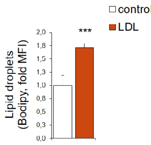
1. The increase in lipid droplets content in LDL exposed MDA-MD-231 cells
We verified a significant increase in lipid droplets content in MDA-MB-231 exposed to LDL for 48 hours.
Figure 1.: Lipid droplets content of LDL exposed MDA-MB-231 cells
Lipid droplets content obtained by Bodipy staining, quantified by flow cytometry, of MDA-MB-231 cells control or exposed to LDL for 48 hours (n=5/each, data from a representative experiment). Data are presented as mean ± standard deviation. Statistical significance was measured using the Student t test (*** p <0.001).
21 2. LDL-cholesterol exposed MDA-MB-231 cells have an increased mitochondrial mass
By using Mitotracker staining with flow cytometry quantification (Figure 2.A.) and quantification of Transmission Electron Microscopy imaged sections (Figure 2.B.), it was possible to confirm the increase in mitochondrial mass and number in LDL exposed cells.
A
B
Figure 2: Mitochondrial mass of LDL exposed MDA-MB-231 cells
22
MitoTracker Deep Red staining analysed by flow cytometry (APC channel: control n= 5; LDL n= 4; data from a representative experiment; left: relative median fluorescent intensity MFI; right: representative flow cytometry histogram). (B) Quantification of the number of mitochondria per cell within the imaged sections and representative TEM images (control n=18; LDL n=15, independent experiments-pool; 1500x, M = Mitochondria). Data are presented as mean ± s.d., Two-tailed Student’s t-test (* p<0.05 and ** p<0.01.).
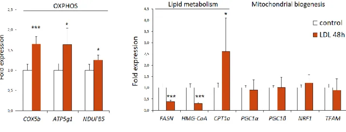
23 3. Effect of LDL-cholesterol in the expression of metabolic enzymes and
mitochondrial biogenesis factors in MDA-MB-231 cells
Quantitative PCR analysis demonstrates an increased expression of genes encoding respiratory chain proteins (such as Cox5b, ATP5g1 and NDUFB5) in LDL exposed MDA-MB-231 cells.
The results also demonstrate a decreased expression of lipid metabolism genes (as HMG-CoA reductase and FASN genes, which are lipid synthesis genes), except for CPT1a (FAO gene) which expression is increased.
There are no significant differences in the expression of genes associated with mitochondrial biogenesis (PGC1 alpha, PCG1, NRF1 or TFAM).
Figure 3: Gene expression signature induced by LDL-exposure in MDA-MB-231 cells.
qPCR analysis of the relative expression of the indicated genes in MDA-MB-231 cells control or exposed to LDL during 48h (control n=4; LDL n=5; data from a representative experiment). Data are presented as mean ± s.d., two-tailed Student’s t-test (*p<0.05 and *** p<0.001).
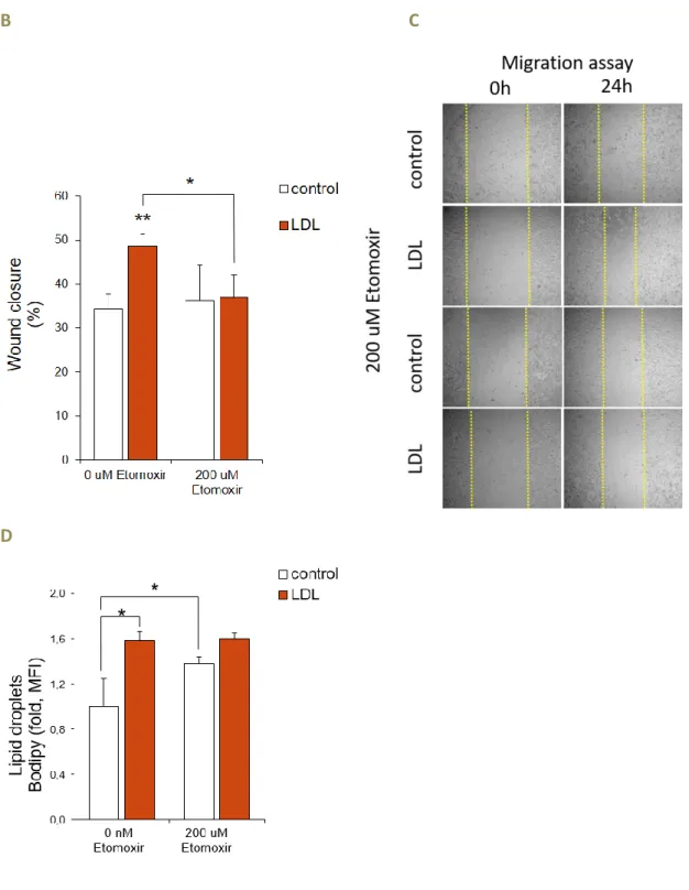
24 4. CPT1a inhibition (Etomoxir) does not revert the increased proliferation,
but reverts the increased migratory capacity of LDL exposed MDA-MB-231 cells
LDL exposed MDA-MB-231 cells have greater proliferation than the control group.
This proliferative pattern is maintained even when exposed to increasing concentrations of Etomoxir (Figure 4.A.).
LDL exposed MDA-MB-231 cells have higher migratory potential compared to the control group. However, when Etomoxir is added, the phenotype is reverted as Etomoxir abolish of the increased migratory capability of the LDL exposed cells, which migration equals the control group (Figure 4.B and 4.C).
Moreover, there is an increased lipid droplets content in LDL exposed cells compared to the control. In the presence of Etomoxir 200 uM, the control group show an increase in lipid droplets whereas lipid droplets content of LDL exposed MBA-MD-231 cells remained unchanged upon Etomoxir treatment (Figure 4.D).
25
B C
D
Figure 4: Effect of the CPT1a inhibitor Etomoxir in the proliferation, migratory capacity and lipid droplets content of LDL exposed MDA-MB-231 cells
(A) Effect of the CPT1a inhibitor Etomoxir (100 and 200 uM) in the proliferation of
MDA-MB-231 cells control or exposed to LDL during 48h (control n=6; LDL n=7; independent experiments-pool).
(B-C) Effect of the CPT1a inhibitor Etomoxir (200 uM) in the migratory capacity (B;
26
images). (D) Effect of the CPT1a inhibitor Etomoxir (200 uM) in lipid droplet content assessed by Bodipy staining by flow cytometry (D; depicted as relative median fluorescent intensity MFI) of MDA-MB-231 cells control or exposed to LDL (n=3/each, data from a representative experiment). Data presented as mean ± s.d., two-tailed Student’s t-test. (* p<0.05 and ** p<0.01).
27
Discussion
The autonomous metabolic reprogramming of rapidly proliferating cancer cells promotes self-sustaining signal transduction mechanisms to foster growth and survival [21]. It is known that cancer cells are able to switch from a proliferative state (characterized by high de novo lipid biosynthesis and rapid cell growth) to a migratory state in which FA uptake are thought to contribute to the formation of signalling molecules that promote cell migration [2]. Upon uptake, intracellular FA are destined for two fates: FAO or cytoplasmic storage as glycerolipids in lipid droplets [10]. Lipid droplets stored TAG are available for energy production by FAO according to the cell needs. The mobilization of FA from lipid droplets is regulated by perilipins, proteins localized in surface of lipid droplets that regulate access of lipases to stored TAG [34].
In our study, we verified an increase in lipid droplets content in LDL exposed MDA-MD-231 cells. Previously, it was also shown that MDA-MB-231 cells exhibited depletion of lipid droplets when incubated in lipid-free medium for 48 hours [32].An increased level of FAs can lead to an increase in lipid droplets (the storage organelle for excess lipids) and this increase in FA stores in vivo suggest that mammary glands are preferentially deriving cellular energy from OXPHOS [17]. This may be explained by the “nutrient sufficient extracellular milieu”, as the majority of substrates are derived from exogenous FA that are actively transported into the cell [10]. FA uptake is also modulated by hypoxia that increases lipid uptake and storage into lipid droplets through induction of Perilipin in breast cancer cells [2]. In TNBC cells, Perilipin overexpression led to a consequent increase in lipid droplet formation, which are associated with increased TNBC cell survival in vitro [34]. In addition to Bodipy and Nile Red, radioactive palmitic acid and stearic acid incorporation studies could be used as alternatives methods for studying the intercellular lipid content in LDL exposed TNBC cells (even under hypoxia and unfavourable conditions) and its consequences in cell phenotype.
Previous results also suggested that LDL exposed MDA-MB-231 cells had more mitochondria, as they presented an increased mitochondrial DNA (mtDNA)/ nuclear DNA ratio, which indirectly expresses an increased mitochondria mass [Teixeira, Nóbrega-Pereira et al., Unpublished]. Although the copy number of mtDNA per cell is maintained within a constant range according to the cell type and energy need, mtDNA
28
copy number tends to be increased in breast cancer patients and it is inversely associated with the concentration of antioxidants in blood [35]. Thus, an increased mtDNA content is associated with an increased in oxidative metabolism and ROS production and TNBC cells have higher ROS compared to other breast cancer cell lines [36].
However, until now only indirect data had demonstrated the potential increase in mitochondria content in LDL exposed TNBC. By using MitoTracker staining with flow cytometry quantification and quantification of mitochondria in Transmission Electron Microscopy imaged sections, it was possible to confirm the increase in mitochondrial mass and number in LDL exposed TNBC cells. Mitochondrial mass is considered a metabolic biomarker and key determinant of mammosphere-forming activity, as high mitochondrial mass seems to be critical for the successful propagation of stem-like cancer cells [37]. It was observed that the “newly-synthesized” mitochondria are concentrated in stem cells during asymmetric cell division, while “old” mitochondria tend to be segregated into daughter cells [37]. These findings may explain why increased mitochondrial mass confers LDL exposed TNBC cells a stem-like phenotype and chemo-resistance [37][38][9].
Besides quantifying mitochondria, MitoTracker Deep Red staining also increases proportionally to mitochondrial membrane potential, suggesting that LDL exposed cells have an increased mitochondrial activity and function. To directly demonstrate an increased mitochondrial function, it would be important to measure Oxygen Consumption Rate (OCR) and Extracellular Acidification Rate (ECAR) in LDL exposed TNBC cells comparing to the control group. Aberrant lipid metabolism drives oxidative DNA damage following oncogene inactivation and oxidative stress has long been implicated in tumorigenesis (driving residual cells toward tumour recurrence) [17]. Mitochondrial proficiency and ROS detoxification are critical for cancer cell viability, because both ATP generation and antioxidant production ensure cancer cell survival when detaching from their basement membrane [21]. Previous reports also show that mitochondrial metabolic markers such as OXPHOS, mitochondrial membrane potential, mitochondrial superoxide and ROS are elevated in TNBC stem-like cells and tumours [9]. FAO, in particular, provides a potent ATP source to fuel highly energy-demanding processes of cell invasion and metabolic reprogramming of mitochondrial bioenergetics influences tumour cell motility in vivo [5][7]. It is conceivable that invading cancer cells
29
enhance their mitochondrial content and up-regulate OXPHOS as they approach more oxygenated areas of the tumour, which ensures the increased production of ATP required for trafficking to distal tissues [21]. By blocking mitochondrial respiration (and OXPHOS in consequence), it would be possible to prevent adaptive mitochondrial trafficking, impair membrane dynamics and suppress tumour cell invasion [7].
Mitochondrial dynamics has recently emerged as a driver of malignancy, contributing to oncogenic-directed transformation, tumour progression and cell invasion [39]. Mitochondrial metabolism couples to mechanisms of organelle/mitochondria dynamics (shape, size and topography) that determine the balance between mitochondrial energy production and cell death programs [39][40]. During the respiratory phase, mitochondria appear elongated and their cristae compartments are enlarged; in the glycolytic phase, however, the cristae appear round and fragmented [9]. MDA-MB-231 cells mitochondria tend to be cleaved into short tubular segments in contrast to the mitochondrial pattern verified in other type of BC cells [41]. MDA-MB-231 cells also have an increased concentration of Drp1 protein and lower amounts of Mfn1 in comparison to ER+ cells, and silencing Drp1 or overexpressing of Mfn1 results in mitochondria elongation or clusters respectively, and suppresses metastatic abilities [41]. In addition, migrating cancer cells tend to have an increased mitochondrial content at the leading edge where lamellipodia and F-actin assembly, high energy processes, are taking place during cell migration [40]. This mitochondrial “spatiotemporal” model was initially described in energy-intensive processes in neurons and may contribute to directional migration of tumour cells as well [7]. Hypoxia and chronic nutrient deprivation also reprogram mitochondrial dynamics to support cell motility and ‘escape’ from an unfavourable environment (as hypoxia, acidosis or therapy-induced environmental stress) [7][39][38]. Both hypoxia and elevated ROS production through OXPHOS activate hypoxia inducible factor (HIF) signalling, which promotes CSCs and tumorigenesis [9]. Pharmacological inhibition of HIF-1a reduces cancer cell stemness, chemotherapy resistance and tumour initiation in TNBC, suggesting that targeting mitochondrial respiration and HIF-1a may reverse chemotherapy resistance in TNBC [9].
MitoTracker staining is able to identify mitochondrial functional changes in chemo-resistant cells (mito-high) and could be employed to screen the tumour
30
sensitivity to current or new therapies [38]. In current ongoing projects, we are using live imaging microscopy to identify modifications of mitochondrial localization and morphology (including mitochondrial fusion and fission processes) and understand if LDL exposure modifies the mitochondrial dynamics and interaction with cytoskeleton components during cell migration and invasion, as previously suggested for TNBC cells [7][39][38]. As functional EMT program seems to be synergistically coupled with mitochondrial biogenesis and respiration, altering mitochondrial function may impact fundamental cellular processes, via retrograde mitochondria-nucleus signalling and contribute to changes in nuclear transcriptome associated with survival and acquisition of cancer stem cell properties [21].
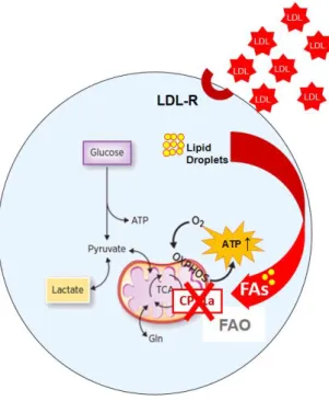
Figure 5: LDL exposition seems to induce the mitochondrial metabolism as LDL
exposed TNBC cells have increased mitochondrial mass and number, produce more ATP and have decreased lactate production. This metabolic signature occurs simultaneously with the acquisition of a more aggressive phenotype since LDL exposed
TNBC cells proliferate two time more and have an increased migratory capability. (Graphic illustration adapted from Viale et al., 2015 Cancer Research Review)
31
The majority of TNBC cells exhibits a metabolic profile characterized by low OXPHOS rate and increased glycolysis compared to other breast cancer cells [36]. However, MDA-MB-231 cells were an exception and exhibit resistance to this metabolic adaptation [36]. Thus, TNBC is not an unique/homogeneous pathology and it is important to notice that much like the genetic and signalling heterogeneity found in TNBC, metabolic heterogeneity is also likely to exist in TNBC patients and models [6][42].
This heterogeneity is verified between LDL exposed MB-231 and MDA-MB-231 under basal conditions (without LDL-cholesterol exposure). Although previous studies described MDA-MB-231 basal metabolic profile to harbour high glycolytic flux and low OXPHOS activity [42]; gene set analysis by microarray revealed that LDL exposed MDA-MB-231 cells have an induction of FAO genes (such as CPT1a) and a repression of lipid synthesis genes (such as HMG-CoA reductase) [see Appendix 1]. Quantitative PCR (qPCR) analysis confirms these results, demonstrating that LDL exposed MDA-MB-231 cells have a decreased expression of genes responsible for lipid synthesis (HMG-CoA reductase and FASN) and increased expression of the FAO enzyme CPT1a. This was expected since FA metabolism is controlled by nutrient-dependent genetic and epigenetic changes [43]. These results confirm that LDL exposure increases FA catabolism and decreases the endogenous lipid synthesis anabolic pathway, probably through the activation of regulatory pathways in response to lipid abundance in the environment. Additionally, gene expression seems to be a dynamic process since previous PCR analyses studies showed a specific up-regulation of OXPHOS genes in circulating cancer cells compared to primary tumour cancer cells and a reversible expression of these genes when circulating cells are retained in their preferred site of metastasis [21].
Gene expression analysis by qPCR further revealed no significant differences in the expression of genes associated with mitochondrial biogenesis (PGC-1α, PCG1, NRF1 or TFAM). However, there is an increased expression of genes encoding respiratory chain proteins (such as Cox5b, ATP5g1 and NDUFB5). Further studies will be needed to understand the regulation of OXPHOS genes transcription in LDL exposed MDA-MB-231 cells, which seem not require an increased expression of PGC-1α and associated mitochondrial biogenesis factors. Is known that PGC-1α can be pro- and
anti-32
tumorigenic in different tumours and one possibility is that PGC-1α interacts with different tissue specific transcription factors, driving distinct genetic programs in different cancer types [21]. However, increased mitochondrial biogenesis appears to be a ubiquitous response to PGC-1α expression regardless of the cancer type or outcome of PGC-1α expression [21]. Therefore, PGC-1α heterogeneous response in different tumours does not fully explains the reason why LDL exposed MDA-MB-231 cells do not require an increased expression of PGC-1α to increase their respiratory chain proteins levels. Adding to the complexity of metabolic regulation, these factors can be controlled by micro-RNAs that act as regulators of post-transcription [43]. Alternatively, there are other two major non-mutually exclusive upstream regulators of FA metabolism that interconnect this pathway to tumour growth: mTORC1 and AKT. Activated AKT signalling increases overall levels of FA synthesis by upregulating the expression of the master lipid transcription factor sterol regulatory element-binding protein (SREBP) [10]. SREBPs control the transcription of enzymes required for cholesterol and fatty acid synthesis in response to modulations of sterol levels [2][44]. In this process, SREBP elements may interact with PGC-1α and increase its bioavailability for the transcription of OXPHOS genes without an increased expression of PGC-1α. Additionally, apart from SREBP, HIF-1α and p53 also contribute to the stabilization of FASN [4].
33
Figure 6: The molecular regulation of de novo fatty acid synthesis and the fate of
saturated long-chain fatty acids and its connection with mitochondrial metabolism. Activated AKT signalling upregulates SREBP expression, increasing PGC-1α bioavailability and OXPHOS genes transition. HIF-1α and p53 also contribute to FASN
stabilization. (Graphic illustration adapted from S. Balaban, et al, 2015, Biomed Res and from Viale et al., 2015, Cancer Research Review)
However, low SREBP expression has also been clustered with high FASN levels, which is an unexpected phenomena since SREBP1 is known to induce the expression of this enzyme [29]. Thus, as the regulation of breast cancer lipid metabolism may differ from that of normal physiology [29], it would be interesting to explore western blotting (studying proteins: the final product of transcription and transduction processes), enzyme activity and procedure the quantification of the metabolic intermediates that regulate the lipid metabolism enzymes under hypercholesterolemia in TNBC cells.
The concept of precision oncology is founded on the presumption that knowing the genomic basis of a patient’s cancer will guide choice of targeted therapies likely to be efficacious [45]. The observation that lipid and mitochondrial metabolism play a key role in tumorigenesis has driven efforts to identify cancer therapeutics that function by targeting oxidative metabolism [40]. Inhibition of either cellular fatty acid synthesis or fatty acid transport into mitochondria reduced cellular ROS levels and DNA damage, attenuating tumour recurrence in vivo [17]. There are two key FAO enzymes that are particularly interesting as potential targets for pharmacological intervention: 3-ketoacylthiolase (3-KAT), that catalysis the final step in FAO, and CPT1a, the rate-limiting enzyme in FAO [46]. CTP1a was further investigated since its expression increases significantly in the LDL exposed MDA-MB-231 cells in contrast to all other lipid metabolism genes. Unlike short-chain FA, which can freely diffuse into mitochondria, long-chain FA enter the mitochondria by the carnitine shuttle system (CPT1a). CPT1a is an integral membrane protein located on the mitochondrial outer membrane and catalyses the limiting step of FAO. Therefore, the rate of mitochondrial FAO is regulated by CPT1a which expression is sensitive to the microenvironment [10]. Moreover, CPT1a activity is upregulated in metastatic TNBC cells [6] and it is essential
34
in TNBC progression and its inhibition leads to decreased colony formation capacity, decreased migration and increased intracellular lipids storage [8][14][34]. Additionally, the inhibition of FA transport into mitochondria reduces cellular ROS levels and DNA damage in in vivo models, which are hallmarks of minimal residual disease cancer cells, responsible for tumour regression [17]. Thus, specific pharmacological inhibition of CPT1a with Etomoxir was here used to determine whether FAO is an essential metabolic determinant in LDL-exposed MDA-MB-231 cells.
Previous studies using Etomoxir described a sharp decrease in Oxygen consumption rate (OCR) after treatment and simultaneously increased the glycolysis, as seen by the high extracellular acidification rate (ECAR) [6].
Figure 6: Specific pharmacological inhibition of CPT1a was here used to determine
whether FAO is an essential metabolic determinant in LDL-exposed MDA-MB-231 cells (Graphic illustration adapted from Viale et al., 2015 Cancer Research Review)
As previously reported, LDL exposed MDA-MB-231 cells have greater proliferation than the control group [26]. This proliferative pattern is maintained even when exposed to increasing concentrations of Etomoxir. Therefore, the inhibition of CPT1a does not impact the increased proliferation induced by hypercholesterolemia and FAO metabolism is not essential for the proliferation of LDL exposed MDA-MB-231
35
cells. Therefore, although MDA-MB-231 cells in “basal conditions” (without LDL-cholesterol exposure) relies in FAO for proliferation, as suggested by decreased colony formation potential upon Etomoxir exposure [13], it does not seem to represent an advantage for the increase proliferation obtained under hypercholesterolemia.
Previous reports have shown an increased migratory potential of LDL exposed MDA-MB-231 [26]. Previous work about LDL influence on MDA-MB-231 evidenced that LDL impacts the migration capability in in vitro models [32]. The EMT process and migratory potential depend (in part) on LDL exposure or availability since lipid composition modifications are required for increasing membrane fluidity [2]. LDL exposure may also increase migration by increasing both lipid droplets and mitochondria, which per se can potentiate cell motility [18]. Lipid droplets are attached to microtubules and mitochondria move to cytoplasmic protrusions in order to fuel chemotaxis, as well as cell invasion [32] [39].
It is known that Etomoxir treatment and CPT1a knockdown decrease MDA-MD-231 migration under basal conditions [13]. In the present study we demonstrate that the increased mobility of LDL exposed MDA-MB-231 cells is also reverted using Etomoxir. This result demonstrates that even when exposed to LDL, migration of MDA-MB-231 cells continue to be sensitive to Etomoxir. Therefore, CPT1a inhibition with Etomoxir does not reverse proliferation, but abolished the increased migratory capacity of MDA-MB-231 cells exposed to LDL. This result reinforces the importance of OXPHOS in driving tumour growth and fuelling disease progression [39].
It is also known that LDL exposed MDA-MB-231 cells benefit from the “lipid accumulating phenotype”. While in other breast cancer cell subtypes, the cholesterol concentration in the endoplasmic reticulum membrane acts as a sensor and influences LDLR transcription and LDL uptake by negative feedback; in TNBC cells occurs the “lipid accumulating phenotype” [32]. This positive energetic balance driven by usage of circulating FA and cholesterol as a substitute for de novo biosynthesis drives (in part) the increased migratory phenotype [32]. In the present study, it was verified an increased in lipid droplets content in LDL exposed cells compared to the control group. As increasing concentrations of Etomoxir are added (100 to 200 μM), the control group show an increase in lipid droplets (as described in previous studies [13]). This
36
phenomena was expected since the regulation of intracellular lipids is perturbed in the direction of lipid accumulation in MDA-MB-231 cells [32].
Nevertheless, the “lipid accumulating phenotype” was not verified when Etomoxir was added to LDL exposed cells. When Etomoxir is used, CPT1a is blocked and FA are less channelled as subtract for mitochondrial respiration; however, the lipid droplets content of LDL exposed cells do not increase (apparently there is no lipid accumulation). We hypnotise that LDL exposed cells continue to use lipids as subtracts for alternative metabolic pathways. It is also relevant to mention that Etomoxir also present selectivity to other metabolic enzymes; it does not only inhibit CPT1a, but also inhibits DGAT, an enzyme that catalysis the final step of Triacyclglycerol (TAG) synthesis [10]. Alongside the endoplasmic reticulum pathway, there is evidence that DGAT can also catalyse the conversion of DAG to TAG [10]. This could lead to bias when interpreting a decrease in lipid droplets as TAG formation is inhibited by Etomoxir, therefore our results should be corroborated using alternative FAO or CPT1a inhibition.
Moreover, it is important to notice that Etomoxir maximal effective concentration (EC50) varies greatly from species to species and depends on cell type. Additionally, CPT1a inhibitors exhibited toxic side effects in clinical trials (Etomoxir treatment in patients results in hepatotoxicity) [46]. For these reasons, the results obtained with Etomoxir should be confirmed with other alternatives. The PPAR inhibition or AMPK inhibition are not recommended as there is a wide variety of pathways in which they are involved, leading to difficulties in interpretation of the results [46]. Tigecycline (a mitochondrial biogenesis inhibitor) and Olygomycin (ATP synthase I inhibitor that impacts OXPHOS) could be used instead of Etomoxir when mitochondrial biogenesis and OXPHOS are augmented along with FAO. A previous report showed that the treatment with a mitochondrial uncoupling agent or ATP synthesis inhibitor (as Olygomycin) reduced lamellipodia formation and decreased breast cancer cell migration and invasion [41]. Treatment of TNBCs with Oligomycin reduced the OCR and mammosphere formation [9]. However, Olygomycin does not specifically target FAO, as it blocks OXPHOS independently of the substrate; therefore it will not be more informative about the specific FAO implication in the acquisition of aggressiveness. Alternatively, we could use shRNA knockdown strategies that allow a
37
specific inhibition, but this method implies electing a target gene and there are multiple genes involved in FAO metabolism. Finally, the inhibition of the LDL receptor may also be interesting to understand both lipid and protein effects of LDL. In addition, we could think about LDLR as a future target for TNBC target therapy as LDLR is overexpressed in breast cancer cells and is expressed at low levels in others cell types. Therefore, LDLR could be a potential target for new therapies such as LDL-mimicking nanocarriers which selectively delivery of antineoplastic agents into TNBC [16]. However, LDLR expression seems to decrease in LDL exposed cells by negative feedback [Monteiro, Nóbrega-Pereira et al., Unpublished].
In summary, the present work shows novel evidence that LDL exposed TNBC MDA-MD-231 cells have increased lipid droplets content, increased mitochondrial number and transmembrane potential and increased expression of FAO (CPT1a) and OXPHOS genes. CPT1a inhibition by Etomoxir does not reverte proliferation, but abolished the increased migratory capacity of MDA-MB-231 cells exposed to LDL. We anticipate that our findings could contribute to the development of new strategies for cancer treatment involving FAO inhibitors, especially for the treatment of patients with hypercholesterolemia. Further studies are in progress in order to clarify the importance of mitochondrial metabolism for cell division, loss of adhesion, apoptosis and drug resistance induced by LDL-cholesterol exposure – both in TNBC cells and other subtypes of breast cancer.
38
Acknowledgments
I would like to express my deepest appreciation to Professor Sérgio Dias, whose guidance was essential for the present work. Thank you for the golden opportunity that is to work in Sergio Dias Lab in Instituto de Medicina Molecular. I am grateful for all the time spent teaching me how to structure my thinking.
My very profound gratitude to Sandrina, who welcomed me in this project since the first day and was crucial in my integration in the lab routine - thank you for all teachings and support.
Thanks to Mafalda, who performed the prior project, for all the guidance throughout this journey. To Susana, who contributed to the present work by performing the proliferation and migration assays using Etomoxir.
I would also like to acknowledge Vanessa Morais Lab for the expertise and discernments about mitochondrial metabolism included in the discussion of the present work. I would also like to express my gratitude to Flow Cytometry Unit and Histology and Comparative Pathology Unit for the technical collaboration. Thanks to Gabinete de Apoio à Investigação Científica, Tecnológica e Inovação (GAPIC) for project financing and for the opportunity to present my work publicly at “Dia da Investigação”.
A deep thanks to all my closest friends. To Catarina, for being my example of determination and commitment over the past 6 years. To Rita and Patrícia, for overcoming adversities together since Anatomy classes in early days at college.
To Ana, who taught me that in life not everything is black or white and who offered me encouragement through long hours of conversation, listening me talking about my work, my worries and dreams.
I would like to thank my loved ones, who have supported me during the entire process. Special thanks to Diogo for always being with me, supporting me throughout this work as in Life in general. Most importantly, thanks to my parents for the constant support throughout my years of study. Thank you for providing me unfailing strength and for turning the world upside down just to help me. To my grandparents, os meus anjos da guarda e da Guarda, I am eternally grateful to you.
39
Bibliography
[1] P. S. Ward and C. B. Thompson, “Metabolic Reprogramming: A Cancer
Hallmark Even Warburg Did Not Anticipate,” Cold Spring Harb. Perspect. Biol., vol. 11, no. 3, pp. 1–15, 2009.
[2] F. Röhrig and A. Schulze, “The multifaceted roles of fatty acid synthesis in cancer,” Nat. Rev. Cancer, vol. 16, no. 11, pp. 732–749, 2016.
[3] A. Deshmukh, K. Deshpande, F. Arfuso, P. Newsholme, and A. Dharmarajan, “Cancer stem cell metabolism: a potential target for cancer therapy,” Mol. Cancer, vol. 15, no. 1, p. 69, 2016.
[4] S. Biswas, J. Lunec, and K. Bartlett, “Non-glucose metabolism in cancer cells-is it all in the fat?,” Cancer Metastasis Rev., vol. 31, no. 3–4, pp. 689–698, 2012. [5] M. C. Caino et al., “Metabolic stress regulates cytoskeletal dynamics and
metastasis of cancer cells,” J. Clin. Invest., vol. 123, no. 7, pp. 2907–2920, 2013. [6] V. Putluri et al., “HHS Public Access,” vol. 14, no. 9, pp. 2154–2165, 2016. [7] M. C. Caino et al., “PI3K therapy reprograms mitochondrial trafficking to fuel
tumor cell invasion,” Proc. Natl. Acad. Sci., vol. 112, no. 28, pp. 8638–8643, 2015.
[8] A. Viale et al., “Oncogene ablation-resistant pancreatic cancer cells depend on mitochondrial function,” Nature, vol. 514, no. 7524, pp. 628–632, 2014.
[9] K. Lee et al., “MYC and MCL1 Cooperatively Promote Chemotherapy- Resistant Breast Cancer Stem Cells via Regulation of MYC and MCL1 Cooperatively Promote Chemotherapy- Resistant Breast Cancer Stem Cells via Regulation of Mitochondrial Oxidative Phosphorylation,” pp. 633–647, 2017.
[10] S. Balaban, L. S. Lee, M. Schreuder, and A. J. Hoy, “Obesity and cancer progression: Is there a role of fatty acid metabolism?,” Biomed Res. Int., vol. 2015, no. Table 1, 2015.
[11] S. Rodríguez-Enríquez et al., “Mitochondrial free fatty acid β-oxidation supports oxidative phosphorylation and proliferation in cancer cells,” Int. J. Biochem. Cell Biol., vol. 65, no. 1, pp. 209–221, 2015.
[12] D. Dai et al., “Pretreatment TG/HDL-C Ratio Is Superior to Triacylglycerol Level as an Independent Prognostic Factor for the Survival of Triple Negative Breast Cancer Patients,” J. Cancer, vol. 7, no. 12, pp. 1747–1754, 2016.
[13] J. H. Park et al., “Fatty Acid Oxidation-Driven Src Links Mitochondrial Energy Reprogramming and Oncogenic Properties in Triple-Negative Breast Cancer,” Cell Rep., vol. 14, no. 9, pp. 2154–2165, 2016.
[14] R. Camarda et al., “Inhibition of fatty acid oxidation as a therapy for MYC-overexpressing triple-negative breast cancer.,” Nat. Med., vol. 22, no. 4, pp. 427–
40
432, 2016.
[15] B. Maiti, M. N. Kundranda, T. P. Spiro, and H. A. Daw, “The association of metabolic syndrome with triple-negative breast cancer,” Breast Cancer Res. Treat., vol. 121, no. 2, pp. 479–483, 2010.
[16] J. Ye et al., “Cellular uptake mechanism and comparative evaluation of
antineoplastic effects of paclitaxel-cholesterol lipid emulsion on triple-negative and non-triple-negative breast cancer cell lines.,” Int. J. Nanomedicine, vol. 11, pp. 4125–4140, 2016.
[17] K. M. Havas et al., “Metabolic shifts in residual breast cancer drive tumor recurrence,” J. Clin. Invest., vol. 127, no. 6, pp. 2091–2105, 2017.
[18] V. S. LeBleu et al., “PGC-1α mediates mitochondrial biogenesis and oxidative phosphorylation in cancer cells to promote metastasis.,” Nat. Cell Biol., vol. 16, no. 10, pp. 992–1003, 1–15, 2014.
[19] I. Samudio et al., “Pharmacologic inhibition of fatty acid oxidation sensitizes human leukemia cells to apoptosis induction,” Leukemia, vol. 120, no. 1, 2010. [20] E. L. LaGory et al., “Suppression of PGC-1?? Is Critical for Reprogramming
Oxidative Metabolism in Renal Cell Carcinoma,” Cell Rep., vol. 12, no. 1, pp. 116–127, 2015.
[21] K. Pantel, M. C. Haigis, and F. M. De Carvalho, “Phosphorylation To Promote Metastasis,” vol. 16, no. 10, pp. 1–32, 2015.
[22] J. Kowalski et al., “NIH Public Access,” vol. 50, no. 4, pp. 381–389, 2010. [23] P. Sancho et al., “MYC/PGC-1α balance determines the metabolic phenotype and
plasticity of pancreatic cancer stem cells,” Cell Metab., vol. 22, no. 4, pp. 590– 605, 2015.
[24] É. Audet-Walsh et al., “The PGC-1α/ERRα Axis Represses One-Carbon Metabolism and Promotes Sensitivity to Anti-folate Therapy in Breast Cancer,” Cell Rep., vol. 14, no. 4, pp. 920–931, 2016.
[25] Y. Wu et al., “Aberrant phosphorylation of SMAD4 Thr277-mediated USP9x-SMAD4 interaction by free fatty acids promotes breast cancer metastasis,” Cancer Res., vol. 77, no. 6, pp. 1383–1394, 2017.
[26] C. Rodrigues dos Santos et al., “LDL-cholesterol signaling induces breast cancer proliferation and invasion.,” Lipids Health Dis., vol. 13, no. 1, p. 16, 2014. [27] A. M. Lorincz and S. Sukumar, “Molecular links between obesity and breast
cancer,” Endocr. Relat. Cancer, vol. 13, no. 2, pp. 279–292, 2006.
[28] M. Pierobon and C. L. Frankenfeld, “Obesity as a risk factor for triple-negative breast cancers: A systematic review and meta-analysis,” Breast Cancer Res. Treat., vol. 137, no. 1, pp. 307–314, 2013.