Budesonide reverses lung hyperinflation in childhood asthma: a
controlled study
N. Neuparth
a,b,*, T. Gamboa
a, C. Pereira
c, J. Rusado Pinto
b, A. Rendas
aaDepartamento Uni6ersita´rio de Fisiopatologia, Faculdade de Cieˆncias Me´dicas, Uni6ersidade No6a de Lisboa, Campo de Santana,
130, 1198Lisboa CODEX, Portugal
bSer6ic¸o de Imunoalergologia, Hospital de Dona Estefaˆnia, Lisboa, Portugal
cSer6ic¸o Uni6ersita´rio de Fisiologia, Faculdade de Cieˆncias Me´dicas, Uni6ersidade No6a de Lisboa, Campo de Santana,130,
1198Lisboa CODEX, Portugal
Received 26 February 1999; accepted 9 August 1999
Abstract
It was investigated whether inhaled budesonide reduces lung volumes in a group of asthmatic children with lung hyperinflation. Budesonide (800 mg bid, for 2 months) was administered to 12 asthmatic children (mean age, 11.293.3 years) with lung hyperinflation (TGV]130% predicted and/or RV]140% predicted) in a randomised, placebo controlled, double blind, crossover study. Body plethysmography (panting frequency controlled at 1·s− 1) was performed at the beginning, 2 months afterwards
(before crossover) and at the end of the study. Budesonide significantly reduced TGV (2.3590.90 l BTPS or 126924% predicted) compared with placebo (2.5491.08 l BTPS, P=0.014 or 140921% predicted, PB0.05). In addition, budesonide significantly increased mean specific conductance (0.0690.02 cm H2O− 1l s− 1to 0.0790.01 cm H2O− 1l s− 1, PB0.05). It was concluded
that budesonide reduced lung hyperinflation most likely by decreasing airway inflammation. © 1999 Elsevier Science Ireland Ltd. All rights reserved.
Keywords:Asthma; Lung hyperinflation; Bronchial inflammation; Budesonide
www.elsevier.com/locate/pathophys
1. Introduction
In a clinical context, hyperinflation implies an abnor-mal increase in the volume of gas in the lungs at the end of tidal expiration [1]. However, when it was at-tempted to relate hyperinflation with the airway nar-rowing occurring in asthma, the picture is much less defined as suggested in a recent review [2] which states that, in this context, there is no clear definition of hyperinflation and of its underlying mechanisms. The authors suggest that both altered properties of airways and lung parenchyma are involved in the development of hyperinflation and point to the importance of mea-suring residual volume (RV), thoracic gas volume
(TVG) and total lung capacity (TLC) to understand the underlying pathophysiological mechanisms.
During an acute attack of asthma [3] there is an increase in all lung volumes which is due not only to the expiratory flow limitation but also to the persistent inspiratory muscle contraction throughout expiration, caused by a reflex mechanism from the constricted airways, which increases end-expiratory volume and produces hyperinflation [4 – 7]. On the other hand, chronic increases of functional residual capacity (FRC), RV and occasionally of TLC can be observed in some asthmatic patients away from the exacerbations of asthma [2,8] but the mechanisms responsible for these changes, such as loss of bronchial-to-parenchyma inter-dependence, are still under discussion. A prior study performed in seven asthmatic patients, found a decrease in FRC after a week of systemic steroids that was attributed to an improvement of lung elastic recoil [9] as was later demonstrated [10].
* Corresponding author. Tel.: + 3000; fax: + 351-1-885-1920.
E-mail address:nneuparth.fpat@fcm.unl.pt (N. Neuparth)
0928-4680/99/$ - see front matter © 1999 Elsevier Science Ireland Ltd. All rights reserved. PII: S 0 9 2 8 - 4 6 8 0 ( 9 9 ) 0 0 0 1 7 - 6
Although it is accepted that the treatment with steroids reduces inflammation both in the airways and in the parenchyma [11,12], the authors were not aware of any study relating such treatment with measurements of lung volumes that are dependent, amongst other factors, upon lung elastic recoil. In order to do that, an inhaled steroid was given to a group of hyperinflated asthmatic children and lung volumes were monitored in a controlled study.
2. Material and methods
2.1. Study subjects
Patients were selected from all asthmatics that have performed lung function tests in an Allergy Clinic during 1 year. From these, 13 children with asthma and lung hyperinflation, aged 6 – 18 years old were included. The diagnosis of asthma was made according to ac-cepted criteria [13]. All patients were free from inhaled and systemic steroids and from an exacerbation of asthma for at least 6 weeks before the beginning of the study. During the study period all patients were only taking inhaled budesonide regularly and a short-acting b2 agonist in a prn basis.
According to their clinical symptoms and/or to daily medication required, 11 patients were classified as hav-ing moderate persistent asthma, whereas two were la-belled as mild persistent [14]. Symptoms of asthma were detected for the first time, in average, since 8.594.2 years ago, with a range between 3.1 and 17.3 years. Chronic lung hyperinflation was considered whenever TGV was repeatedly (in three different occasions dur-ing the previous year) equal or higher than 130% predicted [15] and/or RV was equal or higher than 140% predicted [16]. The predicted values were calcu-lated from Zapletal equations [17].
2.2. Study design
The study was performed in two phases (Phase 1 and Phase 2), with a duration of 4 months. It was a controlled study, double blind, against placebo accord-ing to randomisation in two groups (G1 and G2) of six patients each. A crossover was done at the end of Phase 1. Budesonide (400mg dose− 1) or placebo was
adminis-tered to each patient through a Turbohaler® device
twice a day. The content of the inhaler was blinded both to patients and investigators. Lung volumes were measured three times during the study — before start-ing budesonide (day 0), at crossover (day 57) and at the end of the study (day 113). Patients were instructed to avoid takingb2agonists during the 12-h period
preced-ing the lung function measurements. An informed con-sent was obtained from the tutors in every case and the study was approved by the Hospital Ethics Committee.
2.3. Methods
A body plethysmograph MasterLab Jaeger (Wu¨rzburg, Germany) was used to measure lung vol-umes (TGV, RV and TLC) and specific conductance (sGaw) according to the method described by Ref. [18].
The procedure followed during measurements was that recommended by the European Respiratory Society [19]. Nevertheless, panting frequency was settled at 1 Hz, using a tachometer as a biofeedback signal to the patient, because lung volumes measured by body plethysmography with high panting frequencies are overestimated in asthma [20,21].
2.4. Analysis
The key questions addressed in this study were: did budesonide modify lung volumes (TGV, RV and TLC) and sGaw, compared with placebo? Did lung volumes
correlate with airway calibre before and after treatment with budesonide?
Analysis of variance was used to evaluate the effects of budesonide and placebo according to the methods described in Ref. [22]. Each analysis was tested for period effect and for the presence of carry-over. Delta lung volumes (lung volumes after budesonide — lung volumes after placebo) were correlated with delta con-ductance (Gaw).
3. Results
3.1. Patient characteristics
Distribution by age and sex of the children included in this study is shown on Table 1. One of the patients was excluded because he had an asthma attack treated with oral steroids during placebo phase. For this reason only 12 patients concluded the study. This group had the following mean baseline volumes (Table 2): TGV, 2.791.10 l (141921% pred.); RV, 1.5990.80 l (170953% pred.); and TLC, 4.5291.62 l (118913% pred.).
3.2. Effect of budesonide on lung 6olumes
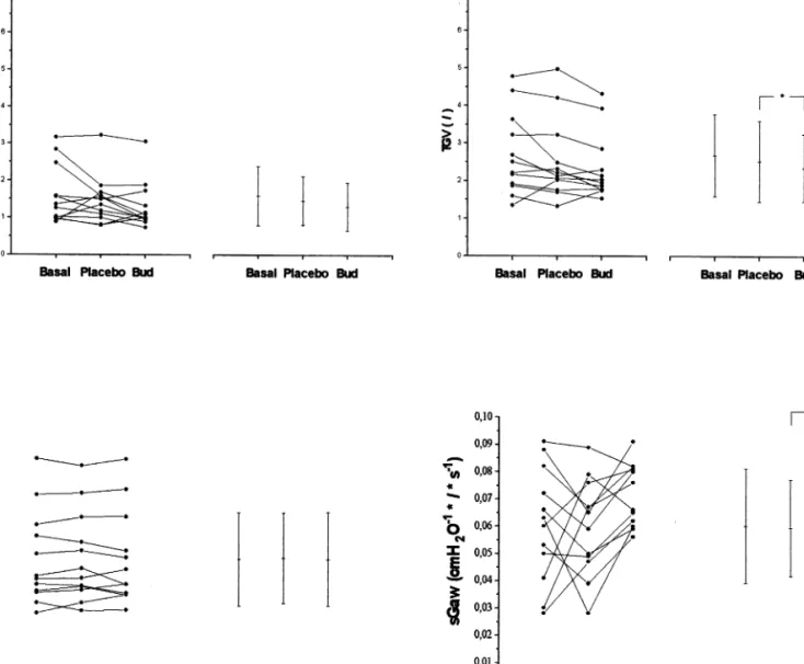
When compared with placebo, only TGV was signifi-cantly reduced (PB0.05) after 2 months of inhaled budesonide, from 2.5491.08 to 2.3591.08 l. There were no differences between basal volumes and volumes after placebo (Fig. 1).
3.3. Effect of budesonide on airway calibre
As patients were evaluated through body plethysmog-raphy, only resistance (or its reciprocal-conductance) was measured. Specific conductance (sGaw) after 2 months of budesonide increased significantly (PB0.05) when compared with sGaw after placebo (from 0.069
0.02 to 0.0790.01 cm H2O− 1·1·s− 1, Fig. 1). This
increase corresponds to a 17% variation.
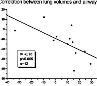
3.4. Relation between lung 6olumes and airway calibre Delta TGV (as defined in Section 2) was significantly correlated with delta Gaw(r = − 0.78, PB0.005, Fig. 2).
4. Discussion
With this study it has been shown, in a group of 12 young asthmatics with lung hyperinflation, that TGV but not RV or TLC decreased significantly after treat-ment with an inhaled steroid. This was shown through a double blind, placebo controlled, crossover design. The reduction of TGV after treatment with budes-onide implies that the anti-inflammatory medication reduced end-expiratory volume. Based on the inflamma-tory theory of asthma, it was hypothesised that this was due, not only to an increase in airway calibre [23 – 25], but also to a reduction in peribronchial and alveolar inflammation [26] which, when present, causes Table 1
Characteristics of the asthmatic children with lung hyperinflation studieda
Age Sex No. TGV RV TLC % Pred l % Pred l % Pred l 1.36 144 3.93 105 F 2.22 1 11 120 124 6.74 244 2.84 163 2 15 M 4.40 1.03 142 3.42 118 M 1.87 3 8 133 13 F 3.64 181 4 2.47 243 5.31 130 116 4.69 168 1.56 127 5 10 M 2.51 10 M 2.68 146 6 0.97 110 3.83 102 1.26 161 3.64 120 7 8 F 1.92 129 0.96 146 3.01 119 8 7 M 1.60 130 3.17 177 4.78 272 147 M 7.99 9 18 117 0.88 122 147 2.65 1.35 F 9 10 0.99 132 2.17 M 122 11 11 3.36 99 5.70 14 M 3.22 131 1.58 146 114 12 12 9M/4F 2.70 141 170 4.52 118 Mean 1.59 53 3 – 1.10 21 0.80 1.62 13 S.D.
aM, male; F, female; % Pred, % predicted values; S.D., standard deviation.
Table 2
sGawevolution of asthmatic children with lung hyperinflation during the studya
Basal After budesonide
No. After placebo
% Pred cm H2O−1l s−1 % Pred cm H2O−1l s−1 % Pred cm H2O−1l s−1 90 0.082 90 1 0.082 0.067 74 2 0.03 48 0.067 108 0.076 123 0.081 63 0.076 68 50 0.06 3 0.047 56 4 0.028 33 0.062 74 0.079 92 5 0.041 48 0.066 77 89 0.082 97 0.089 6 0.091 99 7 0.05 44 0.049 43 0.065 57 8 0.072 52 0.059 43 0.08 58 0.056 63 0.039 90 85 0.053 9 10 0.063 41 0.028 18 0.06 39 0.066 64 0.05 11 49 0.059 57 94 0.065 128 0.088 12 0.091 132 79* 0.072* 67 0.060 65 Mean 0.060 0.021 29 0.018 S.D. 27 0.012 27
a% Pred, % predicted values; S.D., standard deviation.
Fig. 1. Comparison of lung volumes (residual volume (RV), upper left panel; thoracic gas volume (TGV), upper right panel; total lung capacity (TLC), lower left panel) and of specific conductance (sGaw) before budesonide or placebo (basal), after placebo (placebo) and after budesonide
(Bud) treatment of asthmatic children with lung hyperinflation. On the left hand side of each graphic is represented the individual variation. On the right hand side is represented the mean variation (9standard variation); * PB0.05.
a temporary loss of the relationship between small bronchi and parenchyma, thus reducing elastic recoil [27,28]. This is suggested in this study by the fact that a small improvement in airway calibre after budesonide, demonstrated by the increase in sGaw, was not
accom-panied by a change in RV, which remained higher and not different from placebo. This probably means that a higher RV depends not only on airway calibre but also on persistent parenchymal changes such as a reduction in lung elastic recoil that also contributes to the expira-tory flow limitation. Similar observations were made by S¸ekerel et al. [8] who found a reduction of TLC after 8 weeks of treatment with budesonide, 400 mg day− 1.
A recent study [29] has shown that, in chronic stable asthma, lung volumes assessed by measurements of TLC, can be larger than predicted at the start of
adolescence and suggested that this change was due to the mitogenic effect of inflammatory mediators which stimulated alveolar multiplication during childhood. This hypothesis is different from a previous one [30] which explained the hyperinflation in chronic asthmat-ics by a process of alveolar destruction because of the stretching caused by the trapped gas. In this regard it is interesting to note that the younger cases, also at the beginning of adolescence, had higher baseline values of TLC than the older patients, a finding which agrees with the above mentioned hypothesis. The authors are aware that the measurement of TLC by plethysmogra-phy can produce methodological errors, however, they were minimised by a panting frequency B1 Hz. So it was confirmed, as others, that TLC is increased in some of these patients with asthma, whether this is caused by
Fig. 2. Graphic representation of the statistically significant correla-tion between delta thoracic gas volume (TGV) and delta Gaw(before
and after budesonide) of asthmatic children with lung hyperinflation.
References
[1] G.J. Gibson, Pulmonary hyperinflation: a clinical overview, Eur. Respir. J. 9 (1996) 2640 – 2649.
[2] R. Pellegrino, V. Brusasco, On the causes of lung hyperinflation during bronchoconstriction, Eur. Respir. J. 10 (1997) 468 – 475. [3] A.J. Woolcock, J. Read, Lung volumes in exacerbations of
asthma, Am. J. Med. 41 (1966) 259 – 273.
[4] J. Martin, E. Powell, S. Shore, J. Emrich, L.A. Engel, The role of respiratory muscles in the hyperinflation of bronchial asthma, Am. Rev. Respir. Dis. 121 (1980) 441 – 447.
[5] N. Mu¨ller, A.C. Bryan, N. Zamel, Tonic inspiratory muscle activity as a cause of hyperinflation in histamine-induced asthma, J. Appl. Physiol. 49 (1980) 869 – 874.
[6] R. Pellegrino, B. Violante, S. Nava, C. Rampulla, V. Brusasco, J.R. Rodarte, Relationship between expiratory airflow limitation and hyperinflation during methacholine-induced bronchocon-striction, J. Appl. Physiol. 75 (1993a) 1720 – 1727.
[7] P.W. Collet, T. Brancatisano, L.A. Engel, Changes in the glottic aperture during bronchial asthma, Am. Rev. Respir. Dis. 128 (1983) 719 – 723.
[8] B.E. S¸ekerel, A. Tuncer, Y. Sarac¸lar, G. Adaliulu, Inhaled budesonide reduces lung hyperinflation in children with asthma, Acta Paediatr. 86 (1997) 932 – 936.
[9] W.M. Gold, H.S. Kaufman, J.A. Nadel, Elastic recoil of the lungs in chronic asthmatic patients before and after therapy, J. Appl. Physiol. 23 (1967) 433 – 438.
[10] A.J. Woolkock, J. Read, The static elastic properties of the lungs in asthma, Am. Rev. Respir. Dis. 98 (1968) 788 – 794.
[11] S. Pedersen, Can steroids cause asthma to remit? Am. J. Crit. Care Med. 153 (1996) S31
[12] V. Keatings, A. Jatakanon, Y. Worsdell, P. Barnes, Effects of inhaled and oral glucocorticoids on inflammatory indices in asthma and COPD, Am. J. Crit. Care Med. 155 (1997) 542 – 548. [13] International Consensus Report on Diagnosis and Treatment of
Asthma, Eur. Respir. J. 5 (1992) 601 – 641.
[14] National Institutes of Health (NHLBI), Global strategy for asthma management and prevention. Publication no 95 – 3659 (1995).
[15] S.P. Blackie, S. Al-Majed, C.A. Staples, C. Hilliam, P.D. Pare´, Changes in total lung capacity during acute spontaneous asthma, Am. Rev. Respir. Dis. 142 (1990) 79 – 83.
[16] B.E. Pennock, J.J. Cottrell, R.M. Rogers, Pulmonary function testing. What is normal?, Arch. Intern. Med. 143 (1983) 2123 – 2127.
[17] Working Group Paediatrics, Standardisation of Lung Function in Paediatrics. Compilation of reference values for lung function measurements in children, Eur. Respir. J. 2 (Suppl. 4) (1989) 184 – 264.
[18] A.B. DuBois, S.Y. Botelho, G.N. Bedell, J.H.J.r. Comroe, A new method for measuring airway resistance in man using a body plethysmograph: values in normal subjects and in patients with respiratory disease, J. Clin. Invest. 35 (1956) 322 – 326. [19] Official Statement of the European Respiratory Society,
Stan-dardised Lung Function Testing. Lung volumes and forced ventilatory flows, Eur. Respir. J. 6 (Suppl. 16) (1993) 5 – 40. [20] D.O. Rodenstein, D.C. Stanescu, Frequency dependence of
plethysmograph volume in heathy and asthmatic subjects, J. Appl. Physiol. 52 (1982) 159 – 165.
[21] S.A. Shore, O. Huk, S. Mannix, J.G. Martin, Effect of panting frequency on the pletysmographic determination of thoracic gas volume in chronic obstructive pulmonary disease, Am. Rev. Respir. Dis. 128 (1983) 54 – 59.
[22] D.A. Ratkowsky, M.A. Evans, J.R. Alldredge, Crossover exper-iments. Design: analysis and application, in: D.A. Ratkoswsky, M.A. Evans, J.R. Alldredge (Eds.), Statistics: Textbooks and Monographs, Dekker, New York, 1993, pp. 77 – 120.
a higher rate of alveolar multiplication or by remod-elling of lung tissues or both it is not yet clear.
Another explanation for the increase in TLC could be related to an increase in respiratory muscle strength in chronic asthmatics due to the persistent tonic activity of the inspiratory muscle during expiration. Since res-piratory pressures have not been measured, the involve-ment of the respiratory muscles on the genesis of hyperinflation cannot be excluded, although these changes are considered less likely than those of lung parenchyma. However, the authors’ research findings [31], have shown that, at least maximal mouth pressures are similar in children and adolescents with asthma and a group of age matched controls.
The results show that an inhaled steroid, budesonide, can decrease lung volumes in a group of young asth-matics with lung hyperinflation. This reduction in lung volumes was closely related with the increase of airway calibre thus suggesting a major role of airway inflam-mation in the genesis of chronic lung hyperinflation in asthma.
Acknowledgements
The authors would like to acknowledge Astra Por-tuguesa for the logistic support given to this study. We would also like to acknowledge Mrs Carole Caswell from Astra Charnwood (GB) for all the help with statistical analysis. This work was supported by a grant from PRAXIS XXI, PSAU/P/SAU/92/96. Drugs sup-plied by Astra Portuguesa.
[23] A. Mansell, C. Dubrawsky, H. Levison, A.C. Bryan, H. Langer, C. Collins-Williams, R.P. Orange, Lung mechanics in antigen-in-duced asthma, J. Appl. Physiol. 37 (1974) 297 – 301.
[24] S.A. Freedman, A.E. Tattersfield, N.B. Pride, Changes in lung me-chanics during asthma induced by exercise, J. Appl. Physiol. 38 (1975) 974 – 982.
[25] R. Pellegrino, B. Violante, E. Crimi, V. Brusasco, Effects of aero-sol methacholine and histamine on airways and lung parenchyma in healthy humans, J. Appl. Physiol. 74 (1993b) 2681 – 2686. [26] M. Kraft, R. Djukanovic, S. Wilso, S.T. Holgate, R. Martin,
Alve-olar tissue inflammation in asthma, Am. J. Crit. Care Med. 154 (1996) 1505 – 1510.
[27] R. Marshall, J.G. Widdicombe, Stress relaxation of the human lung, Clin. Sci. 20 (1961) 19 – 31.
[28] K.E. Finnucane, H.J.H. Colebatch, Elastic behaviour of the lung in patients with airway obstruction, J. Appl. Physiol. 26 (1969) 330 – 338.
[29] P.J. Merkus, E.M. van Essen-Zandvliet, J.M. Kouwenberg, E.J. Duiverman, H.C. Van Houwelingen, K.F. Kerrebijn, P.H. Quan-jer, Large lungs after childhood asthma: a case-control study, Am. Rev. Respir. Dis. 148 (1993) 1484 – 1489.
[30] I.A. Greaves, H.J.H. Colebatch, Large lungs after childhood asthma: a consequence of enlarged aispaces, Aust. N. Z. J. Med. 15 (1985) 427 – 434.
[31] N. Neuparth, Hiperinsuflac¸a˜o pulmonar cro´nica: um estudo na crianc¸a asma´tica. PhD Thesis, Faculdade de Cieˆncias Me´dicas, Universidade Nova de Lisboa, 1995.

