Faculdade de Ciências
Departamento de Biologia Animal
Osteogenic Differentiation of Human Umbilical
Cord-derived Stromal Cells on Human Amniotic
Membrane
Ana Rita Almeida de Oliveira
Mestrado em Biologia Evolutiva e do Desenvolvimento
2011
Faculdade de Ciências
Departamento de Biologia Animal
Osteogenic Differentiation of Human Umbilical
Cord-derived Stromal Cells on Human Amniotic
Membrane
Ana Rita Almeida de Oliveira
Dissertação orientada por:
Doutora Maria Margarida Gouveia Sancho
Doutora Maria Gabriela Gomes de Figueiredo Rodrigues
Mestrado em Biologia Evolutiva e do Desenvolvimento
2011
Muitas pessoas contribuíram de diferentes maneiras para a realização desta tese. Em primeiro lugar agradeço ao Dr Helder Trindade por me receber no CHSul e claro à Dra Margarida Sancho (Margaraides) que me proporcionou esta oportunidade de realizar este projecto, agradeço todo o seu empenho em me acompanhar e ensinar as mais variadas técnicas laboratoriais, sua amizade e aqueles cafés duplos que me ajudaram a sobreviver até ao fim da tese. O meu muito obrigado.
Agradeço também à Professora Gabriela Rodrigues pelos seus conselhos e pela sua sempre pronta disponibilidade em me ajudar.
Agradeço à Cristiana Teixeira (Cristaiane) pela ajuda em tudo o que precisei no laboratório, pela companhia no autocarro das dez para as nove e pela amizade que demonstrou ter para comigo desde que entrei no laboratório. Agradeço à Sofia (Sofs) pelos grandes almoços na sala de refeições e no japonês. Agradeço ainda à Dra Josefina Oliveira e a todos os outros membros do banco de tecido por me ajudarem a obter os meus cordões, à Dra Alice pela ajuda fornecida no laboratório de citometria de fluxo, ao Dr Artur por todas as vezes que me forneceu material fora do dia previsto e ao Dr Dário Ligeiro, por me facilitar o acesso à área pós-PCR.
Agradeço a todos os meus amigos pelo incentivo e apoio nos momentos mais difíceis. Por último e mais importante agradeço à minha família e ao Daniel pelo apoio e carinho que me deram. Em particular, agradeço aos meus pais que me proporcionaram esta oportunidade de efectuar o mestrado e pelo apoio constante em tudo o que precisei. O meu muito obrigado a todos.
Abbreviations . . . 6
Abstract . . . 8
Sumário . . . 9
Aims of the thesis . . . 12
1. Introduction . . . 13
1.1 Mesenchymal stromal cells . . . 13
1.2 The umbilical cord as a stem cell source . . . 15
1.3 Clinical application of MSCs . . . 17
1.4 MSC multi-lineage differentiation . . . 19
1.5 Human amniotic membrane . . . 23
2. Materials and Methods . . . 25
2.1. Isolation of MSCs from different parts of UC . . . 25
2.2. MSC in vitro culture . . . 25
2.3. Colony Forming Units (CFUs) . . . 26
2.4. Immunofluorescence . . . 26
2.4.1. Flow cytometry . . . 26
2.4.2. Fluorescence Activated Cell Sorting (FACS) . . . 27
2.5. Gene expression analysis . . . 27
2.6. Xeno-free culture of UC-MSCs . . . 28
2.7. Multilineage differentiation and detection methods . . . 29
2.8. Osteogenic differentiation of MSCs on hAM . . . 30
2.9. Data analysis . . . 31
2.9.1 Statistical analysis . . . 31
2.9.2 Image Acquisition . . . 31
3. Results . . . 32
3.1. MSCs can be successfully isolated from different parts of UC . . . 32
3.2. Differences in the number of MSCs isolated from WJ-UC and W-UC, but similar MSCs morphology and immunophenotype . . . 34
3.3. MSCs proliferation change from the early to the late passages for different tissue source . . . 36
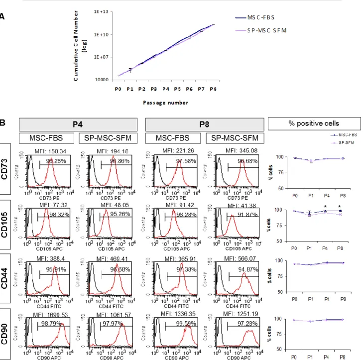
3.4. StemPro MSC SFM induced a decrease in CD105 antigen . . . 39
3.5. hABS is a good xeno-free alternative culture supplement for FBS replacement . . . 41
3.6. Differentiation potential varies between donors and tissue source . . . 43
3.7. hUC-MSC osteogenic ability is related to the cell density, expression levels of CD105 and may be dependent on cell-cell interactions . . . 47
3.8. hABS supplemented expansion medium is capable of maintaining MSC osteogenic potential . . . 52
substrate for a MSC-derived osteoblastic matrix . . . 53
4. Discussion and Future Work . . . 55
References . . . 63
Abbreviations
AM Amniotic membrane AP Alkaline phosphatase Asc Ascorbic acid
Atf4 Activating transcription factor 4 ATMP Advanced therapy medicinal product β-GP β-glycerophosphate
BM Bone marrow
BMP Bone morphogenic proteins BSP Bone sialoprotein
CFU Colony-forming units CR Chemokine receptors Dex Dexamethasone ECM Extracellular cell matrix EGF Epidermal growth factor FBS Fetal Bovine Serum FC Flow cytometry
FGF Fibroblast growth factor GMP Good Manufacturing Practice GVHD Graft-versus-host disease hABS Human AB serum
HGF Hepatocyte growth factor hP Human plasma
hPL Human platelet lysate in medium hPLP Human platelet lysate in plasma hS Human serum
ICAM Intracellular adhesion molecule IGF Insulin-like growth factor IL Interleukin
ISCT International Society for Cellular Therapy KGF Keratinocyte Growth Factor
MFI Mean fluorescence intensity MSC Mesenchymal stromal cells Osx Osterix
PDGF Platelet-derived growth factor
PDEGF Platelet-derived epidermal growth factor PF-4 Platelet factor 4
Amniotic membrane Alkaline phasphatase Ascorbic acid
Activating transcription factor 4 Advanced therapy medicinal product β-glycerophosphate
Bone marrow
Bone morphogenic proteins Bone sialoprotein
Colony-forming units Chemokine receptors Dexamethasone Extracellular cell matrix Epidermal growth factor Fetal Bovine Serum Flow cytometry
Fibroblast growth factor Good Manufacturing Practice Graft-versus-host disease Human AB serum
Hepatocyte growth factor Human plasma
Human platelet lysate in medium Human platelet lysate in plasma Human serum
Intracellular adhesion molecule Insulin-like growth factor Interleukin
International Society for Cellular Therapy Keratinocyte Growth Factor
Mean fluorescence intensity Mesenchymal stromal cells Osterix
Platelet-derived growth factor
Platelet-derived epidermal growth factor Platelet factor 4
_____________________________________________________________________________ _
pWJ Peripheral Wharton’s jelly Runx2/Cbfa1 Core binding factor α1 SFM Serum free medium
TGF-β Transforming growth factor-β
tPRP Thrombin-activated platelet releasate in plasma tWJ Total Wharton’s jelly
UC Umbilical cord UCA Umbilical cord arteries UCB Umbilical cord blood UCV Umbilical cord vein V-UC Umbilical cord vessels WJ Wharton’s Jelly W-UC Whole umbilical cord
Transforming growth factor-β
Thrombin-activated platelet releasate in plasma Total Wharton’s jelly
Umbilical cord
Umbilical cord arteries Umbilical cord blood Umbilical cord vein Umbilical cord vessels Wharton’s Jelly Whole umbilical cord
_____________________________________________________________________________ _
Abstract
Mesenchymal stromal cells (MSC) from the umbilical cord (UC) are a promising tool for regenerative medicine due to their easy acquisition, high expansion potential and the ability to differentiate into multiple lineages. In particular, the capacity for osteogenic differentiation makes them of great interest for clinical application in bone repair.
In this work, 51 MSC lines were isolated from different parts of 26 UC and successfully expanded in fetal bovine serum (FBS)-containing medium. These cells complied with the criteria for MSC classification as they were adherent to plastic, capable of multilineage differentiation, and expressed CD73, CD90, CD44 and CD105 while lacking hematopoietc and endothelial markers. Differentiation of the different MSC lines along the osteogenic lineage was variable depending mostly on the tissue source and isolation method. Moreover, osteogenic differentiation could be modulated by selecting for cells with high expression levels of CD105 (Endoglin) or by the co-culture of MSCs with different osteogenic potential. With the aim of establishing xeno-free clinically compatible culture conditions, StemPro MSCs serum free medium (SP-MSC SFM), human platelet lysate (in medium or in plasma), human plasma and AB serum (hABS) were tested. We have observed that only hABS supplemented medium could efficiently sustain MSC expansion, morphology, immunophenotype and multi-lineage differentiation similarly to FBS-containing medium. Finally, preliminary data indicates that frozen immunologically inert amniotic membrane is a suitable substrate for osteogenic differentiation of MSCs and, therefore, a promising scaffold for producing an osseous matrix for application in regenerative medicine.
Keywords: Amniotic Membrane; Endoglin; human AB serum; Mesenchymal Stromal Cells;
Osteogenic Differentiation; Umbilical Cord.
_____________________________________________________________________________ _
Sumário
A medicina regenerativa é uma área em grande expansão que tenta ultrapassar a escassez e curto tempo de vida dos órgãos e tecidos doados para transplantação e os problemas decorrentes da histocompatibilidade (Chai e Leong 2007). Para tal, células estaminais são expandidas e diferenciadas in vitro num sistema de suporte criado artificialmente. As células estaminais têm o potencial de se auto-renovarem por divisão mitótica mantendo a capacidade de se diferenciarem em vários tipos celulares e deste modo de repovoarem um tecido in vivo. As células mesenquimais do estroma (MSCs, “mesenchymal stromal cells”) representam uma população de células estaminais adultas multipotentes de grande interesse de estudo na área da regeneração de tecidos (Kode et al., 2009). Estas células foram inicialmente identificadas como uma população rara de células estaminais não hematopoiéticas presente na medula óssea (Friedenstein et al., 1966; Friedenstein et al., 1987; Kode et al., 2009; Owen e Friedenstein, 1988). Contudo, tem vindo a ser descrito o isolamento destas células de variados tecidos, entre eles o cordão umbilical humano (UC, “umbilical cord”) (Lu et al., 2006; Nekanti et al., 2010; Shetty et al., 2010). O UC é constituído por duas artérias, uma veia, e uma matriz circundante de tecido conectivo designada por geleia de Wharton (WJ “Wharton’s jelly”), composta por células com morfologia fibroblástica e alguns mastócitos numa substância amorfa rica em proteoglicanos, principalmente ácido hialurónico (Can e Karahuseyinoglu, 2007). MSCs foram já isoladas a partir de todos estes constituintes do UC e constituem uma ferramenta promissora em medicina regenerativa, devido à sua fácil aquisição, elevado potencial de expansão e capacidade de se diferenciar em várias linhagens, nomeadamente osteogénica, adipogénica e condrogénica. Em particular, a sua capacidade de diferenciação osteogénica é de grande interesse para a aplicação clínica destas células em reparação óssea.
Neste trabalho MSCs foram isoladas de diferentes partes de 26 UCs tendo 51 linhas celulares sido obtidas. Em particular, foram isoladas células da geleia de Wharton periférica (pWJ) e total (tWJ), apenas dos vasos (V-UC), e de todo o cordão (W-UC). Para isto aperfeiçoámos um método composto por uma fragmentação mecânica da WJ, V-UC e W-UC em pequenos pedaços seguida de uma digestão enzimática de 3 a 4 horas com colagenase e hialuronidase, deste modo obtendo um bom equilíbrio entre a dissociação do tecido e a viabilidade celular. Para a obtenção de tWJ-UC e V-UC foi necessário efectuar uma digestão enzimática inicial da parte interna do cordão para permitir a remoção dos vasos. As células isoladas foram então expandidas num meio de cultura composto pelo meio basal Dulbecco's Modified Eagle's Medium F12 (DMEM-F12), glutamina e soro fetal bovino (FBS). Estas células preencheram os critérios de classificação para MSCs estabelecidos pela Sociedade Internacional para a Terapia Celular (ISCT), uma vez que: eram aderentes ao plástico; auto-renovaram-se em cultura ao longo de várias passagens; expressam os marcadores de _____________________________________________________________________________ _
superfície específicos CD73, CD90, CD44 e CD105, e não marcadores hematopoiéticos nem vasculares como CD45 e CD34; e foram capazes de se diferenciar nas linhagens osteogénica, adipogénica e condrogénica.
Com o objectivo de obter condições de cultura compatíveis com uma aplicação clinica, ou seja sem produtos animais tais como o FBS, foram testados como candidatos o meio de cultura comercial StemPro (SP-MSC SFM), lisado de plaquetas humanas (ressuspendidas em meio- hPL ou em plasma-hPLP), plasma humano (hP) e soro AB humano (hABS). Um meio de cultura apropriado para esta aplicação, deverá ser capaz de sustentar a expansão de MSCs mantendo a sua morfologia, fenótipo e o potencial de diferenciação em diferentes linhagens. Neste estudo, o meio de cultura com hPLP, hPL ou hP apenas permitiu a sobrevivência das MSCs por 3 passagens, ainda que usando placas de cultura revestidas com fibronectina humana e adicionado 250µM de ácido ascórbico (asc), condições que deveriam facilitar a expansão de MSCs (Choi KM et al., 2008; Potdar e d’Sousa, 2010), como foi possível verificar no meio com FBS.
No meio de cultura SP-MSC SFM e no meio suplementado com hSAB foi possível expandir as células durante pelo menos 8 passagens. Contudo, em SP-MSC SFM as células sofreram algumas alterações morfológicas e uma diminuição da expressão de CD105 ao longo do tempo em cultura. Por seu lado, MSCs expandidas em hSAB (um pool de soro humano AB alogénico) exibiram o comportamento desejado uma vez que nem a sua morfologia nem o imunofenótipo foi alterado quando comparado com células em meio com FBS. De grande importância foi também a observação que, enquanto em MSCs expandidas em SP-MSC SFM houve uma redução na capacidade de diferenciação osteogénica das células, em meio com hSAB esta capacidade foi mantida. Bons resultados no uso de hSAB como suplemento do meio de cultura de MSCs tinham já sido documentados em outros estudos (Yamaguchi et al., 2002; Kocaoermer, Kern e Kluter et al., 2007).
Para a análise do potencial osteogénico das MSCs foram testados diversos meios basais de cultura (com diferentes concentrações de glucose e aminoácidos) e combinações dos osteoindutores ácido ascórbico, dexametasona e β-glicerofosfato. No entanto, observámos que melhores resultados foram obtidos com o meio de cultura comercial MesenCult osteogenic
stimulatory kit. A diferenciação osteogénica das células foi avaliada através da análise da
expressão génica, imunofenotipagem por citometria de fluxo, marcação para a actividade da fosfatase alcalina (AP), característica da fase de deposição de matriz orgânica (aproximadamente aos 14 dias de diferenciação), e numa fase mais tardia (de mineralização) pela deposição de cálcio detectada com vermelho de Alizarina ou marcação von Kossa. Assim, foi possível observar que durante a osteogénese há uma diminuição inicial da expressão de CD105, CD73 e de CD90 (tanto RNA como proteína), seguida do aparecimento de fosfatase alcalina e osteocalcina, e finalmente um aumento gradual da deposição de cálcio a partir dos 21 dias de diferenciação.
Neste estudo foi observada uma grande variabilidade na capacidade de diferenciação das diferentes linhas de MSCs obtidas. Esta variabilidade parece estar de alguma forma _
relacionada com o tecido e método de isolamento utilizado. No entanto, outros factores como a densidade celular no início da cultura, o nível de expressão de CD105 e a interacção entre células parecem também ter uma importante influência na aptidão osteogénica das MSCs
Em diversos aspectos deste estudo o antigénio CD105, pareceu ter um papel relevante, uma vez que a diminuição da sua expressão em SP-MSC-SFM parece inibir a osteogenése, e as células seleccionadas para elevados níveis desta proteína provocam o aumento de diferenciação das MSCs. O CD105, ou Endoglina, é uma glicoproteina membranar de tipo I que faz parte do complexo do receptor TGF-β (Barbara et al., 1999; Blanco et al., 2005). Assim, a regulação dos níveis de expressão de CD105 pode estar relacionada com a modelação da sinalização em resposta ao TGF-β e esta pode determinar aspectos cruciais da diferenciação osteogénica. Este aspecto deverá ser alvo de estudos futuros.
Outro aspecto importante da diferenciação é a matriz ou substrato onde sobre a qual se encontram as células (Salasznyk et al., 2004; Cool e Nurcombe, 2005; Chastain et al.,2006). Neste estudo propusemos a utilização de membrana amniótica (AM “Amniotic membrane”) como um substrato adequado para promover a diferenciação osteogénica de MSCs, o que ajudaria a ultrapassar a necessidade de dissociação das células e disrupção de uma matriz óssea para transplantação num paciente. Os nossos dados preliminares indicam que a AM congelada imunologicamente inerte é um substrato adequado para diferenciação osteogénica de MSCs, apesar de dificuldades em prevenir o enrolamento e enrugamento da membrana durante a diferenciação e de esta diferenciação ser menos eficiente que a de células cultivadas em plástico. Um outro aspecto interessante foi a detecção de um nível basal de AP em MSCs sobre AM em meio de expansão. Isto sugere que a AM tem propriedades intrínsecas que lhe permitem induzir osteogénese, como sejam factores de crescimento presentes na AM (Koizumi
et al., 2000; Valladares et al., 2010). Apesar de estudos adicionais serem necessários de modo
a melhorar as condições de cultura das MSCs na AM, este parece ser um substrato promissor para a produção de uma matriz óssea para aplicação em medicina regenerativa.
Palavras-chave: Células Mesênquimais do Estroma; Cordão Umbilical; Diferenciação
Osteogénica; Endoglina; Membrana Amniotica; Soro AB Humano. _
Aims of the thesis
In this study, with the propose improve the knowledge of the biology of UC-MSCs for future application in bone tissue regeneration, we aim to establish culture conditions for the expansion and differentiation of UC-MSCs on amniotic membrane, in order to produce a bone matrix for application in regenerative medicine. With this purpose, the following objectives were defined:
1. To establish an efficient technique for the isolation of MSCs from different parts of umbilical cord (Wharton’s jelly, vessels and whole cord). For this, both mechanical and enzymatic methods (combinations of collagenase, hyaluronidase and trypsin) were tested in search for the most reliable and efficient one.
2. To determine appropriate culture conditions for the expansion and differentiation of UC-MSC, compatible with a clinical application. To accomplish this, we tried to replace the animal and chemically undefined media components. A commercially available serum-free medium containing the growth factors PDGF-BB, bFGF and TGF-β1(StemPro MSC serum free medium), human platelet lysate (in medium and plasma), human plasma and human AB serum were tested and the cells grown in this medium were compared to FBS-containing standard medium.
3. To characterise UC-derived MSCs in expansion and differentiation conditions. With this purpose, the cell proliferation, gene expression and MSC surface phenotype were assessed for cells grown in expansion conditions (for various periods of time in culture) and after induction of differentiation. Moreover, some culture conditions and characteristics of these cells that could be related to or help predict the osteogenic potential were analysed.
4. To test the amniotic membrane as substrate for the production of a bone matrix. This step aimed at finding a safe and quickly adapted vehicle for transplantation.
_____________________________________________________________________________ _
1. Introduction
1.1. Mesenchymal Stromal Cells (MSCs)
Over the years, much progress has been made in the use of biological resources to reconstruct, replace, or repair diseased cells, tissues and organs in order to improve the quality of life of millions of people around the world (Chai and Leong, 2007; Lutolf and Blau, 2009). The emerging field of Regenerative Medicine attempts to overcome the shortage in donor organs and tissues for transplantation (Chai and Leong 2007) by growing and differentiating cells outside the patient in artificially-created support systems.
In the human body an intrinsic stem cell-based regeneration and repair system that uses stem cells is believed to be found in almost in every tissue type (Bajada et al., 2008). Stem cells have the potential to self-renew by mitotic division while maintaining the ability to differentiate into many cell types, therefore being able to repopulate a tissue in vivo. There are stem cells of several types and origins. Pluripotent stem cells can give rise to all cell types in a body and typically include the following (Xu et al., 2008): embryonic stem cells, derived from the inner cell mass of pre-implantation embryos; epiblast stem cells derived from post-implantation epiblast-stage embryos; germ line stem cells and induced pluripotent stem (iPS) cells obtained by genetic manipulation from somatic cells. Adult stem cells have a more restricted potential and generally give rise only to the differentiated lineages of the tissue in which they reside, being thus referred to as multipotent or unipotent (Watt and Driskell, 2010).
Mesenchymal stromal cells (MSCs) represent a multipotent adult stem cell population (Kode et al., 2009) that has been shown to be capable of differentiating in vitro and in vivo along multiple lineages including those that give rise to bone, cartilage, cardiac and skeletal muscle, neural cells, tendon, adipose, and connective tissue (Nekanti et al., 2010), depending on the surrounding environment. These mesenchymal stromal cells, whose existence was first suggested by Cohnheim in 1867, were later identified in the bone marrow (BM) by Friedenstein and colleagues as a rare population of adherent non-hematopoietic stem cells (Friedenstein et
al., 1966; Friedenstein et al., 1987; Kode et al., 2009; Owen and Friedenstein, 1988). It was
subsequently shown that they can actually be isolated from a variety of other tissues including adipose tissue (Zuk et al., 2002), skeletal muscle (Young et al., 2001), blood (Zvaifler et al., 2000), dental pulp (Gronthos et al., 2000), skin (Lorenz et al.,2008), amniotic fluid (Tsai et al., 2004), umbilical cord blood (Lu et al.,2006) and stroma (McElreavey et al., 1991; Romanov et
al., 2003; Seshareddy et al. 2008; Troyer and Weiss, 2008; Wang et al, 2004), among others
(Chamberlain et al., 2007; da Silva Meirelles et al., 2006; Musina et al., 2005; Riekstina et al., 2009).
Several studies have focused on the characterization of MSCs, revealing important aspects of their biology and defining molecular markers that allow their identification. As the defining characteristics of MSCs are inconsistent among investigators, The International Society
for Cellular Therapy (ISCT) has proposed three major criteria to define human MSCs (Table 1) : (1) adherence to plastic, (2) a specific surface phenotype, and (3) the ability to differentiate in
vitro into cells of the osteoblastic, adipogenic and chondrogenic lineages (Dominici et al., 2006).
Table1: Summary of criteria to identify MSCs (reproduced from Dominici et al., 2006)
1. Adherence to plastic in standard culture conditions
2. Phenotype Positive (≥95% positive) Negative (≤2% positive)
3. In vitro differentiation: osteoblasts, adipocytes, chondroblasts (demonstrated by staining of in vitro cell culture)
MSCs express a large number of adhesion molecules, extracellular matrix proteins, cytokines and growth factors, which are associated with their function and interactions within the various tissues where they are present (Bobis et al., 2006). These cells do not have a unique phenotypic marker but they express a characteristic array of surface proteins including CD73 (5'-nucleotidase), CD90 (Thy-1 cell surface antigen) and the matrix receptors CD44 and CD105 (Endoglin) (Bobis et al., 2006; Chamberlain et al.,2007). MSCs also express adhesion-related antigens (integrins αvβ3, αvβ5, integrin subunit α4, α5 and β1, intracellular adhesion molecule-1 (ICAM-1) and CD44H), and produce cytokines, chemokines and growth factors (interleukin (IL)-6, IL-7, IL-8, IL-11, IL-12, IL-14, IL-15, leukemia inhibitory factor, granulocyte colony stimulating factor (G-CSF), granulocyte macrophage colony-stimulating factor (GM-CSF), stem cell factor, macrophage colony-stimulating factor (M-CSF) and fms-like tyrosine kinase-3 ligand (flk-3L)) (Chamberlain et al., 2007; Bobis et al., 2006; Fox et al., 2007). Additionally, they also express cytokine (IL-1R, IL-3R, IL-4R, IL-6R and IL-7R) and chemokine receptors (CCR1, CCR4, CCR7, CXCR5 and CCR10), which may be involved in the directed migration of MSCs for tissue repair (Fox et al., 2007; Sordi, 2009). On the other hand, MSCs lack the endothelial and hematopoietic stem cell marker CD34, the pan-leukocyte marker CD45, the endothelial markers von Willebrand factor and CD31 (platelet/endothelial cell adhesion molecule), and the monocyte marker CD14 (monocyte differentiation antigen) (Bobis et al., 2006; Chamberlain et al., 2007; Dominici et al., 2006).
Depending on the tissue source, method of isolation, culture conditions and species differences, MSCs present variable morphology, expression levels of many of the markers mentioned, and proliferative and differentiation potentials (Barry et al., 2004; Musina et al., 2005; Riekstina et al., 2009). Human MSC cultures are morphologically heterogeneous ranging from narrow spindle-shaped to large and flattened polygonal cells and some cuboidal one in confluent cultures, even though there is a predominance of the fibroblastic morphology (Javazon et al., 2004; Chamberlain et al., 2007).
CD45 CD34 CD14 or CD11b CD79α or CD19 HLA-DR CD105 CD73 CD90 _____________________________________________________________________________ _
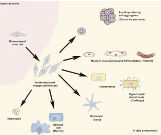
As previously mentioned, MSCs are able to differentiate in vitro and in vivo (Figure 1), mostly along, but not restricted to, the osteogenic, chondrogenic and adipogenic lineages (Chamberlain et al., 2007; Pittenger et al., 1999; Kode et al., 2009). Additionally, MSCs have been reported to home to areas of tissue injury, where they set-up a regenerative microenvironment and participate in tissue repair (Chamberlain et al., 2007; Fox et al., 2007; Sordi, 2009). MSC homing follows a mechanism similar to the one of leukocyte trafficking, even though not involving exactly the same cytokines, chemokines and adhesion molecules (Chamberlain et al., 2007; Fox et al., 2007). MSCs have also been described to possess anti-inflammatory and immunomodulatory properties that can affect multiple arms of the immune system (Chamberlain et al., 2007; Kode et al., 2009; Nauta and Fibe, 2007). These features increase the clinical interest in these cells, particularly in regenerative medicine and for tolerance induction in allogeneic transplantation.
Figure 1. Differentiation potential of MSCs. Pictorial representation of MSCs expanded in vitro can be
induced to differentiate into several lineages following exposure to specific differentiation factors. Image from Kode et al., Cytotherapy 2009, 11:377-91.
1.2. The Umbilical Cord as a stem cell source
Different MSC tissue sources may be especially suited for specific clinical applications as culture-expanded MSCs exhibit heterogeneity in their differentiation potential (Chamberlain
et al., 2007; Nekanti et al., 2010). Although BM-MSCs have been most commonly used in
differentiation studies, they are limited by the fact that the collection process is invasive, the number of cells obtained is limited and the quality of the cells in terms of proliferative and _____________________________________________________________________________ _
differentiation abilities decreases in aged donors (Kim et al., 2010).Therefore, in recent years there has been a lot of interest in the search for novel stem cell sources for specific clinical applications and the umbilical cord is one such source (Nekanti et al., 2010).
The Umbilical Cord (UC) constitutes the link between mother and fetus during pregnancy (Can and Karahuseyinoglu, 2007) and is usually discarded after delivery, thus providing an abundant stem cells source in a noninvasive and ethical way (Nekanti et al., 2010). The UC inner architecture (Figure 2) is composed of a set of two arteries (UCA) and one vein (UCV) and a surrounding matrix of mucous connective tissue, first described by Thomas Wharton in 1656, and thus named Wharton’s Jelly (WJ) (Can and Karahuseyinoglu, 2007). The WJ, which acts as a protective tissue of the UC vessels, comprises specialized fibroblast-like cells and occasional mast cells embedded in an amorphous ground substance rich in proteoglycans, mainly hyaluronic acid (Can and Karahuseyinoglu, 2007).
Figure 2. Pictorial representation of different regions of within the umbilical cord. The cord is divided
in 5 compartments: 1. umbilical cord blood, 2. umbilical vein subendothelium, 3. perivascular, 4. intervascular and 5. subamnion. Wharton’s jelly includes zones 3 through 5. Image from Troyer and Weiss. Stem Cells 2008, 26(3):591-9.
MSCs have been successfully isolated from UC WJ explants (Wang et al., 2004; Friedman et al., 2007; Karahuseyinoglu et al., 2007; Chen et al., 2009; Ishige et al., 2009; Fong
et al., 2011; Hsieh et al., 2010;), UC vascular endothelial surface (Romanov et al., 2003;
Sarugaser, et al., 2005;), UC vessels (Panepucci et al 2004; Ishige et al., 2009) and UC blood (Secco et al., 2009; Martins et al., 2009; Chang et al., 2006; Zeddou et al., 2010). They can also be obtained by processing the whole UC tissue (Zeddou et al., 2010, Secco et al., 2009). Two main methods have been described for isolating UC-MSCs: exclusively mechanical (Mitchell et
al., 2003; Chen et al., 2009; Ishige et al., 2009), or the combination of mechanical and one or
more enzymatic steps (Wang et al., 2004; Seshareddy et al. 2008; Karahuseyinoglu et al., 2007; Fong et al., 2007; Hsieh et al., 2010). Regarding UC blood (UCB), some researchers have successfully isolated MSCs from this source although there is great controversy in the literature regarding its efficiency and feasibility (Kim et al., 2010; Stronk et al., 2007; Martins et _____________________________________________________________________________ _
al., 2009; Choi et al., 2009). Despite the low proportion of MSC obtained from UCB when
compared to BM-MSCs, UCB-MSCs have been shown to exhibit a better performance in culture (Bieback et al., 2004; Yang et al., 2004; Sanchez-Ramos 2006). Nevertheless, more recently, Secco et al. (2008) have showed that UC stroma is much richer in MSCs than UCB. UC stroma MSCs have been reported to have several advantages compared to UCB MSCs and BM-MSCs (Troyer et al., 2008) such as a higher frequency of colony forming units (CFUs), which can be due to the expression of telomerase by UC stroma cells (Mitchell et al., 2003), and a lower immunogenicity (Weiss et al., 2008). Comparing UC stroma, UCA and UCV it was not conclusive which one was a better source (Ishige et al., 2009). Nevertheless, UCV cells appear to have a higher frequency of CFUs than UC and UCA. All these three tissue sources seem to have the ability to differentiate into chondrocytes, adipocytes and osteoblasts, but UC stroma was described by Ishige and colleagues as the one with the least osteogenic potential and UCA with the greatest (Ishige et al., 2009).
When compared with MSCs from BM, UC stroma and UC perivascular MSCs seem to proliferate better than the ones from the BM (Baksh et al., 2007; Hsieh et al., 2010). Regarding differentiation, UC perivascular MSCs were described to differentiate into osteoblasts more rapidly than BM-MSCs (Baskh et al., 2007). When compared to BM-MSCs, UC stroma cells were reported to be more committed to angiogenesis while BM-MSC preferentially undergo osteogenesis (Hsieh et al., 2010; Panepucci et al., 2004; Chen et al., 2009).
1.3. Clinical application of MSCs
As previously mentioned, thanks to their easy attainment, extensive in vitro proliferation, wide-range of differentiation potential, homing ability and immunological properties, MSCs are a promising tool in cell therapy, tolerance induction in allogeneic transplantation, and regenerative medicine (Bernardo et al., 2011; Kode et al., 2009; Krampera et al., 2006). For their application in regenerative medicine, stem cells must follow several criteria. These include: production of substantial numbers of cells, harvesting using a minimally invasive procedure, cell multilineage differentiation in a regulated and reproducible manner, safe and effective transplantation to either an autologous or allogeneic host, and manufacture in accordance with current Good Manufacturing Practice guidelines (Gimble et al., 2007).
Despite being a great promise for the future, until this date, there are few examples of clinical applicability of stem cells in the field of regenerative medicine. This is due to (Bajada et
al., 2008): difficulties with extensive in vitro cell expansion; cell apoptosis following implantation;
problems with vascularisation of the growing tissue; problems in finding viable scaffold biomaterials; high costs associated with prolonged maintenance of the cells in culture; and moral concerns or ethical issues that delay the transition for clinical use.
The increasing interest in the use of MSCs in tissue regeneration and the treatment of immune disorders leads to the need of developing production processes that are in accordance with Good Manufacturing Practices (GMPs). These regulations have the aim of ensuring the _____________________________________________________________________________ _
delivery of safe, reproducible, and effective products (Kode et al., 2009; Sensebé et al., 2011). And therefore cover all aspects of manufacturing (Sensebé et al., 2011). In Europe, MSCs are considered advanced therapy medicinal products (ATMPs), as defined by the European Regulation (Sensebé et al., 2011).
Regarding the procedures for the production of MCSs for therapeutical purposes, the main parameters to be considered are the sources and collection methods, extent of manipulation (preferentially in closed systems), methods for MSC enrichment, purity, culture medium, cell plating density, passaging, and time in culture, as all these may affect the quality of MSCs (Bourin et al., 2008; Sensebé et al., 2010; Sensebé et al., 2011). For these reasons, finding the optimal culture conditions has been of great interest to researchers.
In particular, the culture medium is crucial for the efficacy and safety of MSC cultures. The media used in culture processes should maintain the phenotype, genotypic stability, proliferation and functions of MSCs during multiple passages. Consensus is lacking on the ideal medium for MSC expansion, but the standard culture condition has been a basal medium supplemented with animal serum, typically at a concentration of 10%, with or without added growth factors. Numerous types of basal media have been tested, and one of the main issues, the glucose content, remains controversial as its effect might be dependent on the cell source or differ between short-term and long-term culture (Sotiropoulou et al., 2006; Weil et al., 2009; Pal
et al., 2009; Nekanti et al., 2010). Besides glucose, various other factors are important in the
culture medium such as additional aminoacids, vitamins, inorganic salts, etc. (Nekanti et al., 2010).
Regarding the serum, Fetal Bovine Serum (FBS) is the most commonly and successfully used. However, the animal and poorly defined nature of FBS, the batch to batch variability, the risk of sensitisation due to FBS proteins retained in the cytoplasm of MSCs, and the fact that it is a possible source of bovine prion, viral and zoonose contamination (Schallmoser et al., 2007; Chase et al., 2010) make it an undesirable component of the culture medium. Despite these hindrances, FBS can be used for GMP production of MSCs as long as it is carefully tested before use and a Transportation Security Administration certificate is obtained to ensure safety in terms of the risk of infectious disease transmission (Sensebé et al., 2011). Nevertheless, there still is an increased risk associated with the use of FBS as reported by Selvaggi and collegues that when it was infused into patients lymphocytes expanded in FBS, these patients developed hypersensitivity reactions (Selvaggi et al., 1997).
Nevertheless, there still is an increased risk associated with the use of FBS and some examples have previously been reported: alteration of cell behaviour and morphology in the presence of FBS in culture (Hung and Young 2006) and the development of hypersensitivity, malignant ventricular arrhythmias and in some cases sudden death in patients who received cells cultivated in FBS (Selvaggi et al., 1997; Chachques et al., 2004). Therefore, safety requirements imply replacing FBS with more secure products such as human components or a fully defined serum-free medium.
_____________________________________________________________________________ _
Replacing FBS with human or humanised components is one of the most challenging aspects of GMP translation in MSC production. The reduced efficiency of animal serum-free culture conditions may be overcome by supplementing the medium with growth factors and cytokines that activate signalling pathways crucial for MSC expansion while preserving their differentiation potential. Some of the growth factors already reported to have an essential role are the platelet-derived growth factor (PDGF), epidermal growth factor (EGF), transforming growth factor (TGF)-β, insulin-like growth factor (IGF), and fibroblast growth factor (FGF)-2 (Kilian et al., 2004; Choi SC et al., 2008; Ng et al., 2008; Chase et al., 2010; Tamama et al., 2010). However, the complete growth factor requirements for MSC expansion are still unknown. In other studies, human serum (hS), or in particular human AB serum (hABS), plasma (hP), platelet lysates (hPL) or thrombin-activated platelet releasate in plasma (tPRP) have also been tested as FBS replacements (Kocaoemer et al., 2007; Bieback et al., 2009; Doucet et al., 2005; Shahdadfar et al., 2005; Schallmoser et al., 2007; Strunk et al., 2007; Avanzini et al., 2009; Turnovcova et al., 2009). The most promising and most often used is the platelet lysate, human plasma enriched by growth factors released from platelets. Platelet granules contain many growth factor proteins, including PDGF, fibroblast growth factor (FGF), IGF, TGF-β, platelet factor 4 (PF-4), and platelet-derived epidermal growth factor (PDEGF) (van den Dolder et al., 2006; Doucet et al., 2005; Chevallier et al., 2010). These growth factors, which are released from platelet concentrates when subjected to freeze-thaw cycles, have been shown to successfully support MSC expansion in vitro (Doucet et al., 2005; Schallmoser et al., 2007; Müller et al., 2006). Regarding human serum, its successful use seems to be restricted to autologous serum (Yamamoto et al. 2003; Lin et al., 2005; Shahdadfar et al., 2005; Mizuno et
al., 2006), as allogeneic serum has produced contradictory results (Shahdadfar et al., 2005;
Kocaoemer et al., 2007). However, pooled human allogeneic serum would provide a more abundant and immediately available source and would thus be a more advantageous alternative than autologous serum to expand MSCs for clinical application (Kocaoemer et al., 2007; Mannello et al., 2006). Nevertheless, there still is a need to test the proliferation and functionality of MSCs grown in these media.
1.4. MSC multi-lineage differentiation
As defined by the ISCT, to be classified as MSCs, the cells must differentiate into osteoblasts, chondrocytes and adipocytes under appropriated conditions.
One of the most promising applications of MSCs in regenerative medicine is in bone repair (Krampera et al., 2006; Sensebé et al., 2010). The bone is a mesenchymal-derived tissue with various functions, including mechanical, metabolic and protective of vital organs (Clarke, 2008; Duplomb et al., 2007). It is composed of an organic mineralised connective tissue with two main cell lineages: osteoblasts and osteoclasts (Duplomb et al., 2007). Osteoblasts are mesenchymal-derived cells specialised in the production of organic matrix and in the _____________________________________________________________________________ _
mineralisation process (Jayakumar and Di Silvio, 2010). Osteoblast cells can progressively differentiate into osteocytes and these two cell types together constitute a cellular network that allows the communication between bone surface and mineral matrix (Clarke, 2008; Jayakumar and Di Silvio, 2010). Bone resorption is controlled by osteoclasts, which differentiate from hematopoietic precursors (Clarke, 2008; Duplomb et al., 2007; Jayakumar and Di Silvio, 2010). A balance between osseous tissue formation by osteoblasts and bone resorption by osteoclasts is required for bone homeostasis (Clarke, 2008; Jethva et al., 2009). The osseous organic matrix (the osteoid), produced by the osteoblasts, is mainly composed of collagen type I and II, fibronectin and growth factors. This matrix subsequently gets mineralised forming an inorganic matrix that consists of hydroxyapatite crystals (Clarke, 2008).
A good understanding of the osteoblast differentiation process is important for two main reasons: to allow the application of osteoblast committed-cells in regenerative medicine, and to identify new key genes that could be deregulated during pathological processes (bone tumour, osteoporosis, etc). Osteoblast differentiation is controlled by a hierarchy of transcription factors that are expressed in a defined temporal sequence, both in vivo and in vitro (Franceschi et al., 2009; Komori, 2006, Kalajzic et al., 2005) (Figure 3). During embryonic development the transcription factor Runx2/Cbfa1 (core binding factor α1) first appears with the formation of mesenchymal condensations in areas destined to become bone, and persists through subsequent stages of bone formation (Franceschi et al., 2009). Osterix (Osx) is a zinc finger-containing transcription factor, which is important for the commitment of osteoblast progenitors into the osteoblast lineage (Nakashima et al., 2002). In the absence of Osx, no cortical bone and no bone trabeculae were formed through either intramembranous or endochondral ossification (Nakashima et al., 2002). Activating transcription factor 4 (Atf4) appears to be important in later phases, and is expressed in mature osteoblasts (Franceschi et al., 2009). The activation of these transcription factors is accompanied by the expression of other specific proteins, which can be used as markers to identify the differentiation stage of osteoblasts (Kalajzic et al., 2005; Franceschi et al., 2009). Type I collagen and osteonectin (a calcium-binding glycoprotein) start being transcribed at the phase of bone matrix formation, following Atf4 activation. When the mineralization phase is initiated a large increase in osteocalcin (bone gamma-carboxyglutamic acid-containing protein) expression is observed (Franceschi et al., 2009). Additionally, bone sialoprotein (BSP) and alkaline phosphatase (AP) have also been described to be involved in the mineralisation process (Fauran-Clavel and Oustrin, 1986; Franceschi et al., 2009). Osteogenic assessement is usually performed by staining for AP activity, and in a later phase by Alizarin Red or von Kossa techniques that specifically detect for calcium deposition.
Figure 3. Hierarchy of transcription factors that control osteogenic lineage. Adapted from Franceschi
et al. Cell Tissues Organs 2009, 189:144-152.
The standard method to induce osteogenic differentiation has been to add combinations of dexamethasone (dex), β-glycerophosphate (β-GP), ascorbic acid (asc), and occasionally 1α,25-dihydroxy vitamin D3, to the culture medium in the presence of FBS (Jaiswal et al., 1997; Vater et al., 2011).
Dex is a synthetic glucocorticoid essential for osteoprogenitor cell differentiation (Vater
et al., 2010) but also to enhance cell growth both alone and in combination with β-GP and asc
(Coelho and Fernandes 2000). At an initial phase, this compound seems to induce AP activity and later, in the mineralising phase, increases the deposition of calcium (Coelho and Fernandes 2000; Porter et al., 2003). However, glucocorticoids have been described to cause bone fragility and bone loss, via the inhibition of bone matrix proteins such as type I collagens and osteocalcin (Iu et al., 2005). These contradictory observations suggest that the dex effect on osteogenic differentiation is dependent on both the dosage and the timing of treatment (Porter
et al., 2003; Iu et al., 2005, Park et al., 2010) and that dex is not required continuously, but only
at particular stages, to generate the osteoblast phenotype (Beloti and Rosa 2005; Park 2010). β-GP is enzymatically hydrolysed by alkaline phosphatase, serving as a crucial source of inorganic phosphate during matrix mineralisation (Chang et al., 2000) and it is almost entirely degraded during the first two weeks of culture (Coelho and Fernandes, 2000). When β-GP is added in combination with asc and dex causes an increase in AP activity (Park, 2010; Vater et
al., 2010), but on its own leads to a decrease in the activity of this enzyme (Park, 2010).
Additionally, β-GP seems to be the only of the three factors that by its own has the ability to form mineral deposits (Coelho and Fernandes, 2000).
Asc, or the more stable form, ascorbic acid 2-phosphate, has a mitogenic effect and is a cofactor for collagen hydroxylation (Vater et al., 2011), which is important for its synthesis and maturation (Takamizawa et al., 2004). In addition, asc appears to stimulate cell growth at any concentration (Takamizawa et al., 2004) with optimal maximum proliferation rate when at 250µM (Choi KM et al., 2008).
The osteogenic factors dex, β-GP and asc are generally used in combination with FBS and, as previously discussed, the use of FBS presents several disadvantages when aiming for a clinicalapplication. Therefore, there also is the need for replacing FBS by recombinant growth factors or human components during osteogenic differentiation. Several signalling pathways have been involved in osteoblast differentiation, including the Bone morphogenetic proteins (BMP), FGF, TGF-β, Notch and canonical Wnt pathways (Canalis, 2008; Engin and Lee, 2010; Komori, 2006; Rawadi et al., 2003; Valta et al., 2006; Yamaguchi et al., 1991; Zamurovic et al., 2004). In a number of studies, activators and inhibitors of these pathways have been added to the culture medium as a mean to induce or repress differentiation, hence allowing a better understanding of the differentiation process and revealing candidate growth factors to replace FBS.
In a different approach, Chevallier et al. (2010), have used platelet lysate, which contains a mixture of growth factors (including PDGF, FGF, IGF, TGF-β, PF-4 and PDEGF), and showed that it enhanced MSC expansion and replaced osteogenic agents during pre-osteoblastic induction. In this study, MSC cultured in platelet lysate showed spontaneous osteoblastic gene expression and enhanced in vivo osteogenic capacity during ectopic bone formation (Chevallier et al., 2010). Moreover, cells expanded in other animal serum-free supplements, such as with AB hS, autologous hS and hPLP, seem to maintain their osteogenic potential (Kocaoemer et al.,2007; Doucet et al., 2005; Lange et al., 2007; Yamamoto et al., 2003; Bernardo et al., 2006). Shahdadfar and colleagues observed that osteogenesis was sucessful in cells expanded with autologous hS, even though taking longer for this to be achieved (Shahdadfar et al., 2005).
Pre-clinical and clinical studies have been performed for several bone disorders, including the genetic disorders Osteogenesis Imperfecta, infantile hypophosphatasia and osteopetrosis, as well as the acquired disease osteoporosis (Horwitz et al., 1999; Arthur et al., 2009; Jethva et al., 2009). On the other hand, in vivo studies on animal model transplantation showed that MSCs represent a good tool to improve bone regeneration (Jones and Yang, 2011) having, for example, increased bone formation in rat femur (Bruder et al., 1998) and osteoporotic rabbit (Wang et al., 2006) models. Also, a clinical trial involving the injection of autologous osteoblasts cultured from bone-marrow precursors showed a significant fracture healing improvement (Kim et al., 2009a).
MSCs also have the ability to differentiate into chondrocytes. For chondrogenesis to be achieved it requires close cell-to-cell contact and the addition of chemical supplements. The close cell-to-cell contact is generally achieved by promoting MSC concentration forming a three-dimensional a micromass pellet. The cell pellet enhances the upregulation of extracellular cell matrix (ECM) interations, leading to the formation of pre-chondrocytes. After condensation, the cells become highly proliferative and change the ECM composition until reaching a mature chondroblast. Collagen type II is the most abundant fiber in this ECM, which is typical of articular cartilage. During this process, MSC fibroblastic shape changes to a large and round morphology. The three most important factors added to promote chondrogenesis are dex, asc _
(which are commonly use in osteogenesis) and TGF- β, but BMPs, FGF and IGF are also commonly added. In animal serum free conditions, chondrogenic differentiation seems to be also maintained in the presence of autologous hS and hPLP, as it was for osteogenesis (Shahdadfar et al., 2005; Lange et al., 2007; Doucet et al., 2005). MSC differentiation into chondrocytes would allow their use in the treatment of several pathologies such as pain in the joints, which is mostly due to cartilage degeneration (Vater et al., 2010; Nesic et al., 2006) and avoid the need for orthopaedic surgery with all its complications and side effects (Koga et al., 2009; Vater et al., 2010).
Finally, MSC are also able to differentiate into adipocytes. Adipocyte differentiation begins with the commitment of MSCs into pre-adipocytes that contain small lipid-rich vacuoles (Frith and Genever 2008; Can and Karahuseyionoglu et al., 2007). Subsequently, these cells develop into mature adipocytes, when, eventually, the lipid vacuoles fuse and fill the cells (Chamberlain et al., 2007). Along this process fibroblastic MSCs are converted to a spherical morphology (Vater et al., 2010.) To induce adipogenesis it is normally use dex, 3-isobutyl-1-methyl-xanthine (IBMX), insulin and indomethacin are normally used. Some researchers additionally use triiodothyinine (T3), Asc-2-P and bFGF-2 (Vater et al., 2010). Upon induction the cells express specific adipocyte markers such as peroxisome proliferation-activated receptor γ2, lipoprotein lipase, and the fatty acid-binding protein aP2 (Pittenger et al., 1999; Chamberlain
et al., 2007). Adipogenesis evaluation is performed histologically using the Oil Red dye, which
marks the lipids in the vacuoles (Can and Karahuseyionoglu et al., 2007; Vater et al., 2010). The adipogenic differentiation potential of MSCs expanded in animal serum-free conditions is controversial as some authors argue that MSCs growing hPLP supplemented in culture media retain adipogenesis ability (Bernado et al., 2006; Doucet et al., 2005), whereas others show contradictory results (Lange et al., 2007). As for the other mesenchymal lineages, adipocyte differentiation from MSCs would have important clinical applications following procedures or pathologies where large volumes of adipose tissue are lost (Gomillion and Burg 2006; Vater et
al., 2010).
1.5. Human Amniotic Membrane
The promising use of MSCs in the field of regenerative medicine is undeniable, either through the local implantation of in vitro expanded autologous MSCs or of a pre-differentiated tissue matrix loaded onto suitable scaffolds.
The ideal scaffold is expected to be biocompatible and should mimic the structure and biological function of native extracellular matrix. Active research is under way for the development of improved scaffolds, using a variety of natural and synthetic materials and advanced technologies (Sittinger et al., 2004; Tsigkou et al., 2009; Nöth et al., 2010). The human amniotic membrane (AM) is a good candidate for this application. The AM has as primary function the protection of the embryo during its development. Therapeutically, this membrane has been successfully used as a “biological dressing” over more than one hundred _____________________________________________________________________________ _
years in cutaneous wounds and burns (Lo and Pope, 2009). However, it was only later that the great potential of the AM in ophthalmological, urological, gynaecological and neurological fields was realised (Tan et al., 2009; Burman et al., 2004; Sharifiaghdas et al., 2009; Ward et al., 1989). The AM has recently been used in some studies as a substrate for MSC expansion and differentiation along different lineages with the aim of subsequent transplantation. Among other studies, Tan et al. (2011) have reported the successful chondrogenic differentiation of MSCs on AM for use as a cell delivery vehicle, Kim and colleagues showed that grafts of AM loaded with MSCs play an effective role during the healing of skin defects in rabbits (Kim et al., 2009b) and, in another report, a layer of somatic epidermal adult stem cells was cultivated and carried on AM into the ocular surface of a goat model of limbal stem cell deficiency (Yang et al., 2008).
Importantly, a variant of the AM that is not rejected after allogeneic transplantation is currently prepared at the Tissue Bank of the Centro de Histocompatibilidade do Sul. During the preparation of this AM, apoptosis of the cells in the membrane is induced turning it immunologically inert, while preserving the tissue integrity. Also, the AM contains a variety of cytokines and growth factors (Koizumi et al., 2000) which may be responsible for its intrinsic regenerative and antiinflamatory properties (Fernandes et al., 2005; Insausti et al., 2010; Velez
et al., 2010; Samandari et al., 2011). The high levels of growth factors such as EGF, bFGF,
keratinocyte growth factor (KGF) and hepatocyte growth factor (HGF), support epithelialization and regulation of fibrosis and neovascularization process (López-Valladares et al., 2010). López-Valladares and colleagues have shown that there are growth factor variations depending on the age of AM donors and the gestational stage, as mothers under 35 years old with 38 to 39 weeks of pregnancy have higher levels of growth factors (López-Valladares et al., 2010). Although it has not been proven, it is expected that higher levels of growth factors produce better clinical results (Koisumi et al., 2000; López-Valladares et al., 2010). The AM is thus a promising substrate for MSC attachment and differentiation and a safe and effective vehicle for transplantation.
_____________________________________________________________________________ _
2. Materials and Methods
2.1. Isolation of MSCs from different parts of UC
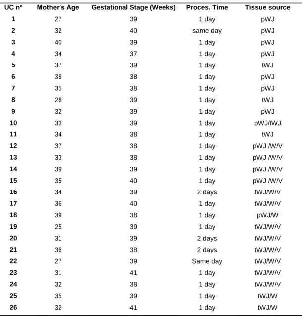
In this study, MSCs were isolated from different parts of 26 human umbilical cords using a variety of methodologies. The UCs were obtained with written informed consent of the parturients from elective caesareans. Human umbilical cords were first washed in Hanks’ balanced salt solution (GIBCO; 14175-129) with antibiotics (Penicillin-Streptomycin and Gentamicin) and then processed for MSC isolation. This was accomplished using different combinations of mechanical and/or enzymatic and mechanical methods in search for an optimal one (see supplementary Table 1). MSCs were obtained from WJ (peripheral and total), vessels and whole cord. For the isolation of peripheral Wharton’ Jelly (pWJ) ), pieces of mesenchymal tissue were cut around the vessels from the outside part of the UC. MSCs from total Wharton’s Jelly (tWJ) were obtained by cutting open UC and subjecting the inner part to an initial 1h of enzymatic digestion for removal of the arteries and veins to isolate the WJ. The pWJ and tWJ mesenchymal tissue was then cut into small fragments and directly plated or submitted to a 4 to 16h enzymatic digestion. The arteries and veins obtained by this method were also cut into small pieces and submitted to a 4h enzymatic digestion to isolate MSCs from vessels (V-UC). To obtain the MSCs from whole parts of umbilical cord (W-UC) a cord piece with 2-3cm was cut into small fragments and submitted to a 4h enzymatic digestion. The enzymatic solution used for tissue digestions was composed of 0.1-2 mg/ml collagenase XI (Sigma; C9697) alone or with 100U/ml hyaluronidase (Sigma; H2126). In some cases, a subsequent digestion with Tryple Express® (a trypsin replacement from Invitrogen; 12604-013) was performed. The dissociated cells were then washed in Dulbecco’s phosphate buffered saline (DPBS - GIBCO; 14190-169), resuspended in MSC/FBS medium or in StemPro® MSC SFM medium (GIBCO; A10332-01) and cultured on tissue culture plastic plates at 37ºC in a 5% CO2 humidified
incubator.
2.2. MSC in vitro culture
For routine in vitro MSC expansion, cells were cultured in MSC-FBS medium on tissue culture plastic flasks (Greiner; C6481-200EA) at 37ºC in a 5% CO2 humidified incubator. MSC/FBS medium was composed of DMEM-F12 (1:1) (GIBCO; 21331-020), 2mM L-Glutamine (GIBCO; 25030-024), 15mM HEPES (GIBCO;15630-056), 100U/ml penicillin (GIBCO; 15140-122), 100μg/ml streptomycin (GIBCO; 15140-15140-122), 5 μg/ml Gentamicin (GIBCO; 15710-049) and 10% MSC-qualified Fetal Bovine Serum (FBS – GIBCO; 12662-029). Every 2-3 days, approximately half of the medium was replaced by fresh medium and when reaching approximately 80% confluence the cells were passaged by dissociation using Tryple. The cells were then counted using a Neubauer hemocytometer and replated at 0.5 or 1x10⁴cells/cm2. For low confluence experiences cells were replated at 200cells/cm2 in 75-cm2 flasks (Greiner; _____________________________________________________________________________ _
C7106-120EA) MSC-FBS medium. Low passage MSCs were also frozen for later use in DMEM-F12 containing 10% tissue-culture grade dimethyl sulfoxide (DMSO, Merck; K28452252-101) and 10% FBS, kept at -80ºC for a few days and then stored in liquid nitrogen.
2.3. Colony forming units (CFUs) assay
For CFUs analysis, cells were plated at 200cells/cm2 and cultured in petri dish (Nunc; 150350) in MSC-FBS medium. The medium was replaced twice a week and cultures were stopped at day 14. Colonies were washed with PBS, fixed with 0.05% glutaraldehyde (Sigma; G6257-10X10ML) for 5min and stained with 0.5% aqueous crystal violet (Sigma; C0775-25G). The colonies were then washed 3 times with PBS and the colonies were counted. All experiments were performed in triplicates.
2.4. Immunofluorescence 2.4.1. Flow Cytometry
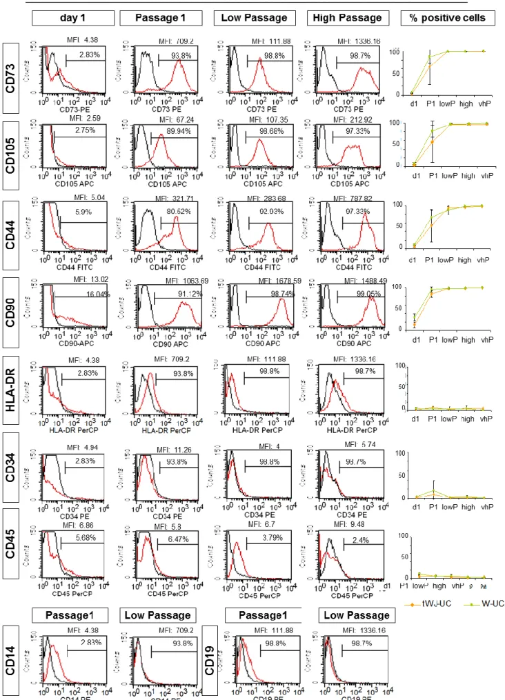
For the immunophenotypic characterization of UC MSCs, the cells were analysed for the presence of the surface markers CD90, CD73, CD105, CD44, CD34, CD45, CD19, CD14 and HLA-DR using the specific fluorochrome-conjugated monoclonal antibodies listed on table 2. Briefly, the cells were dissociated into a single cell suspension and approximately 1x10⁵ cells were incubated with the appropriate monoclonal antibodies (see table 2) in 0.5%BSA/PBS (Sigma;A7284-50ML) for 1 hour in the dark. Following this incubation, the cells were washed in 2mL of PBS and centrifuged. The pellet was then resuspended in PBS and analysed by flow cytometry in a FACSCalibur instrument with CellQuest software (both BD Biosciences).
Table 2: List of monoclonal antibodies used for MSC immunophenotyping.
Antigen Antibody Clone Isotype Fluorescence Brand
CD34 8G12 Mouse IgG1, κ PerCP-Cy5.5 BD Biosc.
CD34 8G12 Mouse IgG1, κ PE BD Biosc.
CD19 HD37 Mouse IgG1, κ PE BD Biosc.
CD14 MφP9 Mouse IgG1, κ PE BD Biosc.
HLA-DR L243 Mouse IgG1, κ PerCP BD Biosc.
CD44 IM7 Rat IgG2b, κ FITC BioLegend
CD44 IM7 Rat IgG2b, κ PE-Cy7 BioLegend
CD45 HI30 Mouse IgG1, κ PerCP BioLegend
CD73 AD2 Mouse IgG1, κ PE BioLegend
CD90 5E10 Mouse IgG1, κ APC BioLegend
CD105 43A3 Mouse IgG1, κ FITC BioLegend
CD105 43A3 Mouse IgG1, κ APC BioLegend
bAP 0.G.2 Mouse IgG1, κ FITC Abcam
TNAP B4-78 Mouse IgG1, κ FITC Santa Cruz
Biotechnology, inc
_____________________________________________________________________________ _
Osteocalcin 190125 Mouse IgG1, κ PE R&D System
2.4.2. Fluorescence Activated Cell Sorting (FACS)
The population enrichment in for CD105low and CD105high or CD105 low /CD90low and CD105high/CD90high MSCs was performed by FACS sorting. The cells expanded in MSC/FBS medium were dissociated using Tryple and the cell suspension were passed through a 50µm cell strainer (Partec; 04-004-2327) to obtain a single cell suspension and approximately 1x10⁶ cells were incubated with CD105 and CD90 monoclonal antibody (see Table 2) in PBS/0.5%BSA for 1 hour in the dark in a sterile environment. Following this incubation, the cells were washed in 2mL of PBS and centrifuged at 1400rpm. The resuspended pellet in PBS was passed through FACSAria instrument (with FACS Diva software) (both BD Biosciences) for sorting of the populatons of interest. Then, CD105low(/CD90)low and CD105high(/CD90)high MSC populations were gated and sorted directly to fresh culture medium containing antibiotics. The sorted populations were then plated into tissue culture plates with 1mL of MSC/FBS medium.
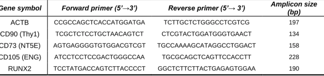
2.5. Gene Expression Analysis
The gene expression of markers of MSCs and of different stages of osteoblastic differentiation was analysed by RT-PCR. With this purpose, cells were dissociated, pelleted and immediately stored at -80ºC until RNA extraction. RNA was extracted using the TRIzol reagent (Invitrogen; 15596-026) or the RNeasy mini kit (Qiagen; 74104) in combination with the in column DNase digestion set (Qiagen; 79254), and quantified using a Beckman Coulter DU 530 UV/Vis spectrophotometer or NanoDrop® 2000 spectrophotometer. The extracted RNA was run on a 1.8% agarose gel to verify its quality and integrity. The RNA was then reverse transcribed using the SuperScript III Reverse Transcriptase system (Invitrogen; 18080-129). 1μg of total RNA, 1 μl of 50 μM random primers (Applied Biosystems) and RNase free water (Sigma; 95284-100ML) were mixed to a final volume of 10 μl. After a 5 minute incubation at 65°C the samples were put on ice and a mix containing 0.5mM Deoxynucleotide set (Sigma; DNTP100-1K), 5 mM DTT, 2 U/μl RNaseOUT RNase inhibitor (Invitrogen; 10777-019) and 10 U/μl Superscript III in first strand buffer was added. A reaction mixture without the enzyme was also set up as a negative control (designated “-RT”). The mixture was incubated at 25°C for 5 min, 50°C for 1h and at 70°C for 15 min. The cDNAs were diluted 1:10 in dH2O and kept at -20°C until PCR analysis.
For RT-PCR amplification a reaction mix was set up as follows: 100 ng of cDNA were added to 10 μmol of each primer (Eurofins MWG Operon), 0.2 mM dNTPs, 1 unit of AmpliTaq DNA Polymerase and PCR Buffer (Applied Biosystems; N8080166), in a total volume of 30 μl. Negative controls included the –RT samples and the non-template control (containing dH2O
instead of cDNA). PCR was performed on a GeneAmp PCR System 2720 (Applied Biosystems) and PCR conditions included an initial denaturation step at 94ºC for 5 minutes, followed by 35 _____________________________________________________________________________ _