ACTA RADIOLÓGICA PORTUGUESA May-August 2019 Vol 31 nº2 5-9
Breast Cancer in the Eldery
O Cancro da Mama em Idade Avançada
João Abrantes1, Carolina Carneiro2, Bernardete Rodrigues3, Raquel Lameiras4,
José Carlos Marques5
1Centro Hospitalar Barreiro Montijo - Hospital
Nossa Senhora do Rosário, Barreiro, Portugal
2Centro Hospitalar Universitário do Algarve,
Portugal
3Centro Hospitalar Tondela-Viseu, Portugal 4Hospital Garcia de Orta
5Instituto Português de Oncologia de Lisboa
Francisco Gentil, Portugal
Address João Abrantes Serviço de Imagiologia
Centro Hospitalar Barreiro Montijo - Hospital Nossa Senhora do Rosário
Av. Movimento das Forças Armadas 79C 2834-003 Barreiro, Portugal
email: [email protected]
Resumo
Introdução: O cancro da mama representa uma
das principais causas de morte a nível mundial, porém, apesar da tendência de aumento da incidência desta patologia com o avançar da idade, persiste uma lacuna na adequada caracterização desta doença nas mulheres com idades mais avançadas. Objetivo: Caracterização clínica, imagiológica e histológica da patologia tumoral mamária em mulheres de idade igual ou superior a 80 anos e comparação com uma população representativa de idades inferiores.
Métodos: Estudo unicêntrico retrospetivo de
dados clinico-epidemiológicos, imagiológicos e histopatológicos de mulheres com idade igual ou superior a 80 anos, submetidas a estadiamento por ressonância magnética mamária e comparação com uma população representativa de mulheres com idades inferiores a 80 anos.
Resultados: As doentes mais idosas
apresentam-se com doença em estádios mais avançados, existindo uma diferença estatisticamente significativa em relação ao estadiamento clínico entre os dois grupos (P-Value = 0.004). As mulheres com idades inferiores a 80 anos têm maior prevalência de carcinoma ductal in situ (P-Value = 0.02), com maior extensão de doença nestes casos (P-Value = 0.025). Os casos de CDIS em mulheres com idade avançada estão mais frequemente associados com positividade para os recetores de estrogénio (P-Value = 0.03). Existe uma concordância moderada entre o estadiamento por ressonância magnética e o estádio patológico tanto no grupo das mulheres com idade superior ou igual a 80 anos (Kappa=0.50) como no grupo das mulheres em faixas etárias inferiores (Kappa=0.55).
Conclusão: O cancro da mama nas mulheres
com idade avançada apresenta características diferentes da patologia tumoral mamária em doentes de faixas etárias inferiores.
Palavras-chave
Neoplasias da mama; Carcinoma ductal in situ; Carcinoma ductal; Imagem por ressonância magnética.
Abstract
Background: Breast cancer is a leading cause
of death worldwide, and despite consistent observation that cancer incidence increases with advancing age, there still remains a gap in the adequate characterization of this disease in elderly women. Purpose: Characterization of the clinical, imaging and histological features of breast cancer in women aged 80 years or older and comparison with features from a representative group of younger women.
Methods: Single-center retrospective analysis of
clinical, imaging and histopathological findings of women aged 80 years or older submitted to magnetic resonance imaging and comparison with a representative population of women under 80.
Results: Older patients have more advanced
disease at presentation, with a significant difference in the clinical stage distribution between the two groups (P-Value = 0.004). Younger patients have a higher prevalence of ductal carcinoma in situ (P-Value = 0.02), with more extensive DCIS disease (P-Value = 0.025). DCIS in patients of 80 years or older is more frequently associated with positive estrogen receptors (P-Value = 0.03). Moderate agreement was found in the concordance between the clinical stage according to breast MR imaging and the pathological stage in patients of 80 years or over (Kappa = 0.50) and in younger aged patients’ group (Kappa = 0.55).
Conclusion: Breast cancer in elderly women
presents different characteristics from those in younger aged patients.
Keywords
Breast neoplasms; Carcinoma intraductal noninfiltrating; Carcinoma ductal; Magnetic resonance imaging.
Original Article / Artigo Original
Introduction
Cancer is a leading cause of death worldwide, with breast cancer being the most commonly occurring cancer in women globally. Despite consistent observation that the cancer incidence increases with advancing age, there still remains a gap in the adequate characterization of this disease in elderly women.
This study aims to characterize the clinical, imaging and histological features of breast cancer in women aged 80 years or older and comparing them with features from a representative group of younger women.
Material and methods
Inclusion criteria for the study were patients with histologically confirmed breast cancer under staging by
6
magnetic resonance (MR) imaging. This retrospective study included a total of 240 women, comprising 87 women of 80 years or older who performed MR staging between January 2016 and December 2017 and 153 women under the age of 80, corresponding to all the female patients in this age group who underwent cancer staging by breast MR, in the first semester of 2016.
The following clinical, imaging and histopathological findings were analyzed and compared between the two groups of patients: age, tumor size and extension, lymph node status, presence of metastatic disease, multifocality and multicentricity, histological type, degree of differentiation, immunohistochemical characteristics, molecular subtype and cellular proliferation status. The tumor staging determined by MR was compared with the pathological staging (in patients submitted to radical or conservative surgery).
Statistical analysis
Statistical analysis was performed using the computing environment R (R Core Team, 2014). Categorical variables were compared using the chi-square test, while numeric variables were compared with a t-test, or with the Wilcoxon test, in case of non-normality. The agreement between radiological and pathological stage was measured using the Cohen’s kappa coefficient.
Results
In the 240 women included in the present study, fourteen patients had bilateral breast tumors at staging, with a total of 254 tumors evaluated. The mean age of the oldest group was 83.6 years (minimum 80, maximum 94) and 57.6 years (minimum 27, maximum 79) in the younger group. In terms of classification by histological type, invasive ductal carcinoma not otherwise specified was the most prevalent in both age groups, representing 76.4% of the cases in women under 80 and 72.2% of the cases in the older group. Specific forms of invasive carcinoma were identified in 12.1% and 20.6% of the cases, among which invasive lobular carcinoma was the most frequent, present in 5.1% of the cases of women under 80 years and 6.2% of the younger population group. Ductal carcinoma in situ (DCIS) was the histological diagnosis in 11.5% and 7.2%, with a statistically significant predominance (P-Value = 0.02) in the population of less than 80 years.
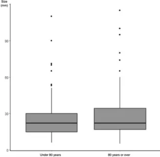
The cases of invasive tumors (Figure 1) had an average size of 28.2 mm in women aged 80 years or older, with the mean tumor size in the group of younger women being 24.3 mm, revealing a trend, albeit not statistically significant for the sample size (P-value = 0.145), of more extensive disease in the older aged group of patients. The tumor extension of DCIS cases (Figure 2) shows a statistically significant difference (P-Value = 0.025), with more extensive disease in the younger group of patients (mean size of 31.3mm) than in the group of women aged 80 years or older (mean size of 15.6mm).
The majority of the cases of older women were in the initial stages of the disease (stage IA 26.7%, stage IIA 44.2%, stage IIB 10.5%) and presented stage III in 12.1% of the cases. Metastatic disease was documented in only 3 cases. In the study group of women younger than 80 years, most cases represented disease in the earliest stages of the disease (IA stage 35.3%, stage IIA 21.6%, stage IIB 15.7%), with a
single case of metastatic disease, representing a statistically significant difference in the distribution of stages between the two groups (P-Value = 0.004).
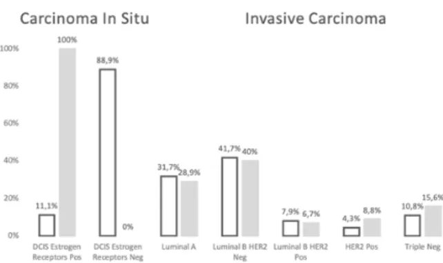
Most cases of invasive tumors were moderately differentiated carcinomas (G2), with undifferentiated disease (G3) representing the second most frequent tumor grade found in both groups. Hormonal positive disease (estrogen and progesterone receptor positivity) was predominant, with no statistically significant differences being documented in the histological and immuno-histochemical characterization of both groups (Table 1). The molecular subtype distribution of invasive and in situ carcinomas according to the 2013 St. Gallen consensus was evaluated (Figure 3), with a significant difference found (P-Value = 0.03) between the two groups regarding the prevalence of DCIS with and without estrogen receptors. In relation to invasive tumors, there is no difference in the distribution between the two groups, and there was an overall predominance of carcinomas with Luminal B HER2 Negative subtype.
Figure 1 – Invasive carcinoma dimensional distribution.
The largest tumor diameter represents a crucial prognostic factor in breast cancer patients,12 and we found that the
older age group had a tendency to present in the clinical staging, by breast MR, larger tumors than younger patients, findings that are supported by the available scientific evidence.13–15
Regarding DCIS size we found the inverse relationship, in patients younger than 80 years, presenting larger carcinoma in situ (31.3mm vs 15.6 mm). The literature does not provide adequate data regarding DCIS extension in elderly women, having most studies focused on the management and treatment of these patients, namely the use of isolated surgery versus surgery and radiotherapy in the cases of low grade and smaller size DCIS.16
Regarding the histologic type, invasive ductal carcinoma not otherwise specified is the most prevalent type of tumor diagnosed (72.2% of the cases in the older age group), with tumors having special histological features identified relatively often (20.6%). However, we found a lower percentage of lobular carcinomas (5.1% of the cases) when compared to the available data in the literature, ranging up to 17% of the cases as described by Fisher et al. in a cohort of 382 women over 70 years of age.6 Papillary carcinoma
was the histological diagnosis in 4 women aged 80 or older in our study (4.5%), representing a rare histological subtype with a favorable prognosis and with a reported higher incidence in the elderly.17,18 Another subtype with favorable
prognosis is the mucinous carcinoma, which was found in 4.5% of the patients in the older age group of our cohort, a lower percentage when compared to the reported incidence in other studies of women in this age group, which ranges from 4.9% to 16%.11,19
The histological grade of the tumor is regarded as a crucial prognostic factor, with correlation to the disease free and overall survival times.3 Our study did not find statistical
differences between both age groups regarding tumor differentiation, the majority of the cases consisting of moderately differentiated carcinomas (G2), representing 64.4% of the cases of older aged women, findings in line with other studies published.11,14 Regarding undifferentiated
disease, we found a total of 21 cases (23.3%) in women aged 80 or older, and the results are not homogeneous in the literature, with some reporting a lower prevalence of higher grade carcinoma (16.8% in the study by Chatzidaki et al.) and others finding a higher number of cases of undifferentiated breast carcinoma, ranging from 28% to 37%.11,13,20
Regarding hormone receptor status, multiple studies have shown that older patients are more likely to have hormone receptor-positive breast carcinoma,5,10,21,22 and
that Estrogen Receptor (ER) positive and Progesterone Receptor (PR) positive disease leads to a lower risk of breast cancer-specific mortality compared to ER-negative and PR-negative tumors.23 In our study, ER and PR status
was evaluated in all patients of the advanced age group with invasive carcinoma, with the majority being ER-positive (76%) and PR-ER-positive (55,6%). Similar incidence In terms of imaging, the statistical analysis of the
concordance between the clinical stage according to breast MR imaging and the pathological stage (in patients submitted to radical or conservative surgery) revealed a moderate agreement in both the group of women with 80 years or older (Kappa = 0.50) and the younger aged patients group (Kappa = 0.55).
Discussion
The Portuguese population is aging and in 2080 the number of Portuguese individuals aged 65 years and older is projected to be 3.3 million (more than 50% of the estimated 2.1 million in 2016).1
Although breast cancer incidence peaks in women aged 70–79 years (452 cases/100,000 women in the United States of America), a fifth of all new breast cancer cases occurs in the older women group (aged 75 years or older). A 75 year old woman of average risk who has been breast cancer free, still has a 5.2% probability of developing breast cancer during the rest of her lifetime.2
Breast cancer represents a heterogeneous disease, with multiple distinct subtypes identified according to the different pathological, biological, and clinical characteristics presented.3,4
The issue of breast cancer characterization in patients of advanced age is a controversial topic, with some authors stating that it has a tendency to show less aggressive features, but the lack of scientific data regarding the clinical, imaging and histological characteristics of breast carcinoma in the very elderly female population (80 years old or older) hinders the ability to produce adequate statistical analysis and conclusions in this setting.5–7
When compared to younger aged women, breast cancer in older women exhibits some different histological and biological characteristics, with a variety of combinations of favorable and less favorable features, as shown in our work and in agreement to other studies already published.8,9
The largest number of tumors in the older aged group were invasive carcinomas (92.8%), while only 7.2% of the cases represented carcinoma in situ, in accordance to reports of reduced incidence of DCIS in elderly women.10,11
Age Group
Tumor differentiation Estrogen Receptor Progesterone Receptor HER2 Ki-67 G1 G2 G3 N/A Pos. Neg. N/A Pos. Neg. N/A Pos. Neg. N/A High Low N/A
< 80 21 88 23 7 114 21 5 82 52 5 17 117 5 81 58 5
> 80 10 58 21 1 68 22 - 50 40 - 14 76 - 56 34
-Figure 3 – Molecular subtypes of invasive carcinoma and DCIS.
8
of ER positivity was previously reported on women aged 75 years or older (74%)24 and ≥80 years (72% and 76%).7,11
Although no statistical difference was found between immunohistochemical characterization of both groups in invasive tumors, a study including more than 10,000 patients, showed a higher incidence of ER-positive tumors in women aged over 50 years (81%) when compared to women under 50 years (63%).25
Many authors reported that older women initially present more advanced tumor stages,26 and we found that the group
of women aged 80 years or older presented higher tumor stages (44.2% - stage IIA) more frequently when compared to the younger population studied (35.3% - stage IA). A study by Lyman et al.21 also showed that women of older
age (65 years or older) were more likely to have advanced stage cancer when compared to female patients with less than 65 years. A crucial factor associated with earlier staging at the time of diagnosis is the use of mammography, whose benefit proves true even for the elderly women.27–29 The use
of screening mammography leads to a reduction of breast cancer mortality by detecting breast cancers when tumors are small and node negative, with pooled estimates from randomized controlled trials revealing a reduction of at least 20% in mortality in women aged 74 years or younger,30
and a meta-analysis of European observational studies showing a decrease in breast cancer mortality by 38–48% in women who actually were screened.31 Regarding older aged
women, a cohort of more than 12000 women found that patients with breast cancer aged 80 years or older were 0.37 times less likely to have an advanced stage at presentation for each previous mammogram performed.27
Regarding the performance of mammography in this group of patients, screening exams have a higher detection rate for invasive cancer when compared to younger women, aspects related both to the normal decrease of breast density with age and higher incidence of breast tumors in the elderly.32
The sensitivity and specificity of screening mammography is significantly higher in older women when compared to the lower aged patients and they experience fewer false-positives in mammography and biopsies.33
Albeit the use of screening programs with mammography is of renown scientific validity, there is a lack of consensus in the multiple professional societies and organizations regarding the optimal interval between examinations, the adequate timing to initiate and when to discontinue the screening.34 The existence of compelling data regarding the
potential to decrease breast cancer morbidity and mortality in older women, with studies showing that screening performance improves with age,35 improves prognosis
and 10-year survival in women 75 years old and older36
and improves life expectancy in older women without severe comorbidity,37 are compelling arguments for the
termination of any specific upper age limit in screening mammography programs.
Regarding preoperative staging of breast cancer, all the patients enrolled in the present study were submitted to breast MR evaluation and statistical analysis revealed a moderate agreement of the concordance between imaging and pathological staging, in both the group of patients of 80 years or older and the group of younger women. The use of MR imaging in the breast evaluation is a well-researched subject, with proven high sensitivity in breast cancer diagnosis and screening, and the use of MR is increasingly being evaluated and used in the preoperative staging of breast cancer. A systematic review and meta-analysis of the accuracy of MR imaging in the detection of breast cancer multifocal (MF) and multicentric (MC) extension showed that MRI detects additional disease in the same breast in 16% of women, leading to an adequate surgical planning, with the pool of studies demonstrating the use of more extensive local surgery in 11.3% of patients and conversion to mastectomy in 8.1% of all patients with MF/MC disease, although the establishment of the clinical value of detecting additional disease in these patients is still under evaluation.38
Adequate evaluation of regional lymph node status is of paramount importance for staging, treatment planning, and prognosis. The use of MR imaging for breast cancer staging allows for evaluation of nodal involvement, not only in the axillary region but also in the detection of internal mammary and supraclavicular adenopathy. The impact of MR staging in the management of these patients is very high, allowing for adequate guidance of biopsy in abnormal axillary lymph nodes and confirmation of internal mammary nodal metastases, leading to an optimal staging and therapeutic planning.39
Conclusion
Women aged 80 years or older are more likely to present more advanced initial stage disease and tend to have larger invasive carcinomas in the staging exams. Ductal carcinoma in situ is less prevalent and shows smaller dimensions in the group of patients of 80 years or older, more often presenting positive estrogen receptors.
There are no significant differences regarding the tumor biological profile/molecular subtypes of invasive carcinomas between the study groups and a moderate agreement was found between the radiological staging by MR imaging and the pathological staging in the women submitted to surgical treatment.
Older patients with breast cancer lack adequate representation in the current medical literature and additional studies are required in order to clarify the different characteristics of breast cancer in this population.
Received /Recebido 28/01/2019 Acceptance / Aceite 11/03/2019 Ethical disclosures / Divulgações Éticas
Conflicts of interest: The authors have no conflicts of interest to declare. Conflitos de interesse: Os autores declaram não possuir conflitos de interesse. Financing Support: This work has not received any contribution, grant or
scholarship.
Suporte financeiro: O presente trabalho não foi suportado por nenhum
subsídio ou bolsa.
Confidentiality of data: The authors declare that they have followed the
protocols of their work center on the publication of data from patients.
Confidencialidade dos dados: Os autores declaram ter seguido os protocolos
do seu centro de trabalho acerca da publicação dos dados de doentes.
Protection of human and animal subjects: The authors declare that the
procedures followed were in accordance with the regulations of the relevant clinical research ethics committee and with those of the Code of Ethics of the World Medical Association (Declaration of Helsinki).
Protecção de pessoas e animais: Os autores declaram que os procedimentos
responsáveis da Comissão de Investigação Clínica e Ética e de acordo com a Declaração de Helsínquia da Associação Médica Mundial.
References
1. Projeções de População Residente 2015-2080. Instituto Nacional de Estatística 2017.
2. Howlander N, Boone A, Krapcho M. SEER cancer statistics review, 1975–2014. National Cancer Institute. 2017.
3. Elston CW, Ellis IO. Pathological prognostic factors in breast cancer. I. The value of histological grade in breast cancer: experience from a large study with long-term follow-up. Histopathology. 2002;41:154-61. 4. Ellis IO, et al. Pathological prognostic factors in breast cancer. II. Histological type. Relationship with survival in a large study with long-term follow-up. Histopathology. 1992;20:479-89.
5. Daidone MG, Coradini D, Martelli G, Veneroni S. Primary breast cancer in elderly women: biological profile and relation with clinical outcome. Crit. Rev. Oncol. Hematol. 2003;45:313-25.
6. Fisher CJ, et al. Histopathology of breast cancer in relation to age. Br. J. Cancer. 1997;75:593-6.
7. Evron E, et al. Breast cancer in octogenarians. Cancer. 2006;106:1664-8. 8. Wildiers H, et al. Management of breast cancer in elderly individuals: recommendations of the International Society of Geriatric Oncology. Lancet Oncol. 2007;8:1101-15.
9. Witherby SM, Muss HB. Update in medical oncology for older patients: focus on breast cancer: management of early breast cancer. Cancer J. Sudbury. 2005;11:506-17.
10. Cheung KL, et al. Pathological features of primary breast cancer in the elderly based on needle core biopsies--a large series from a single centre. Crit. Rev. Oncol. Hematol. 2008;67:263-7.
11. Chatzidaki P, Mellos C, Briese V, Mylonas I. Does primary breast cancer in older women (≥80 years) have unfavorable histological characteristics? Arch. Gynecol. Obstet. 2011;284:705-12.
12. Carter CL, Allen C, Henson DE. Relation of tumor size, lymph node status, and survival in 24,740 breast cancer cases. Cancer.1989;63:181-7. 13. Gennari R, et al. Breast carcinoma in elderly women: features of disease presentation, choice of local and systemic treatments compared with younger postmenopasual patients. Cancer. 2004;101:1302-10. 14. Molino A, et al. Pathological, biological and clinical characteristics, and surgical management, of elderly women with breast cancer. Crit. Rev. Oncol. Hematol. 2006;59:226-33.
15. Rosen PP, Lesser ML, Kinne DW. Breast carcinoma at the extremes of age: a comparison of patients younger than 35 years and older than 75 years. J. Surg. Oncol. 1985;28:90-6.
16. Falco G, et al. Breast conserving treatment for ductal carcinoma in situ in the elderly: Can radiation therapy be avoided? Our experience. Int. J. Surg. 2014;12:S47-9.
17. Li CI, Uribe DJ, Daling JR. Clinical characteristics of different histologic types of breast cancer. Br. J. Cancer. 2005;93:1046-52. 18. Li CI, Moe RE, Daling JR. Risk of mortality by histologic type of breast cancer among women aged 50 to 79 years. Arch. Intern. Med. 2003;163:2149-53.
19. Honma N, et al. Breast carcinoma in women over the age of 85: distinct histological pattern and androgen, oestrogen, and progesterone receptor status. Histopathology. 2003;42:120-7.
20. Giordano SH, Hortobagyi GN, Kau S-WC, Theriault RL, Bondy ML. Breast cancer treatment guidelines in older women. J. Clin. Oncol. Off. J. Am. Soc. Clin. Oncol. 2005;23:783-91.
21. Lyman Null, et al. Age and the risk of breast cancer recurrence. Cancer Control J. Moffitt Cancer Cent. 1996;3:421-7.
22. Diab SG, Elledge RM, Clark GM. Tumor characteristics and clinical outcome of elderly women with breast cancer. J. Natl. Cancer Inst. 2000;92:550-6.
23. Jatoi I, Chen BE, Anderson WF, Rosenberg PS. Breast cancer mortality trends in the United States according to estrogen receptor status and age at diagnosis. J. Clin. Oncol. Off. J. Am. Soc. Clin. Oncol. 2007;25:1683-90. 24. McCarty KS, et al. Relationship of age and menopausal status to estrogen receptor content in primary carcinoma of the breast. Ann. Surg. 1983;197:123-7.
25. Pujol P, Hilsenbeck SG, Chamness GC, Elledge RM. Rising levels of estrogen receptor in breast cancer over 2 decades. Cancer 1994;74:1601-6. 26. Yancik R, Ries LG, Yates JW. Breast cancer in aging women. A population-based study of contrasts in stage, surgery, and survival. Cancer. 1989;63:976-81.
27. Badgwell BD, et al. Mammography before diagnosis among women age 80 years and older with breast cancer. J. Clin. Oncol. Off. J. Am. Soc. Clin. Oncol. 2008;26:2482-8.
28. Randolph WM, Goodwin JS, Mahnken JD, Freeman JL. Regular mammography use is associated with elimination of age-related disparities in size and stage of breast cancer at diagnosis. Ann. Intern. Med. 2002;137:783-90.
29. McCarthy EP, et al. Mammography use, breast cancer stage at diagnosis, and survival among older women. J. Am. Geriatr. Soc. 2000;48:1226-33. 30. Tabár L, et al. Insights from the breast cancer screening trials: how screening affects the natural history of breast cancer and implications for evaluating service screening programs. Breast J. 2015;21:13-20.
31. Broeders M, et al. The impact of mammographic screening on breast cancer mortality in Europe: a review of observational studies. J. Med. Screen. 2012;19:14-25.
32. Carney PA, et al. Individual and combined effects of age, breast density, and hormone replacement therapy use on the accuracy of screening mammography. Ann. Intern. Med. 2003;138:168-75.
33. Nelson HD, et al. Harms of breast cancer screening: systematic review to update the 2009 U.S. preventive services task force recommendation. Ann. Intern. Med. 2016;164:256-67.
34. Lee CS, Moy L, Joe BN, Sickles EA, Niell BL. Screening for breast cancer in women age 75 years and older. AJR Am. J. Roentgenol. 2018;210:256-63.
35. Lee CS, et al. Association of patient age with outcomes of current-era, large-scale screening mammography: analysis of data from the national mammography database. JAMA Oncol. 2017;3:1134-6.
36. Ilenko A, et al. Could patients older than 75 years benefit from a systematic breast cancer screening program? Anticancer Res. 2017;37:903-7.
37. Braithwaite D, Walter LC, Izano M, Kerlikowske K. Benefits and harms of screening mammography by comorbidity and age: a qualitative synthesis of observational studies and decision analyses. J. Gen. Intern. Med. 2016;31:561-72.
38. Houssami N, et al. Accuracy and surgical impact of magnetic resonance imaging in breast cancer staging: systematic review and meta-analysis in detection of multifocal and multicentric cancer. J. Clin. Oncol. 2008;26:3248-58.
39. Lee SC, Jain PA, Jethwa SC, Tripathy D, Yamashita MW. Radiologist’s role in breast cancer staging: providing key information for clinicians. RadioGraphics. 2014;34:330-42.