i
Universidade de Lisboa
Faculdade de Medicina de Lisboa
Analysis of the effect of the over-expression of receptor activator of
NF-κB (RANK) in estrogen receptor-positive breast cancer cells over
tumour behaviour in vivo
Inês André Correia
Orientador(a):
Prof.ª Doutora Sandra Cristina Cara de Anjo Casimiro
Dissertação especialmente elaborada para obtenção do grau de Mestre em Oncobiologia
ii
Universidade de Lisboa
Faculdade de Medicina de Lisboa
Analysis of the effect of the over-expression of receptor activator of
NF-κB (RANK) in estrogen receptor-positive breast cancer cells over
tumour behaviour in vivo
Inês André Correia
Orientador(a):
Prof.ª Doutora Sandra Cristina Cara de Anjo Casimiro
Dissertação especialmente elaborada para obtenção do grau de Mestre em Oncobiologia
iii
Todas as afirmações efetuadas no presente documento são da exclusiva
responsabilidade do seu autor, não cabendo qualquer responsabilidade à Faculdade de Medicina de Lisboa pelos conteúdos nele apresentados.
A impressão desta dissertação foi aprovada pelo Conselho Científico da Faculdade de Medicina de Lisboa em reunião de 20 de Dezembro de 2016.
i
AGRADECIMENTOS
Para dar como concluído com êxito este percurso, não poderia deixar de agradecer a todos aqueles que tanto contribuíram para o meu sucesso.
Em primeiro lugar, quero expressar a minha gratidão à Prof.ª Doutora Sandra Casimiro, que me guiou e orientou desde o primeiro dia, por toda a partilha e transmissão de conhecimentos, pela confiança que sempre depositou em mim e, acima de tudo, pela paciência e amizade com que sempre me acolheu. Ao Prof. Doutor Luís Costa, pela disponibilidade em me receber na sua equipa e por todos os ensinamentos que tanto interesse e motivação me despertaram para querer continuar a contribuir para os avanços em Oncologia. Ao longo da vida profissional que ambiciono perseguir irei com certeza ter a oportunidade de aplicar todos os conhecimentos que me foram transmitidos pelos dois, por isso um grande obrigado.
Um enorme obrigado à Raquel, a minha verdadeira companheira de armas durante este último ano, pela amizade, por todas as conversas e risadas e pela preciosa ajuda, em todos os níveis. Fazer este percurso sem ti não teria, sem dúvida, sido a mesma coisa, minha pequenina. Às minhas colegas da Unidade LCosta, Irina e Marta, por toda a ajuda e partilha de conhecimentos. Um especial agradecimento à Inês Pinto e à Ana Duarte, que também comigo percorreram este caminho, por toda a partilha de ideias e pelo carinho e amizade que já se mantêm há tantos anos.
Aos grandes pilares da minha vida, os meus (gigantescos!) amigos: à Cláudia, Aninhas, Raquel, amizades de longa data que ainda hoje me aquecem o coração; à Margarida, Joana, ao Simão, Gabi, Gil, Bernardo, Gonçalo e tantos outros, por nunca permitirem que o meu sorriso desaparecesse, nem mesmo nos momentos mais difíceis. Um especial agradecimento ainda ao Diogo, pela enorme amizade, pelo apoio e incentivo incomparáveis, por surgir no momento certo e por me devolver o brilho.
Por fim, o maior obrigado de todos é para a minha família, sem os quais nada disto teria sido possível. Aos meus pais, por todo o apoio incondicional, por me incutirem os seus ideais e fazerem de mim a pessoa perseverante, lutadora e ambiciosa que hoje sou. Aos meus irmãos, Ricardo e Filipe, por, indiretamente, fazerem com que nunca desistisse de procurar ser melhor (ou ‘A melhor’). À minha tia Carla e ao meu tio Francisco, por todo o carinho e encorajamento desde sempre. Aos meus avós, mesmo àqueles que já cá não estão, por contribuírem para a minha boa formação. À minha piolha, Filipinha, por involuntariamente fazer com que eu própria me tentasse elevar ao ponto de conseguir estar à altura de ser um exemplo a seguir.
Um final obrigado a todos os que igualmente contribuíram para a conclusão com sucesso desta nova etapa e que não estão aqui discriminados. Estarei eternamente grata pois, sem qualquer um de vós, tudo teria sido tão mais difícil.
ii
INDEX
AGRADECIMENTOS ... i
LIST OF ABREVIATIONS ... iii
LIST OF TABLES AND FIGURES ... v
RESUMO ... vi
ABSTRACT ... ix
INTRODUCTION ...1
1. Breast cancer ... 1
1.1. Etiology and epidemiology ...1
1.2. Classification and prognosis ...1
1.3. Metastization patterns ...4
2. RANK-RANKL signalling pathway ... 4
2.1. RANK signalling in bone remodelling ...5
2.2. RANK signalling in mammary gland development ...6
2.3. RANK signalling in immune system ...7
3. RANK-RANKL signalling pathway in cancer ...8
3.1. RANK signalling in breast cancer ...9
3.2. RANK signalling and bone metastases ...10
3.3. RANK as a therapeutic target in breast cancer ...12
4. Objectives ... 13
MATERIALS AND METHODS ...14
RESULTS ...20
1. RANK overexpression and knockdown ... 20
2. RANK OE decreases tumour growth, but increases EMT, CTCs and desmoplasia... 24
3. RANK OE decreases MCF-7 cells’ proliferation and increases migration in vitro ... 28
DISCUSSION ...30
CONCLUSIONS AND FUTURE PERSPECTIVES ...33
REFERENCES ...34
iii
LIST OF ABREVIATIONS
Akt – Serine/threonine protein kinase BC – Breast cancer
BPs – Bisphosphonates
BRCA1/2 – Breast cancer type 1 susceptibility gene 1/2 cDNA – Complementary deoxyribonucleic acid
CNA – Copy number aberrations CNV – Copy number variations CSC – Cancer stem cell
CTL – Cytotoxic T lymphocyte DC – Dendritic cell
DMEM – Dulbecco´s Modified Eagle Medium DNA – Deoxyribonucleic acid
DFS – Disease-free survival DSS – Disease-specific survival DTC – Disseminated tumour cell ECM – Extracellular matrix EGF – Epidermal growth factor
EGFR – Epidermal growth factor receptor EMT – Epithelial-to-mesenchymal transition ERK – Extracellular signal-regulated kinase FFPE – Formalin-fixed paraffin-embedded GFP – Green fluorescent protein
HRT – Hormone replacement therapy HE – Hematoxylin & Eosin
IGF – Insulin-like growth factor IHC – Immunohistochemistry IL – Interleukin
KD – Knockdown Luc – Luciferase
MCF-7 – Michigan Cancer Foundation-7 (Breast Cancer Cell Line)
MDA-MB-231 – M.D.Anderson – Metastatic Breast 231 (Breast Cancer Cell Line) MaSC – Mammary stem cell
iv MMP – Matrix metalloproteinase
mRNA – Messenger ribonucleic acid NF-κB – Nuclear factor kappa B OE – Overexpression
OPG – Osteoprotegerin OS – Overall survival
PCR – Polymerase chain reaction PDGF – Platelet-derived growth factor
PDGFR – Platelet-derived growth factor receptor PTHrP – Parathyroid hormone-related peptide RANK – Receptor activator of nuclear factor kappa B
RANKL – Receptor activator of nuclear factor kappa B ligand RFP – Red fluorescent protein
RNA – Ribonucleic acid
RT-qPCR – Quantitative reverse transcription PCR SNP – Single nucleotide polymorphism
TF – Transcription factor
TNBC – Triple negative breast cancer
TNFRSF11A – Tumour necrosis factor receptor superfamily member 11 TNFSF11 – Tumour necrosis factor ligand superfamily member 11
v
LIST OF TABLES AND FIGURES
List of Tables:
Table 1 – Molecular classification of breast cancer subtypes. ... 3 Table 2 – List of primers used in qPCR. ... 15
List of Figures:
Figure 1 - Schematic representation of RANK-RANKL signalling pathway. ... 5 Figure 2 - RANK-RANKL signalling pathway in mammary epithelial cells. ... 7 Figure 3 - RANK-RANKL signalling pathway in immune response. ... 7 Figure 4 – RANK-RANKL signalling pathway is involved in all stages of cancer progression. ... 8 Figure 5 – ‘Vicious cycle’ of bone metastases. ... 11 Figure 6 - RANK stable activation and knockdown by lentiviral transduction. ... 21 Figure 7 – Stable RFP expression in MCF-7 RANK OEGFP+Luc+ cells by lentiviral transduction
and cell sorting. ... 22 Figure 8 – RANK overexpression leads to functional activation of RANK signalling pathway. ... 23 Figure 9 – RANK OE tumours have decreased proliferation rate in vivo. ... 25 Figure 10 – RANK OE cells prevail over parental cells, eight weeks post tumour inoculation. ... 26 Figure 11 – RANK OE tumours are more desmoplasic. ... 26 Figure 12 – RANK overexpression increases the number of circulating tumour cells (CTCs). ... 27 Figure 13 – RANK overexpression up-regulates the expression of epithelial-to-mesenchymal transition (EMT)-related genes. ... 27 Figure 14 – MCF-7 RANK OE cells proliferate less, but migrate more than MCF-7 parental cells. ... 28 Figure 15 – MCF-7 RANK OE cells migrate more than MCF-7 parental cells. ... 29
Supplementary Figures:
Figure S1 – RANK signaling pathway has distinct ERK phosphorylation patterns in different BC cell lines. ... 41 Figure S2 – MCF-7 cells have low engraftment rate in BALB/c nude mice. ... 42
vi
RESUMO
O cancro da mama é o segundo tipo de cancro mais frequente no mundo e o mais frequente na mulher. Segundo os últimos dados do GLOBOCAN, quase 2 milhões de casos de cancro da mama foram diagnosticados mundialmente em 2012. Em Portugal, o cancro da mama é o terceiro tipo de cancro com maior incidência e mortalidade, quando considerados ambos os sexos, apenas superado pelo cancro colo-rectal e cancro da próstata, sendo igualmente o mais frequente no sexo feminino. Com o desenvolvimento de novas terapêuticas e métodos de rastreio para deteção precoce, têm havido melhorias significativas nos outcomes clínicos de doentes com cancro da mama. Contudo, este continua a ser o quinto tipo de cancro com maior taxa de mortalidade a nível mundial, tendo sido responsável por 522 000 mortes em 2012. A principal causa de morte por cancro da mama deve-se ao desenvolvimento de metástases em locais secundários, sendo que 20 a 30% das mulheres que são diagnosticadas com cancro da mama em estadio inicial irão desenvolver metástases no curso da sua doença.
O cancro da mama é uma doença bastante heterogénea, quer ao nível clínico, quer ao nível morfológico e molecular. De acordo com características moleculares específicas, nomeadamente a expressão de recetores hormonais (RH) e HER2, o cancro da mama pode ser dividido em três subtipos principais, com diferentes implicações clínicas (prognósticas e terapêuticas): cancro da mama HR-positivo (luminal), que compreende cerca de 60% dos casos; cancro da mama HER2-positivo (HER2+), com
aproximadamente 20% dos casos; cancro da mama triplo negativo (basal ou
Claudin-low), com cerca de 30% dos casos. Entre os diferentes subtipos, o cancro da mama do
tipo luminal apresenta o melhor prognóstico. Contudo, cerca de 30 a 40% das doentes com cancro da mama luminal desenvolvem metástases no prazo de 15 anos após o diagnóstico, sobretudo metástases ósseas (em 70% dos casos).
No contexto do cancro da mama, a via de sinalização do recetor ativador da via do NF-κB (RANK) – ligando do RANK (RANKL) tem sido alvo de inúmeros estudos dado o seu potencial terapêutico. Esta via é extremamente importante em três processos fisiológicos distintos: na remodelação óssea; no desenvolvimento da glândula mamária; e na ativação funcional de células do sistema imunitário, nomeadamente células dendríticas (DCs). Por outro lado, a via RANK-RANKL desempenha um papel fundamental na tumorigénese mamária e progressão tumoral de cancro da mama.
Estudos anteriores in vitro demonstraram que a ativação da via de sinalização RANK-RANKL afeta a capacidade de invasão de células de cancro da mama, induzindo transição epitélio-mesênquima (TEM) e expressão de marcadores de células estaminais tumorais. Em estudos com amostras clínicas de cancro da mama, a sobreexpressão de RANK tem sido principalmente associada a tumores triplo negativos. No entanto, estudos anteriores do nosso grupo permitiram demonstrar que a sobreexpressão do RANK numa linha celular de cancro da mama do tipo luminal (HR+), MCF-7, que expressa níveis
endógenos baixos de RANK, leva à promoção da TEM, migração e invasão celulares, e aquisição de características de estaminalidade, aumentando a população de células CD44+/CD24-/low.
vii
Com base nestes resultados, interrogámo-nos se a sobreexpressão do RANK em células de cancro da mama do tipo luminal poderá condicionar um fenótipo mais agressivo in vivo, sendo a resposta a esta questão o principal objetivo do presente estudo.
Assim, neste estudo foi utilizado um modelo animal de xenoenxerto ortotópico de células de cancro da mama do tipo luminal, por forma a podermos analisar o efeito da sobreexpressão do RANK em células MCF-7 no crescimento tumoral e invasibilidade do tumor. Foi utilizada a linha celular MCF-7GFP+Luc+, que permite seguir o crescimento
tumoral in vivo por análise de bioluminescência, e células MCF-7 com sobreexpressão de RANK (MCF-7 RANK OE) foram derivadas da linha parental por transdução lentiviral. Foi ainda derivada uma linha de células MCF-7 RANK OERFP+, também por transdução
lentiviral, por forma a podermos visualizar simultaneamente as duas populações em co-cultura ou co-inoculação.
As células tumorais, parentais ou RANK OE, foram inoculadas na glândula mamária de ratinhos NSG e, uma vez que o nosso principal objetivo era verificar se as células RANK OE seriam mais agressivas e invasivas, foi ainda inoculada uma mistura (1:1) dos dois tipos de células na mesma glândula mamária (grupo Mix). Cinco semanas após inoculação foi colhido sangue para identificação de células tumorais circulantes (CTCs). Os ratinhos foram sacrificados oito semanas após inoculação, os tumores foram seccionados e congelados ou fixados em formol e incluídos em parafina. Foram ainda colhidos sangue e medula óssea para identificação de CTCs e células tumorais disseminadas (DTCs), respetivamente, bem como órgãos viscerais (pulmões, fígado e baço) para deteção de metástases.
Por análise de bioluminescência verificámos que os tumores RANK OE e Mix apresentaram um menor crescimento do que os tumores do grupo parental (p<0.001), com um crescimento idêntico entre si. O menor crescimento deveu-se a um índice proliferativo inferior, medido pela imunodeteção de Ki67, dado corroborado pelos resultados in vitro, que demonstram que as células com sobreexpressão do RANK têm uma taxa de proliferação inferior à das células parentais.
Após análise das células constituintes dos tumores por citometria de fluxo, observámos que os tumores do grupo Mix eram predominantemente constituídos por células com sobreexpressão do RANK (média ~60% do total de células), o que sugere que células RANK OE apresentam uma vantagem adaptativa sobre células com baixa expressão deste recetor (parentais).
Apesar dos tumores serem mais pequenos, foram identificadas mais CTCs em ratinhos inoculados com tumores RANK OE ou Mix, cinco semanas após inoculação, quando comparando com ratinhos inoculados com células parentais (p<0.05). Para além disso, é de notar que as CTCs identificadas em ratinhos do grupo Mix foram maioritariamente RANK OE, sugerindo uma maior capacidade invasiva neste tipo de células. A hipótese de que as células RANK OE são mais invasivas foi também corroborada in vitro através de ensaios de migração, onde as células MCF-7 RANK OE apresentaram maior capacidade de migração do que células parentais, mesmo em co-cultura (p<0.001). Estes resultados são consistentes com o facto de os tumores com
sobreexpressão do RANK terem expressão aumentada de marcadores
mesenquimatosos (Vimentina, Snail e N-caderina), quando comparando com tumores parentais. Este resultado deverá ser confirmado por análise da expressão dos
viii
marcadores de EMT diretamente no tecido tumoral dos ratinhos (imunohistoquímica ou imunofluorescência).
Por último, observámos ainda que os tumores RANK OE e Mix apresentam um elevado grau de desmoplasia (p<0.05), estroma denso rico em colagénio, avaliado histologicamente por análise de secções coradas com Tricrómio de Masson.
Em suma, os resultados deste estudo sugerem a existência de uma correlação positiva entre a sobreexpressão do RANK em células de cancro da mama do tipo luminal e a aquisição de características mais agressivas, como maior desmoplasia e invasibilidade, para além de possuírem uma menor taxa de proliferação, que lhes poderá conferir resistência à quimioterapia.
Coexistindo num tumor “heterogéneo” com células com baixa expressão de RANK, as células que apresentam sobreexpressão deste recetor demonstram ter uma vantagem adaptativa, um resultado que necessita ainda de ser clarificado. Trabalhos futuros envolverão a co-cultura dos dois tipos de células e medição do crescimento celular específico, ou ainda a análise do crescimento celular em esferoides compostos pelos dois tipos celulares numa matriz 3D. Será ainda efetuado um estudo de sobrevivência e análise de CTCs e taxa metastização em modelo animal, comparando o efeito da inoculação das células RANK OE e parentais na mesma glândula mamária ou em glândulas contra-laterais.
Uma vez que a via de sinalização RANK-RANKL tem emergido como potencial alvo terapêutico em contexto adjuvante em cancro da mama, para além do seu papel como inibidor da reabsorção óssea em contexto metastático, é importante clarificar a contribuição desta via nos diferentes subtipos. Já foi demonstrado que a via RANK-RANKL está correlacionada com piores outcomes clínicos, desde sobrevivência global a doença livre de progressão, contudo está ainda por clarificar se a inibição desta via em cancro da mama poderá ter algum benefício para além do que possui no contexto da doença metastática do osso. Estudos futuros serão necessários para compreender de que forma a sobreexpressão de RANK em cancro da mama do tipo luminal se poderá correlacionar com a progressão tumoral, metastização e resistência à quimioterapia.
Palavras-chave:
Cancro da mama; Via de sinalização RANK-RANKL; Metastização; Células tumorais circulantes (CTCs); Invasibilidade
ix
ABSTRACT
Breast cancer (BC) is the second most common cancer in the world and the most frequent cancer among women. Most frequent BC are hormone receptor positive (HR+),
commonly designated by luminal BC. Bone metastasis (BM) are the most frequent amongst metastatic BC and occur in about 70% of all HR+ BC cases, significantly
decreasing the overall survival.
The receptor activator of NF-κB (RANK) pathway not only controls bone remodelling, mammary gland development and activity of dendritic cells, but is also involved in the BC onset and progression, as well as in the preferential metastasis of BC cells to bone. Despite clinical data points to overexpression of RANK mostly in HR- BC,
previous studies have shown that RANK overexpression (OE) in luminal MCF-7 cells resulted in a mesenchymal transformation and increased stemness and migration potential. Therefore, we hypothesize that RANK+ luminal BC cells could have an
enhanced invasion ability and that metastasis may be enriched for this subpopulation of cells.
In this study we used an orthotopic xenograft mouse model of luminal BC to study how RANK OE correlates with tumour burden and invasiveness. MCF-7 parental, RANK OE or both cell-types were inoculated in the mammary fat pad of NSG mice. RANK OE and Mix tumours exhibited a similar decreased growth rate, but an increase in CTCs, desmoplasia and epithelial-to-mesenchymal transition (EMT)-related genes. Predominant CTCs in the blood of Mix tumour-bearing mice and predominant cells in the Mix tumours were RANK OE cells.
Our results suggest that RANK OE cells present a more aggressive and invasive phenotype and that, when coexisting in the same tumour with cells with low RANK expression, RANK OE cells present an adaptive advantage. Although further studies are still required to elucidate RANK’s contribution in luminal-type BC, these findings can have high relevance since RANK pathway is emerging as a major target in BC treatment.
Key-words:
Breast cancer; RANK-RANKL signalling pathway; Metastization; Circulating Tumour Cells (CTCs); Invasiveness
1
INTRODUCTION
1. Breast cancer
1.1. Etiology and epidemiology
Breast cancer (BC) is the second most common cancer in the world, just after lung cancer, and the most frequent among women, with almost 1,7 million new cases diagnosed worldwide in 20121. Despite improved outcomes, mostly due to earlier
detection and development of new therapies, BC still remains as the fifth most lethal cancer, responsible for 522 000 deaths in 2012, an increase of 18% in comparison to 2008. Current predictions and statistics suggest that BC incidence and mortality will continue to rise.
In Portugal, BC is the third most common cancer, following colorectal and prostate cancers, and the most common in women. In 2012, 6 088 new cases of BC were diagnosed in Portugal, and 1 570 patients died with this malignancy1.
Several features, either genetic or lifestyle-based, have been correlated to BC risk over the years. Age is a major risk factor for BC and the estimated average probability of a 30-year-old woman to develop BC over a 10-year period is 0.43%, about 10 times less than that for a 70-year-old woman (3.74%)2. Overweight has also been associated with
higher risk of developing BC3, which can be correlated to the fact that obesity can lead
to increased circulating levels of estrogen4. In fact, long exposure to endogenous (early
menarche and late menopause) and exogenous (contraceptives and hormone-replacement therapies (HRT)) estrogens is also a risk factor for developing BC5,6.
The majority of BC is sporadic, driven by genetic alterations on somatic cells, but occasionally these alterations can occur in the germline and therefore be inherited, leading to an increase in susceptibility to the disease. In fact, genetic predisposition is known to be one of the most important risk factors for BC, being present in 20% of BC patients who have a positive family history of BC7. Breast cancer susceptibility genes 1
and 2 (BRCA1 and BRCA2) were identified in the ‘90s as associated with inherited susceptibility to both breast and ovarian cancers8–10. Intense studies on this subject have
culminated in the application of such knowledge in the clinical practice, with the implementation of genetic screening for germline mutations in these genes in women with a family history of both breast and ovarian cancers.
It is extremely important to understand and address BC risk factors to improve BC prevention and decrease its incidence and mortality.
1.2. Classification and prognosis
BC is a very heterogeneous disease, both at the molecular and clinical level, and it can be subdivided into different subtypes, according to histopathological and molecular characteristics. BC classification into subtypes not only allows the stratification of patients according to prognosis, but also defines the best therapeutic approach for each one.
2
Histologically, BC can be classified as ductal carcinomas, if originated in the mammary ducts; or lobular carcinomas, if originated in the lobules11,12. Ductal BC are
the most frequent, representing approximately 80% of BC cases, and have decreased disease-specific survival (DSS) when comparing to lobular carcinomas13. Independently
of histological sub-type, BC can be classified as in situ, when the tumour mass is delimitated by the basal membrane, or invasive, if cancer cells already invade the basal membrane. Demonstrating already a more aggressive potential, invasive BC has worse prognosis.
Breast tumours are also classified according to specific molecular characteristics into three major subtypes with clinical implications: hormone receptor (HR) positive BC (HR+ BC); HER2 amplified BC (HER2+ BC); and triple negative BC (TNBC).
However, the molecular portraits of BC have being refined over the last 15 years, essentially due to the huge technologic advances in gene expression-related techniques. In the first seminal work by Perou et al. it was possible to correlate the phenotypic diversity observed in BC and specific gene expression patterns14. Importantly, the
different patterns of gene expression were correlated with biologic features, like variation in growth rate, in the activity of specific signalling pathways, and in the cellular composition of the tumours. It was also shown that the gene expression clusters reflected two different epithelial origins, from basal (and/or myoepithelial) or luminal epithelial cells. The gene expression cluster characteristic of the luminal cells was anchored by the previously noted cluster of transcription factors (TF) that included the estrogen receptor (ER), and segregated from HR-HER2+ tumours or basal-like, mostly
TNBC. This led to the current denomination of BC intrinsic sub-types into three groups that are related to different molecular features of mammary epithelial biology: ER+/luminal-like, basal-like, and HER2+.
The ER-positive (ER+)/luminal group of BC is not only the biggest, representing
almost 60% of all BC, but also the most diverse one, and in general has the better prognosis11,15,16. Patients with ER+ BC are likely to benefit from hormone-based
therapies, which inactivate the ER signalling, either by blocking (e.g. Tamoxifen) or inducing the degradation of ER (e.g. Fulvestrant); or suppress the estrogen biosynthesis (e.g. aromatase inhibitors)17,18. Luminal BC can be subdivided into luminal A and luminal
B. Luminal A breast tumours represent approximately 40% of all BC and are characterized by expression of HRs and low proliferation rate. Luminal B are less common than luminal A (~20% of all BC) and also express HRs, although at lower levels, but present a variable expression of HER2 and a relatively higher proliferation rate15.
These molecular and biologic characteristics reflect a poorer prognosis of luminal B BC, when comparing to luminal A BC16.
The HER2+ BC are the second most prevalent, accounting for approximately 20% of
all BC cases. Although the amplification of HER2 per se is associated with tumour aggressiveness and poor prognosis, the development of targeted anti-HER2 therapies (e.g. Trastuzumab and Pertuzumab), has significantly improved the overall survival (OS) for these patients12,19,20.
TNBCs have the worst prognosis16, due to the higher tumour aggressiveness and
lack of targeted-therapies, rendering chemotherapy as the only possible therapeutics. Two major molecular subtypes are included into the TNBC group: basal-like and claudin-low. Basal-like BC represents about 15% of all BC and is characterized by expression of
3
basal-epithelial cells-related genes, such as keratins 5/6, and no expression of neither HRs or HER221. Claudin-low BC, on the other hand, is characterized by high expression
levels of epithelial-to-mesenchymal transition (EMT)-associated genes, cancer stem cell-like features, immune response genes and low to absent expression of luminal differentiation markers22.
From the work of Perou et al. it also came out a fourth subgroup of BC, named “normal-like BC”, which presented a similar gene expression pattern to the normal breast, with high expression of genes characteristic from non-epithelial cells14.
The characteristics of each molecular BC subtype are resumed in Table 1.
Table 1 – Molecular classification of breast cancer subtypes.
More recently, Curtis et al. analysed the relative influence of single nucleotide polymorphisms (SNPs), copy number variations (CNVs) and copy number aberrations (CNAs) on tumour expression architecture of a collection of over 2,000 primary fresh-frozen BC specimens from tumour biobanks in the UK and Canada23. This study led to
the stratification of BCs into ten integrative subgroups, clustered by well-defined CNAs, presenting different clinical outcomes, opening new doors in BC research.
Molecular subtype Common characteristics Frequent mutations Prevalence
Luminal A ER+ and/or PR+ HER2 -Low Ki67 Ck8/18+ PIK3CA (49%) TP53 (12%) GATA3 (14%) MAP3K1 (14%) 30-40% B ER+ and/or PR+
HER2 overexpression/amplification and/or high Ki67 TP53 (32%) PIK3CA (32%) MAP3K1 (5%) 20% HER2-positive ER- and PR -HER2 amplification High Ki67 TP53 (75%) PIK3CA (42%) PIK3R1 (8%) 20% TNBC Basal-like ER- and PR -HER2 -High Ki67 Ck5/6+ TP53 (84%) PIK3CA (7%) BRCA1 EGFR1 15% Claudin-low
EMT-associated genes (vimentin, fibronectin) Stem-cell like genes (CD44+/CD24-)
CXCL12
Non-applicable 10%
Normal-like High expression of genes from non-epithelial cell types (e.g. Adipose tissue) Non-applicable Unknown
4
1.3. Metastization patterns
BC distant recurrence remains common and incurable, being responsible for 15% of cancer deaths in women1. Most deaths due to cancer still result from the progressive
growth of metastases that are resistant to the current available therapies. Even though many improvements have been made in BC treatment, 20 to 30% of patients diagnosed with early BC will have disease recurrence at secondary sites on the course of their disease24.
BC metastasizes typically to bone, lungs, liver, soft tissue and brain25. Bone is the
most common site of metastization in both luminal and HER2+ BC24. Compared to
luminal A BC, luminal B and HER2+ subtypes show higher rates of lung and liver
metastasis. On the other side, the most common metastatic sites for TNBC are the brain, lung and distant nodes, having low rates of relapse in liver and bone.
Since about 70% of all metastatic luminal BC cases have bone metastases (BM), bone is the most frequent site of relapse in BC. Patients with BM will develop skeletal-related events (SREs), such as pain (and consequent radiotherapy), spinal cord compression, pathologic fractures, and hypercalcemia, that will cause severe morbidity and significantly decrease OS24,26. Patients with BM-only have a more indolent disease
in terms of progression than patients with bone and visceral metastasis27.
Results from previous studies suggest that tumour expression profiles could help to predict the first site of metastasis25,28–32. However, there are still no clear predictive
signatures of organ-specific BC tropism.
2. RANK-RANKL signalling pathway
The receptor activator of NF-κB (RANK) is a transmembrane protein from the tumour necrosis factor receptor (TNFR) superfamily, initially identified in dendritic cells (DC)33. The RANK ligand (RANKL) is expressed by osteoblasts, T cells and stromal cells
and can be present either in its transmembrane or soluble form33–36.
Upon binding to RANKL, RANK triggers the downstream activation of several signalling pathways involved in proliferation, survival and differentiation. RANK-RANKL pathway is known to be involved in DC survival and activity33, osteoclastogenesis37 and
mammary gland development38. It has also been shown that RANK-RANKL pathway is
crucial for proliferation and differentiation of hair follicular cells, thymic modullary cells and microfold intestinal cells39,40.
When RANK signalling is triggered by RANKL, several TNFR-associated factor (TRAF) proteins, namely TRAF2, 5 and 6, interact with different RANK motifs in its cytoplasmic domain: motif 1 (PFQEP369-373), motif 2 (PVQEET559–564) and motif 3 (PVQEQG604–609),
being essential for downstream activation41 (Figure 1). TRAF6 interaction with RANK’s
motif 1 is essential to functional activation of osteoclasts42, leading to activation of
NF-κB and MAPK (JNK, ERK, p38) pathways. Interaction between RANK-TRAF6 strongly induces cFos and NFATc1 transcription in osteoclast precursor cells, which is fundamental to osteoclastogenesis43–45. PI3K/AKT pathway is activated in DCs through
the formation of a complex that includes RANK-RANKL, TRAF6 and c-Src46. Functional
5
2.1. RANK signalling in bone remodelling
Physiologic bone remodelling cycle involves constant bone resorption and formation, a process depending on several factors, but tightly controlled by RANK-RANKL-osteoprotegerin (OPG) axis37,48. RANK is expressed by the osteoclast precursors
and mediates osteoclast differentiation, activation and survival, while RANKL is expressed by osteoblasts and other stromal cells37,49.
Osteoclasts are multinucleated cells that derive from hematopoietic precursors from the monocytic lineage37,50 and are the major resorbing cells in the bone
environment. RANK pathway activation in monocytic precursors leads to TRAF6 recruitment and activation42, which mediates the activation of several downstream
targets, such as c-Src kinase, PI3K, and AKT/PKB, as previously mentioned46.
Downstream NFATc1 and cFos transcription is induced, which is fundamental for osteoclast functional activation. RANK-deficient (RANK-/-) mice are characterized by
profound osteopetrosis, resulting from block in osteoclast differentiation, elucidating the importance of RANK signalling for osteoclast function37.
Figure 1 - Schematic representation of RANK-RANKL signalling pathway.
RANKL, both in its transmembrane or soluble form, binds to RANK, triggering its trimerization. Recruited TRAFs bind to TRAF-binding motifs present in the receptor and downstream targets are activated (PI3K/AKT, NF-κB and MAPK pathways). Osteoprotegerin (OPG) is a decoy receptor for RANKL, controlling RANK-RANKL signalling. Adapted from [47]
6
Bone remodelling process is controlled by OPG, a secreted decoy receptor for RANKL that lacks transmembrane domains51. By blocking RANK-RANKL interaction, it
inhibits osteoclastogenesis, attenuating bone resorption and permitting bone formation. Overexpression of OPG in mice also results in osteopetrosis, due to defective osteolysis51.
2.2. RANK signalling in mammary gland development
Mammary gland morphogenesis is a multistep process that is controlled by sex and pregnancy hormones, beginning with extensive proliferation, ductal elongation and side branching of the mammary epithelium and ending with the formation of complex lobulo-alveolar structures52. Several studies have shown that RANK signalling is involved
in mammary epithelial cell differentiation and mammary stem cells (MaSCs) expansion, being sufficient to elicit ductal side-branching and alveologenesis in the mammary gland of the virgin mouse53.
RANKL mediates the expansion of mammary epithelia that occurs during pregnancy, acting through RANK to deliver proliferative and survival signals that promote the development of lobulo-alveolar structures and, thus, formation of lactating mammary glands38. In fact, RANK expression is strictly regulated in a spatial and temporal manner
during mammary gland development54, and it has been demonstrated that RANK
-/-female mice show disrupted mammary gland development during pregnancy and impaired lactation, with absolute absence of lobulo-alveolar development38,54.
RANK pathway not only interferes with mammary epithelial differentiation, but also mediates the major proliferative response of mammary epithelium to progesterone and progesterone-driven expansion of MaSCs55. RANKL expression is induced by
progesterone, prolactin and parathyroid hormone-related peptide (PTHrP)38 interaction
within luminal epithelial cells that express ER and progesterone receptor (PR), conducting either in an autocrine or paracrine way to the activation of RANK expressed in myoepithelial cells (ER-PR-), leading to their proliferation and survival56,57 (Figure 2).
In mammary epithelia, RANK activation triggers the downstream intermediate NF-κB, whose expression is strongly regulated during mammary gland development57,58.
IKKα and NF-κB activation are required for optimal cyclin D1 expression, a known downstream target of RANK signalling in mammary cells that promotes cell proliferation52,57. Inhibitor of DNA binding protein 2 (ID2) is activated by RANK pathway
and leads to downregulation of the cell cycle inhibitor p2157,59. This suggests that this
7
2.3. RANK signalling in immune system
Several members of the TNFR superfamily are involved in the regulation of the immune response, namely CD40, that increases the functional activity of DCs60,61; and
RANK, a regulator of DCs’ survival and activity33,34,62 (Figure 3). Mature bone marrow and
spleen DCs express high levels of RANK on their surface, suggesting an important role for RANK in the regulation of their activity46. Previous studies have demonstrated that
stimulation of DCs with RANKL leads to enhancement of mature DCs’ survival and allostimulatory activation of T cells33,34,40,62.
RANK-RANKL pathway has been shown to enhance DCs’ tumour antigen immunogenicity, by increasing effector and memory cytotoxic T-lymphocyte (CTL) responses63. In another study, RANKL blockade in vivo has been shown to increase
tumour-specific effector T cells persistence, increasing anti-tumour immunity64.
Figure 2 - RANK-RANKL signalling pathway in mammary epithelial cells.
Activation of progesterone receptor (PR) in luminal epithelial mammary cells, by progesterone/progestin (P), leads to an increase in RANKL expression. This will potentiate paracrine RANK activation in myoepithelial cells, triggering NF-κB pathway and consequently cyclin D1 transcription activation, promoting cell survival and proliferation. Intracellular activation of Id2 is involved in p21 transcription blockage. RANKL also activates RANK pathway in luminal epithelial cells, in an autocrine loop.
Adapted from [57]
Figure 3 - RANK-RANKL signalling pathway in immune response.
Mature DCs express high levels of RANK in their surface, while T cells express its ligand RANKL. Not only is RANKL signalling involved in survival and activation of DCs by itself, but CD40L-CD40 and RANK-RANKL interactions seem to enhance T cell activation by DCs. Adapted from [65]
8
Additionally, it has also been shown that RANK is essential for lymph node organogenesis37,65. RANK-deficient mice lacked all peripheral lymph nodes, retaining
only musical-associated lymphoid tissues, and had impaired B-cell development37.
The crosstalk between cancer cells and the immune system and its impact on tumour progression remains far from being completely understood. It is important to further clarify the role of RANK signalling pathway in this process.
3. RANK-RANKL signalling pathway in cancer
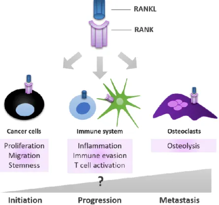
The relevance of RANK-RANKL signalling pathway in different physiologic processes described above, suggests that it may also be implicated and/or dysregulated in cancer. RANK-RANKL relevance in the context of bone metastatic disease has been addressed for long, but in fact, over the past years it was found that RANK is expressed in different types of tumour cells, affecting tumour invasiveness and stemness; and also that it has a key role in mammary tumorigenesis. Therefore, RANK-RANKL pathway may impact all stages of tumour progression, from initiation to metastization, including immune response (Figure 4).
Figure 4 – RANK-RANKL signalling pathway is involved in all stages of cancer progression. RANK signalling is involved in three different contexts during tumour progression: in cancer cells, it influences their capability to proliferate, migrate and invade; in osteoclasts, it enhances their functional activation, contributing to the ‘vicious cycle’ of bone metastasis, where bone resorption is increased; in the immune cells, it can influence immune responses and even be involved in the acquired capability of the tumour cells to evade these responses. Adapted from [47]
9
3.1. RANK signalling in breast cancer
A decade ago, a seminal paper by Jones et al. has shown that different epithelial tissues, human primary breast tumours, and breast and prostate cancer cell lines expressed functional RANK in their surface66. It has been previously shown that several
BC cell lines expressed the receptor but its function was unknown67. Jones et al. have
demonstrated that, in cell lines, RANK activation by RANKL induced downstream activation of PI3K/AKT and ERK pathways, and increased cell migration66. Posteriorly,
different studies have shown that activation of RANK pathway in several in vitro models leads to a more aggressive phenotype, with increased migration and invasion ability66– 70. After that the importance of RANK signalling in BC has been deeply investigated, and
several studies demonstrated its role in BC onset71–74.
It has been demonstrated that RANK pathway activation promotes proliferation and aberrant survival of mammary epithelial cells, by inhibiting their terminal differentiation54. In a transgenic mice model for RANK overexpression, mouse mammary
tumour virus (MMTV)-RANK mice not only have increased mammary epithelial proliferation, impaired differentiation of the lobulo-alveolar structures and deficient lactation54, but also spontaneous development of mammary tumours75. Moreover, mice
with constitutive activation of RANK-RANKL pathway spontaneously developed high-grade invasive tumours with evidence of pulmonary metastasis75. These tumours were
extensively heterogeneous, originated from different populations of basal or luminal cells or alternatively from multipotent progenitors.
RANK pathway is also involved in the development of progestin-driven mammary cancer. Several studies clearly demonstrated that RANKL is involved in the promotion of the expansion of MaSCs by progesterone76,77 and that it may also mediate the critical
role of progesterone in the promotion and growth of breast tumours78.
MMTV-Cre/rankfloxed/Δ mice transgenic model, which lacks RANK in mammary epithelial cells,
show attenuate tumour development when comparing with WT mice79. Additionally,
Beleu et al. demonstrated that a large number of PR- cells proliferate in a
RANKL-dependent mechanism, through paracrine stimulation80. All these results suggest that
RANKL inhibition could not only reduce BC cells’ proliferation and delay tumorigenesis, but also mediate the reduction in MaSCs. Interestingly, it is known that the risk associated with HRT is greater for estrogen–progesterone combination than for estrogen alone, either in continuous or sequential therapy81, which is in accordance to
these findings.
In this context, the use of anti-RANKL therapies may prove to be effective in the prevention of BC. In fact, it has already been shown that inhibition of RANK signalling reduces the accumulation of MaSCs in invasive mammary tumours by inducing tumour cell differentiation, and decreased metastasis-initiating cell pools82.
Importantly, these studies were expanded to the human setting, and it was shown that overexpression of RANK in untransformed MCF10A cells induced changes associated with both stemness and transformation, such as mammary gland reconstitution, EMT, increased migration and anchorage-independent growth66,68. Later
it was also shown that MCF-7 ER+ BC cells that express low endogenous levels of RANK
10
RANK overexpression in MCF-7 cells resulted in increased EMT markers, migration, and CD44+/CD24-/low subpopulation, known markers for MaSC84.
Recently, RANK-RANKL signalling pathway has also been implicated in progenitor cell expansion and tumorigenesis in inherited BC. It was shown that genetic inactivation of RANK in the mammary epithelium markedly delayed onset, reduced incidence, and attenuated progression of BRCA1:p53 mutation-driven mammary cancer85. Additionally,
long-term pharmacological inhibition of RANKL in mice abolished the occurrence of BRCA1 mutation-driven pre-neoplastic lesions. Another study has shown in the clinical setting that the progesterone–RANKL/OPG system is in fact dysregulated in BRCA-mutation carriers, with lower OPG levels in serum and tissue86.
Despite all the evidences from basic and translational studies, clinical evidences for the prognostic role of RANK expression in BC are still sparse72–74,87. High levels of RANK,
either mRNA or protein, were found in human primary BC ER-/PR-, with high pathologic
grade and high Ki6788. In this study, high RANK mRNA expression was significantly
associated with metastatic occurrence. It has also been demonstrated, using 295 primary BC patients microarray datasets, that low levels of RANK and higher OPG expression correlates with both longer OS and disease-free survival (DFS)71.
Altogether these results support the hypothesis that inhibition of RANKL–RANK axis may offer a promising therapeutic target for preventing tumour progression and metastization, beyond its use for management of SREs that emerge with BM.
3.2. RANK signalling and bone metastases
Bone is the most common site of relapse in BC. BM are associated with increased morbidity and decreased OS, urging the need to find therapeutic options that prevent SREs but also that impact on tumour growth.
BM fit perfectly in the 1889 Stephen Paget’s ‘seed and soil’ hypothesis to explain cancer relapse: “When a plant goes seed, it seeds are carried in all directions, but they can only live and grow if they fall on congenial soil”89. Metastatic tumour cells seeded in
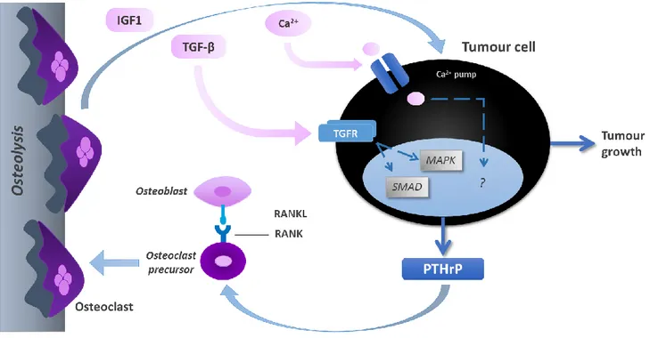
bone secrete growth factors and cytokines that alter the normal balance of bone remodelling, driving further activation of osteoclasts in a ‘vicious cycle’ of bone resorption90,91. PTHrP secreted by tumour cells leads to osteoclasts’ differentiation and
activation, via RANKL up-regulation, and ultimately increases bone resorption. This will lead to the release of mitogenic growth factors that induce tumour growth (Figure 5).
11
RANK and RANKL not only control bone remodelling, as previously mentioned, but are also involved in this preferential metastasis of BC cells to the bone93. In fact,
deregulation of RANK-RANKL signalling pathway is known to be involved in the establishment of BM and consequent pathological conditions, such as osteoporosis94.
Being essential for osteoclast differentiation and activation and also being expressed in many tumour cell types, it was suggested that RANK pathway could play a direct role on tumour cells. Armstrong et al. demonstrated that RANK is expressed in prostate cancer cells, another type of cancer that preferentially metastasizes to bone, and promotes invasion in a RANKL-dependent manner, by activating several downstream events that lead to cellular proliferation, invasion and migration70. Other previous in vitro and in vivo
studies have indicated that RANKL can promote distant metastasis via direct effects on RANK-expressing cells, either tumour or progenitor cells. It was proven that RANKL inhibition not only protects against bone destruction but also inhibits BM progression and delays the formation of de novo BM in cancer models56.
RANK expression was associated with accelerated bone metastasis formation71,93.
In fact, RANK overexpression alone seems to be sufficient to confer a significantly greater metastatic growth in bone93. Previous studies have also elucidated an important
role of RANK signalling in the upregulation of MMP-1, a metalloproteinase that contributes to bone metastases formation and osteoclastogenesis69. High levels of
MMP-1 are significantly associated with decreased OS of patients with BM, and in a mouse model MMP-1 knockdown (KD) resulted in smaller x-ray osteolytic lesions and osteoclastogenesis and decreased tumour burden95.
Figure 5 – ‘Vicious cycle’ of bone metastases.
Cancer cells in bone secrete high amounts of PTHrP, which induces osteoclasts’ differentiation and activation via up-regulation of RANKL, promoting osteolysis. Bone disruption leads to the liberation of several cytokines and growth factors, such as IGF-1 and TGF-β, which in turn enhance tumour proliferation. Adapted from [92]
12
3.3. RANK as a therapeutic target in breast cancer
RANK-RANKL pathway has been implicated in several contexts of BC disease, from early tumorigenesis to progression and metastasis. As already mentioned above, both
in vitro and in vivo evidences point RANK-RANKL axis as a promising therapeutic target
for prevention of BC onset, progression and metastization. But the importance of RANK-RANKL pathway in the context of BM has been addressed for long. The use of denosumab, a fully humanized anti-RANKL monoclonal antibody that decreases bone resorption, was approved by FDA in 2010 for the prevention of SREs in patients with BM; and in 2011 for the treatment of bone loss in patients with BC undergoing hormone ablation therapy. In fact, adjuvant denosumab in postmenopausal BC patients receiving aromatase inhibitors has shown to reduce the risk of clinical fractures96.
Inhibition of bone resorption makes bone a less favourable microenvironment for tumour cells to growth. Since the host microenvironment is essential for cancer colonization at secondary sites, there is a rational to the adjuvant use of therapies that decrease bone resorption (antiresorptive therapies) in patients with BC. Currently, denosumab is being studied on a phase III clinical trial to determine whether adjuvant RANKL inhibition prolongs BM-free survival and DFS in BC97.
Moreover, it has been demonstrated that RANK-RANKL pathway directly contributes to increased proliferation and survival of preneoplastic breast cells, promoting tumour growth, as well as to an increased expansion of MaSCs population75– 79. Further clinical studies are required in order to determine the potential benefit of
RANK-RANKL targeted therapies in prevention of BC in high risk groups. Importantly, RANK pathway seems to be implicated in BRCA1-mutation driven mammary cancers85,86,
which opens doors to the investigation of a new possibility of BC prevention in women that have this inherited mutation.
13
4. Objectives
Over the past years, the role of RANK-RANKL signalling pathway in BC development and progression has been widely assessed. RANK is expressed by cancer cells and is involved in the promotion of mammary tumorigenesis. Several in vitro studies have correlated RANK expression with EMT, increase in migration and invasion potentials, and stemness.
Although RANK overexpression has been mostly associated to TNBC, previous studies suggest that RANK overexpression in luminal BC cells also induces a more invasive and stem-like phenotype. Therefore, we hypothesize that RANK-positive luminal BC cells could have enhanced invasion ability, and so forth metastases may be enriched for this particular population of cells.
The present study aimed to contribute to test this hypothesis by determining the effect of RANK overexpression in luminal BC cells in tumour burden and invasiveness in
14
MATERIALS AND METHODS
Cell Culture
Human breast carcinoma cell lines MDA-MB-231GFP+Luc+ and MCF-7GFP+Luc+ were
obtained by Sérgio Dias Lab (IMM), through lentiviral transduction with GFP-Luciferase lentiviral particles and cell sorting of pure GFP+ cell populations. MDA-MB-231GFP+Luc+
cells were cultured in Dulbecco’s Modified Eagle’s Medium (DMEM, Gibco) supplemented with 10% (v/v) fetal bovine serum (FBS, Gibco), 1% (v/v) Penicillin/Streptomycin (10,000 U/mL Penicillin, 10,000 µg/mL Streptomycin, Gibco). MCF-7 GFP+Luc+ cells were cultured in the same medium, additionally supplemented with
0,01mg/mL insulin (Gibco). Cells were maintained at 37°C with5% CO2.
RANK overexpression and knockdown
For lentiviral transduction, MDA-MB-231GFP+Luc+ and MCF-7GFP+Luc+ cells were seeded
in 24-well plates, at a density of 4×10⁴ cells/well. 24 hours after seeding, medium was
replaced by fresh medium containing 8µg/ml Polybrene (Sigma). RANK lentiviral activation particles (h) (30µl/well; sc-400559-LAC) or control lentiviral particles (15µl/well;sc-437282); and RANK shRNA (h) lentiviral particles (15-20µl/well; sc-42960-V) or control shRNA lentiviral particles (15-20µl/well; sc-108080) were used (Santa Cruz). For RANK activation, cells were selected with 0,5µg/mL Puromycin dihydrochloride (Sigma), 5µg/ml Blasticidin S HCl (Santa Cruz) and 200µg/mL Hygromycin B (Santa Cruz), starting three days after transduction. For RANK KD, cells were selected with 0,5µg/mL Puromycin dihydrochloride (Sigma) starting three days after transduction. RANK overexpression (OE) and KD was confirmed by RT-qPCR and flow cytometry.
RFP lentiviral transduction
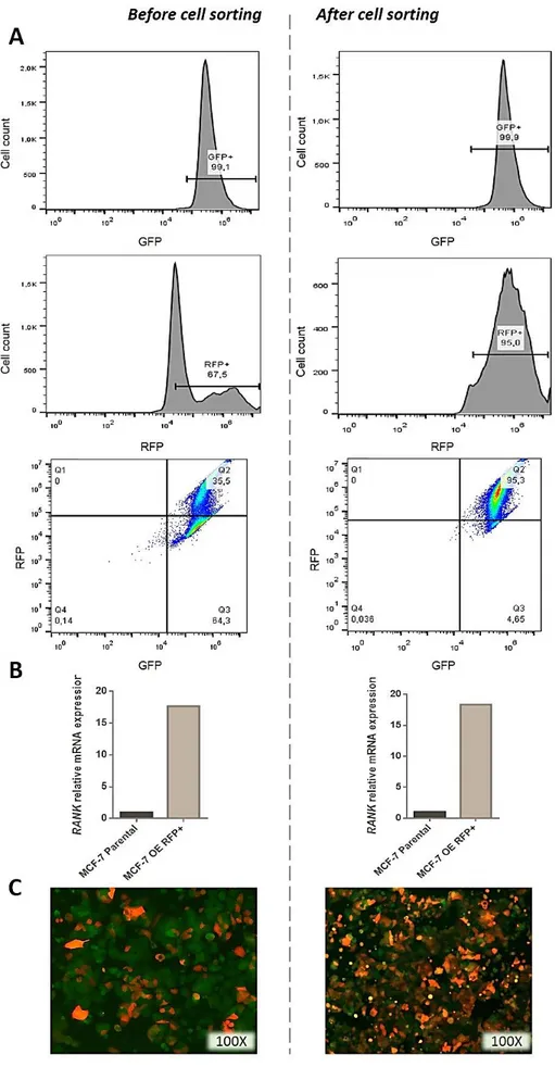
MCF-7GFP+Luc+ cells overexpressing RANK (RANK OEGFP+Luc+) were transduced with
Cignal Lenti Positive Control (RFP) ready-to-transduce lentiviral particles (Quiagen). Cells were seeded in 96-well plates (104cells/well), and 24 hours after seeding, medium was
replaced with fresh medium supplemented with 8µg/ml Polybrene and lentiviral particles were added at a multiplicity of infection (MOI) of 25 or 50. RFP+ cells were selected with 0,5µg/mL Puromycin dihydrochloride (Sigma). RFP positivity was confirmed by flow cytometry in a BD Accuri C6 flow cytometer and RFP+ cells were sorted in a FACS Aria III cell sorter (BD Biosciences) to obtain a RFP+ pure cell population.
RT-qPCR
For RT-qPCR analysis of mRNA expression, cells were grown up to 80-90% confluency and total RNA was extracted using the NZY Total RNA Isolation kit (Nzytech), according to manufacturer’s instructions. Total RNA was quantified in a NanoDrop spectrophotometer (Thermo Ficsher Scientific).
15
Total RNA (500ng to 1µg) was treated with RQ1 RNase-free DNase I (Promega) for 30min at 37°C, according to manufacturer’s instructions. DNase I-treated RNA was reverse transcribed using the NZY M-MuLV First-Strand cDNA Synthesis kit (Nzytech), and Oligo(dT)20 primer, according to manufacturer’s instructions.
cDNAs were amplified by real-time PCR using TaqMan Gene Expression Master Mix (Applied Biosystems), according to manufacturer’s instructions, and specific primers for human RANK, 18S and GAPDH; or Power SYBR Green PCR Master Mix (Applied Biosystems, Life Technologies) and specific primers for Vimentin, Fibronectin,
E-cadherin, N-E-cadherin, Snail, Slug, and Twist; in a Viia7 System (Applied Biosystems)
(Table 2). Gene expression was normalized using the housekeeping genes 18S or GAPDH, and relative mRNA expression was calculated using the 2-∆∆Ct method.
List of primers used is resumed in Table 2.
Table 2 – List of primers used in qPCR.
Gene Common name Primer sequence (5’-3’)
GAPDH GAPDH PPH00150F (SA Biosciences)
TNRFS11A RANK PPH01102C (SA Biosciences)
Vimentin Vimentin F: GAAAACACCCTGCAATCTT R: CCTGGATTTCCTCTTCGTG
FN1 Fibronectin F: CAGTGGGAGACCTCGAGAAG
R: TCCCTCGGAACATCAGAAAC
SNAI1 Snail1 F: CTCTTTCCTCGTCAGGAAGC R: GGCTGCTGGAAGGTAAACTC
SNAI2 Slug F: CCAAACTACAGCGAACTGGA R: GTGGTATGACAGGCATGGAG
TWIST1 Twist F: CCGGAGACCTAGATGTCATTG R: CCACGCCCTGTTTCTTTG
Flow Cytometry
To analyse RANK expression by flow cytometry, cells were grown up to 80-90% confluency, trypsinized, centrifuged at 200 g for 5min, and then ressuspended in FACS buffer (PBS 1X, 2%FBS). Cells were fixed with 1mL ice cold methanol, at -20°C for 10min. After washing with FACS buffer, cells were permeabilized with PBS 1X, 0,5% Tween-20, on ice for 10min. Cells were incubated with a specific mouse monoclonal antibody against human RANK (1μg/ml; clone 9A725; Enzo) for 30min at 4°C in the dark, washed with FACS buffer, and incubated with a goat anti-mouse secondary antibody conjugated with Alexa Fluor 633 (1:400; Life Technologies) for 20min at 4°C. After labelling cells
16
were washed twice with FACS buffer, ressuspended in 1mL FACS buffer and analysed on a BD Accuri C6 flow cytometer (BD Biosciences).
For RFP-positive cell sorting, cells were grown up to 80-90% confluency, trypsinized and ressuspended in FACS buffer. RFP-positive cells were sorted on a FACSorting Instrument – FACSAria III (BD Biosciences), and cultured under standard conditions.
Flow Cytometry data analysis was performed in FlowJo V10 software.
Western Blot
Activation of RANK pathway upon stimuli with RANKL was analysed by Western blot. For this purpose, 4x10⁵ cells were seeded in 6-well plates for 24h, and serum-starved in
low-serum medium (0,1% FBS, 1% Pen/Step) for another 24h. Medium was replaced by fresh low-serum medium containing 1μg/mL human RANKL (PeproTech) and total cell lysates obtained at different time points. Total cell lysates were prepared with 200µl RIPA buffer per well (Santa Cruz), with 1:100 phenylmethane sulfonyl fluoride (PMSF, Santa Cruz), 1:100 protease inhibitor (PI, Santa Cruz), 1:100 sodium orthovanadate (NaOR, Santa Cruz) and 1:100 phosphatase inhibitor cocktail (Sigma), on ice for 30min. Samples were sonicated in a Soniprep 150 equipment (MSE) for approximately 1min and centrifuged at 10,000g for 20min, at 4°C. Supernatants were transferred to new tubes and total protein was quantified using Pierce BCA Protein Assay Kit (ThermoSicentific), according to manufacturer’s instructions. Proteins were resolved by SDS-PAGE, using 10% polyacrylamide gels, and then transferred to nitrocellulose membranes using an iBlot® Gel Transfer Device (Invitrogen), according to manufacturer‘s instruction.
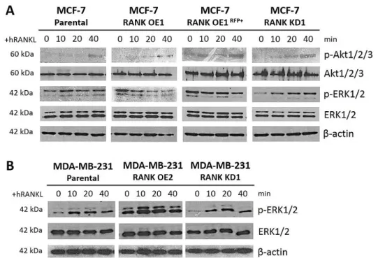
Membranes were blocked for 1h at room temperature (RT) in 5% Non-Fat Dry Milk (NFDM) in PBS-0.1% Tween (PBST) for β-actin; or in 5% bovine serum albumin (BSA) (Santa Cruz) for other antibodies. Membranes were incubated with the following specific antibodies, overnight at 4ºC: mouse anti-β Actin antibody (1:25,000; Ab6276; Abcam), rabbit anti –Akt1/2/3 (1:250; H-136), rabbit anti -pAkt (1:250; Ser-473), rabbit anti –ERK1/2 (1:1,000; C-14) and rabbit anti –pERK1/2 (1:500; Thr-202/Tyr-204), all from Santa Cruz. After washing with PBST, membranes were incubated with horseradish peroxidase-conjugated (HRP) specific secondary antibodies: donkey anti-mouse (1:2,000) or goat anti-rabbit (1:4,000) (Santa Cruz) for 2h at RT. Proteins were detected using a Novex® ECL HRP chemiluminescent substrate reagent kit (Invitrogen) according to the manufacturer‘s instructions, and x-ray films (Fujifilm) developed in a Curix 60 processor (AGFA).
Proliferation Assay
Cells were seeded in 96-well plates, at a density of 104 cells/well. Approximately 4
h after seeding, 1:20 Alamar blue (Invitrogen) was added to each well and fluorescence was measured 24 and 48h after incubation (excitation 560nm; emission 590nm) in an Infinite M200 microplate reader (Tecan).
17
Migration assay
MCF-7GFP+Luc+ and MCF-7 RANK OEGFP+Luc+ cells were grown to confluency in 24-well
plates, and a straight wound was made across each well with a sterile 200µl pipette tip. After washing away loose cells with PBS 1X, cells were incubated with low-serum medium with or without 1μg/mL RANKL. Each well was digitally imaged using an inverted wide-field Zeiss microscope (Jena) at 0h and 24h after stimuli with RANKL.
MCF-7GFP+Luc+ and MCF-7 RANK OEGFP+Luc+RFP+ cells were plated 1:1 at confluence
density in 6-well plates. Cells were left to adhere ON in low-serum medium and a straight wound was made across each well with a sterile 200 pipette tip. After washing loose cells, cells were incubated with low-serum medium for another 24h. Each well was digitally imaged using a Zeiss Axio Observer at 0h and 24h after scratch.
Migrated area was calculated using ImageJ 1.6.0 software.
Orthotopic xenograft model
In all studies involving animals, mice were handled and euthanized in accordance with approved institutional, national and international guidelines, applying the Principle of the 3Rs. This study was approved by the ethical Committee of the Rodents’ facility of IMM and DGAV.
Estradiol supplementationNine days prior to BC cell inoculation (day -9) 4 week-old female BALB/c nude or NOD scid gamma (NSG) mice were subcutaneous implanted with 17β-estradiol pellets (60-day release, 0.36mg/pellet, Innovative Research of America), inserted with a trochar in the mid-scapular region. For this procedure mice were anaesthetized with isoflurane.
Tumour inoculationMCF-7GFP+Luc+, MCF-7 RANK OEGFP+Luc+ and MCF-7 RANK OEGFP+Luc+RFP+ cells were
harvested in the exponential phase of growth, washed with PBS, trypsinized and counted with a haemocytometer. Cells were ressuspended in 50mL ice-cold PBS and washed twice by centrifugation at 200g, for 10min. Cells were ressuspended in ice-cold 50% phenol-red free Matrigel Matrix (in PBS) (BD Biosciences) to a final concentration of 2×10⁵ cells/mL. Mice were injected unilaterally into the fourth abdominal (nude) or
second thoracic (NSG) fat pad with a total of 2×10⁴ MCF-7GFP+Luc+, MCF-7 RANK OEGFP+Luc+,
or MCF-7GFP+Luc+/MCF-7 RANK OEGFP+Luc+RFP+ (1:1) cells.
Tumour visualizationEvery week (or starting at week four in the BALB/c nude mice experience) post tumour inoculation, mice were injected with 100µl/10g body weight (BW) XenoLight D-Luciferin - K+ Salt Bioluminescent Substrate (PerkinElmer) and after 4 min they were anesthetized with 75mg/KgBW Ketamine + 1mg/KgBW Medetomidine.
After approximately 6min luminescence was analysed in an IVIS Lumina, using Living Image 3.0 software (30s of exposure; field of view D 12,5cm; subject height 1,5cm), and mice recovered with 1mg/KgBW Atipamezole.
18
Circulating tumour cells (CTCs) and disseminated tumour cells (DTCs) identification For CTC analysis, venous blood was collected from the submandibular vein five weeks post tumour inoculation, or by cardiac puncture before sacrifice, into 1.5 mL centrifuge tubes with 5µl EDTA 0,5M (pH8.0). Erythrocytes were lysed by incubation with 1X RBC Lysis Buffer Multi-species (eBioscience) for 13min at RT. Cells were washed with FACS buffer and centrifuged for 3min at 2,000 rpm. The supernatant was discarded and cells were ressuspended in FACS buffer.For DTC analysis, tibia and/or femur were removed and the epiphyses of the bones were cut off. Using a 1-cc syringe with a 26G needle (Omnifix® 100 Duo, B.Braun) filled with DMEM, bone marrow cells were flushed from both ends of the bone shafts onto a 15mL Falcon tube fitted with a sterile 70µm Nylon cell strainer (Falcon®).
Samples were analysed for GFP and/or RFP expression in a BD Accuri C6 flow cytometer.
Sacrifice and necropsyMice were sacrificed eight weeks post tumour inoculation by administration of 0,25mg/KgBW Sodium Pentobarbital (Eutasil®). Necropsies were performed to identify macrometastases. Additionally, bone marrow was collected to detect disseminated tumour cells (DTCs) by flow cytometry.
Tumours were harvested and divided into four parts to be frozen in liquid nitrogen embedded in O.C.T. compound (Tissue-Tek, Sakura); frozen in RNA later solution (Ambion); formalin fixed and paraffin embedded (FFPE); and analysed by flow cytometry after cell disruption using a sterile 70µm Nylon cell strainer (Falcon®). Visceral organs (lung, liver, spleen) were collected and FFPE.
Hematoxylin-Eosin (H&E) and Masson’s Trichrome staining were performed by the Histology and Comparative Pathology Service at IMM.
Immunohistochemistry5µm tissue sections from FFPE samples were stained by immunohistochemistry (IHC) for the detection of Ki67, in an AutostainerPlus (Dako Cytomation. Deparaffinization and antigen retrieval was performed in a PT Link Pre-Treatment Module for Tissue Specimens (Dako), using Antigen Retrieval Solution pH9.0, at 94ºC for 20min. Endogenous peroxidase was blocked with 3% H2O2 (Sigma) for 10min at RT, and
total protein was blocked by incubation with Protein Block Solution (Dako), for 20 min at RT. Slides were incubated for 30min with rabbit anti-human Ki67 primary antibody (MIB-1, Dako), 1:100 in Antibody Diluent (Dako). Slides were incubated with EnVisionTM Detection System, rabbit/mouse (Dako), according to manufacturer’s instructions, with 5min of incubation with DAB. Slides were counterstained with hematoxylin, dehydrated, mounted with Quick-D mounting medium (Klinipath) and visualized in a bright field microscope (LeicaDM750 with a Leica ICC50 HD camera). Imunoratio was obtained through the calculi of the percentage of DAB-stained nuclear area over total nuclear area (hematoxylin-stained nuclei regions) (5 fields, 400x, ImageJ software).
19
Statistical Analysis
Data was analysed using GraphPad Prism6 software. The number of replicates performed for each experiment is indicated. Statistics were performed by one-way ANOVA or unpaired t-test (in vitro experiments); two-way ANOVA (tumour burden and mice BW). Results are presented as mean with SEM and p-value <0.05 was considered significant.
20
RESULTS
1. RANK overexpression and knockdown
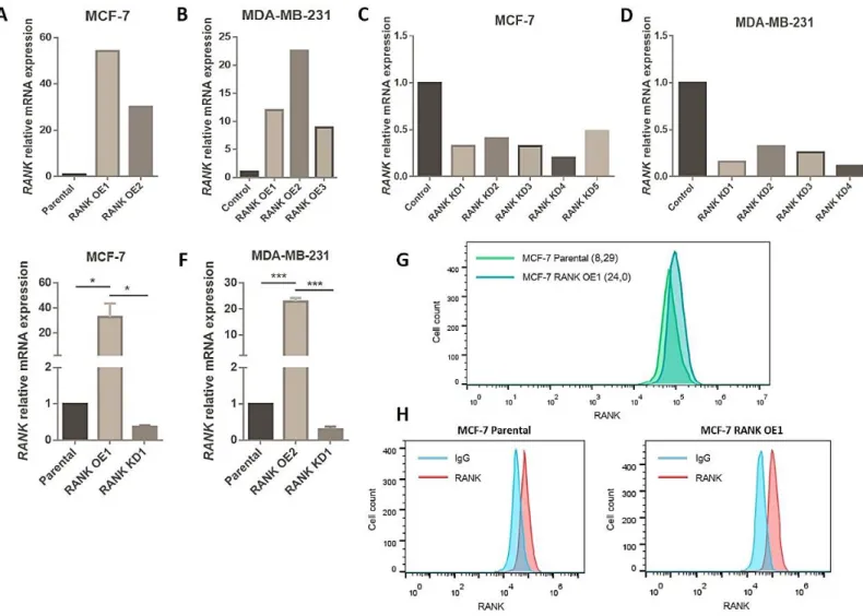
In this study we propose to address the effect of RANK overexpression (OE) in the behaviour of luminal BC cells in vivo. Therefore, we started by generating stable MCF-7 RANK OE cells by lentiviral transduction. We also generated non-luminal BC cells with RANK OE (MDA-MB-231 triple negative BC cells), or the respective counterparts with RANK knockdown (KD), to be used in control assays. To be able to monitor tumour growth in vivo we used GFP+Luc+ parental cells to derivate OE and KD clones.
RANK OE was obtained by transduction with RANK lentiviral activation particles, that consist in a dCas9 nuclease fused to a VP64 activation domain, in conjunction with a target-specific sgRNA. This synergistic activation mediator (SAM) maximizes the activation of endogenous RANK. Upon transduction, activated clones were selected with blasticidin, hygromycin and puromycin, and analysed for RANK mRNA expression (Figure 6A,B). Two MCF-7 OE clones and three MDA-MB-231 OE clones were obtained, with between 9 and 54-fold up-regulation of RANK expression, when comparing to cells transduced with control activation particles.
RANK KD was obtained by transduction of specific shRNA lentiviral particles. Upon transduction, KD clones were selected with puromycin, and analysed for RANK mRNA expression (Figure 6C,D). Five MCF-7 KD clones and four MDA-MB-231 KD clones were obtained, with between 0,1 and 0,4-fold down-regulation of RANK expression, when comparing to cells transduced with control shRNA particles.
We selected the clones MCF-7 OE1, KD1 and MDA-MB-231 OE2 and KD1 to perform the subsequent studies, and analysed RANK mRNA expression at different cell passages to confirm stable RANK OE or KD (Figure 6E,F).
RANK OE in MCF-7GFP+Luc+ cells was also confirmed at the protein level by flow


