Carla Sofia Martins Calçada
outubro de 2013
Use of the Comet Assay to study the role
of MGMT, MMR and p53 in the repair of
alkylating DNA damage
UMinho|20 13 Carla Sof ia Mar tins Calçada Use of t he Come t Assa y to s tudy t he role of MGMT , MMR and p53 in t he repair of alky
lating DNA damage
Carla Sofia Martins Calçada
outubro de 2013
Dissertação de Mestrado
Mestrado em Genética Molecular
Use of the Comet Assay to study the role
of MGMT, MMR and p53 in the repair of
alkylating DNA damage
Escola de Ciências
Trabalho realizado sob orientação da
Nome: Carla Sofia Martins Calçada
Endereço eletrónico: [email protected]
Número do Bilhete de Identidade: 13573982
Título da dissertação:
Use of the Comet Assay to study the role of MGMT, MMR and p53 in the repair of alkylating DNA damage
Orientador:
Doutora Prof.ª Doutora Cristina Pereira-Wilson
Ano de conclusão: 2013
É AUTORIZADA A REPRODUÇÃO INTEGRAL DESTA DISSERTAÇÃO DE MESTRADO APENAS PARA EFEITOS DE INVESTIGAÇÃO, MEDIANTE DECLARAÇÃO ESCRITA DO INTERESSADO, QUE A TAL SE COMPROMETE
Universidade do Minho, outubro de 2013
iii
Começo por agradeço à Prof. Doutora Cristina Pereira-Wilson, orientadora deste projecto, por me ter recebido no seu grupo de investigação, pela orientação, incentivo, ensinamento e acima de tudo pela confiança transmitida durante a realização deste trabalho. Muito obrigada!
Um especial obrigado à Dalila por toda a ajuda, disponibilidade, pela enorme paciência e por me ensinares tudo o que precisava saber para trabalhar neste mundo científico. Mas acima de tudo pela simpatia, amizade, pelos momentos de descontração e de partilha de confidências nos bons e nos maus momentos. Foi um enorme prazer conhecer-te e trabalhar contigo.
À Cristina, Alice e Cristovão por todo o tempo e paciência que tiveram comigo. Obrigada por me terem ajudado a integrar nesta nova fase e por me ajudarem a concretizar este projecto.
A todos os colegas de laboratório, pelo bom ambiente, pelos momentos de boa disposição, pela ajuda, pelos almoços e por estarem sempre lá quando eu precisei! Joana, Luísa e Cristina Mesquita, obrigada pelas brincadeiras, pela vossa energia contagiante, amizade e apoio. Sem dúvida este ano não seria o mesmo sem vocês! Adorei conhecer-vos!
Carla, Tânia, Marisa e Raquel as palavras não são suficientes para vos agradecer! Obrigada pela vossa grande amizade e por me colocarem um sorriso na cara quando mais precisei. Obrigada por estarem sempre presentes e por me transmitirem tanta segurança. Espero que nunca deixem de fazer parte da minha vida!
À minha manita Ana Rita, apesar da distância a nossa grande amizade prevaleceu! Sei que posso sempre contar contigo!
Um obrigado especial às meninas que considero como minha segunda família, Cláudia, Ana Rita, Margarida e Rita. Obrigada por me fazerem lembrar do meu valor quando me sinto insegura e pelos grandes momentos partilhados. É muito bom sentir que posso contar com vocês em qualquer momento e espero que esta nossa grande amizade prevaleça para o resto da vida independentemente do que o futuro nos possa reservar.
Nada disto seria possível sem o apoio incondicional dos meus pais. Obrigada por acreditarem em mim e por me darem força para nunca desistir nos momentos mais complicados. Sempre que precisei vocês estavam lá para me amparar. Agradeço por me receberem sempre com um enorme sorriso e de braços abertos. Obrigado por toda a compreensão e amor!
v
Alkylating agents are one a major class of mutagens contributing to DNA damages and carcinogenesis. However, as alkylating agents are powerful inducers of DNA damage related cell death, and are commonly used in the chemotherapy. O6-methylguanine (O6meG) and O6
-chloroethylguanine (O6ClethG) are the most cytotoxic lesions caused by Temozolomide (TMZ) and
1,3-bis-(2-chloroethyl)-1-nitrosourea (BCNU), respectively. MGMT (O6-methylguanine-DNA
methyltransferase) repair protein directly reverses O6alkylG lesions and, consequently, it is
considered as a prognostic marker of resistance in cancer cells exposed to alkylating agents. Therefore, expression levels of this protein predict cancer cell susceptibility and the success of therapy. However, it has been observed that not all tumors with low MGMT activity respond to alkylating agents with increased cell death. Mismatch repair (MMR) system and functional status of p53 are also two most relevant factors in this response.
The aims of this project were to study the genotoxic effects of alkylating drugs as detected by the CoMeth assay, and to determine the dependence of a functional MMR system and p53 in the sensitivity of cells to treatment. Thus, using a proliferating MMR-proficient cancer cell line Caco-2, we report that CoMeth assay allows the qualitative evaluation of O6meG lesions as well as
O6ClethG, after their conversion to strand breaks. To determine whether the DNA damages
(comet assay) and cell death (nuclear condensation assay) induced by (TMZ) and (BCNU) agents are mediated by MMR we used a MMR-deficient colon HCT116 cell line. We observed that even though MMR status seemed to be determinant for cells’ sensitivity to TMZ, this was not applied for the case of BCNU. Resistance to the O6-chloroethylating agent involves other mechanisms that
are independent of functional MMR system. We found that induction of DNA damages by BCNU was independent of p53 status, however p53-wt cells showed more pronounced apoptosis compared with p53-null cells.
Altogether, our data show that functional MMR system is required for genotoxicity (apoptosis) induced by TMZ. For BCNU, we show that the efficiency on cancer cells is not dependent on the MMR status but only the activity of MGMT. In p53-wt cells, cell death by BCNU was more pronounced. This project also demonstrated the value of the CoMeth assay as a tool for the study of anticancer chemotherapeutic drugs.
vii
RESUMO
Agentes alquilantes são uma das maiores classes de mutagénicos contribuindo para os danos no DNA e para a carcinogénese. Contudo, como os agentes alquilantes são indutores potentes de danos no DNA relacionados com a morte celular eles são frequentemente usados na quimioterapia. O6-metilguanina (O6meG) e O6-cloroetilguanina (O6-cletG) são as lesões mais
citotóxicas causadas pela Temozolomida (TMZ) e 1,3-bis-(2-cloroetil)-1-nitrosureia (BCNU), respetivamente. MGMT (O6-metilguanina-DNA metiltransferase) repara a proteína e reverte
diretamente as lesões O6-alquilG e, consequentemente, é considerado um marcador de
prognóstico de resistência em células cancerígenas expostas a agentes alquilantes.
Desta forma, os níveis de expressão desta proteína preveem a suscetibilidade das células cancerígenas e o sucesso da terapia baseada em agentes alquilantes. Contudo, tem sido observado que nem todos os tumores com baixa actividade da MGMT respondem a agentes alquilantes com morte celular aumentada. O sistema de reparação Mismatch repair (MMR – do inglês mismatch repair) e o estado funcional do p53 são também dois dos fatores mais relevantes nesta resposta.
Os objetivos deste projeto foram estudar os efeitos genotóxicos de drogas alquilantes, sendo detetadas pelo ensaio do Cometa e determinar a dependência do estado funcional do sistema Mismatch repair e do p53 na sensibilidade às drogas alquilantes. Desta forma, usando uma linha celular cancerígena proliferativa Caco-2, com um sistema MMR eficiente, demonstramos que o ensaio do Cometa permite uma avaliação qualitativa das lesões O6meG
bem como as O6-cletG, depois da sua conversão em quebras na cadeia de DNA. Para determinar
se os danos no DNA (ensaio do cometa) e morte celular (ensaio de condensação nuclear) induzidos pelos agentes metilantes (TMZ) e cloroetilantes (BCNU) são mediados por reparação MMR usamos a linha celular HCT116 deficiente no sistema MMR. Observamos que apesar do estado MMR parecer ser determinante para a sensibilidade das células à TMZ, este facto não foi observado para o caso do BCNU. A resistência ao agente O6-cloroetilante envolve outros
mecanismos que são independentes do sistema funcional MMR. Verificamos que a indução dos danos no DNA pelo BCNU foram independentes do estado do p53, contudo as células com p53 ativo apresentaram uma apoptose mais pronunciada comparado com as células com p53 inativo. De um modo geral os nossos resultados mostram que o sistema funcional MMR é necessário para a genotoxicidade (apoptose) induzidos pela TMZ. Para o BCNU, mostramos que a eficiência nas células cancerígenas não é dependente do estado do MMR mas apenas da actividade da MGMT. Nas células com p53 ativo a morte celular induzida pelo BCNU foi mais pronunciada.
viii
Este estudo também demonstrou o papel do ensaio do cometa como ferramenta no estudo de drogas anticancerígenas.
ix
CONTENTS
Agradecimentos ... iii Abstract ... v Resumo ... vii CONTENTS ... ix Abbreviations list ... xi 1. Introduction ... 1 1.1. Colorectal carcinoma (CRC) ... 41.2. DNA damages and genomic stability ... 7
1.3. Alkylating agents ... 8
1.4. Mechanism of DNA repair ...10
1.5. Clinical chemotherapeutic agents – Temozolomide (TMZ) and Carmustine (BCNU) ...20
1.6. Natural compounds and chemoprevention ...21
1.7. p53 – the tumor suppressor ...23
1.8. Apoptosis ...24
2. Research Objectives ...27
3. Material and methods ...31
3.1. Cell lines, culture conditions and reagents ...33
3.2. Cell toxicity by MTT reduction assay ...34
3.3. Nuclear condensation ...35
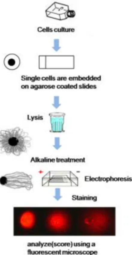
3.4. DNA damage assessment by comet assay ...36
3.5. Protein extraction and Western Blot analysis ...38
3.6. Statistical analysis ...39
4. Results ...41
4.1 Effects of TMZ, BCNU and O6-BG on cell viability ...43
4.2. Genotoxic effects of TMZ, BCNU and O6-BG in Caco-2 cell line ...44
4.3. Effects of TMZ, BCNU and O6-BG on cell death in Caco-2 cells ...46
4.4. Role of DNA mismatch repair on induction of DNA damages and apoptosis by alkylating agents ...48
4.5. Role of p53 tumor suppressor gene on induction of DNA damages and apoptosis by alkylating agents ...51
4.6. Effects of some phytochemicals on MGMT protein expression...54
x
6. References ...73 7. Annexes...83
xi
ABBREVIATIONS LIST
5-FU 5-Fluorouracil
8-oxoG 8-Oxo-7,8-dihydroguanine
ACNU Nimustine
ALKBH AlkB homologue
ALS Alkali-labile sites
AP Abasic sites
APC Adenomatous polyposis coli
APE1 Apurinic/apyrimidinic endonuclease
ASR World age-standardized incidence rate
ATM Ataxia telangiectasia mutated
ATR Ataxia telangiectasia-rad3-related kinase
ATRIP ATR-interacting protein
BCNU 1,3-bis(2-chloroethyl)-1-nitrosourea
BER Base excision repair
BSA Bovine serum albumin
CHK1 Checkpoint kinase 1
CHK2 Checkpoint kinase 2
CIN Chromossomal instability
CRC Colorectal cancer
Curc Curcumin
DDR DNA damage response
DMEM Dulbecco’s modified Eagle’s medium
DMSO Dimethyl sulfoxide
DSB Double strand break
EGCG (-)-epigallocatechin-3-gallate
EndoIII Endonuclease III
FA Fanconi anemia
FAP Familial adenomatous polyposis coli
FBS Fetal bovine serum
FPG Formamidopyrimidine DNA glycosylase
GGR Global genomic repair
hMLH1 human MutL homolog 1
hMSH2 human MutS homolog 2
HNPCC Hereditary non-polyposis colorectal cancer
HR Homologous recombination
ICL Interstrand crosslink
IDL Insertion-deletion loop
xii
L Luteolin
LMP Low melting point agarose
LP-BER Long-patch BER
MGMT O6-methylguanine-methyltransferase
MMR Mismatch repair
MMS Methyl methanesulfonate
MNNG N-methyl-N’-nitro-N-nitrosoguanidine
MNU N-methyl-N-nitrosourea
MPG N-methylpurine-DNA glycosylase
MSI Microsatellite instability
MTT 3-(4,5-dimethylthiazol-2-yl)-2,5-diphenyltetrazolium bromide
N1-O6-ethanoG N1-O6-ethanoguanine
NER Nucleotide excision repair
NHEJ Non-homologous end joining
NMP Normal melting point agarose
O4-meT O4-methylthymine
O6-BG O6-benzylguanine
O6-ClethG O6-chloroethylguanine
O6meG O6-methylguanine
OA Oleanolic acid
OGG1 8-Oxoguanine glycosylase
PBS Phosphate buffered saline
PI3K Phosphatidylinositol 3-kinase
PMSF Phenylmethylsulfonyl fluoride
PTEN Phosphatase and tensin homolog deleted on chromosome 10
PVDF Hybond-P polyvinylidene difluoride membranes
Q Quercetin
R Rutin
RA Rosmarinic acid
Resv Resveratrol
ROS Reactive oxygen species
RPA Replication protein A
SB Strand break
Sil Silibinin
SP-BER Short-patch BER
SSB Single strand break
TCR Transcription coupled repair
TGF β Tumor Growth Factor b
TMZ Temozolomide
TP53 Tumor protein p53
3
Figure 1.1 – The tem hallmarks of cancer proposed in 2011 by Hanahan and Weinberg (Adapted from (Hanahan
and Weinberg, 2011).
Cancer are the most health problem and are the leading cause of death in economically developed countries. This problem arises as a consequence of population aging and adoption of cancer-associated behaviors including smoking, alcohol and diets (Jemal et al., 2011). Even though the advance, over the years, in treatment strategies, such as surgery, radiotherapy and chemotherapy, it is estimated that, in 2008, 12.7 million new cancer cases and 7.6 million cancer related deaths occurred (Ferlay et al., 2010).
The process of carcinogenesis is highly complex and comprise a set of interactions between molecular (genetic and epigenetic) and environmental factors. Tumorigenesis is originated through one or more mutations that occur in a cell, giving them a selective advantage by enabling it to grow under conditions of limitation of nutrients, oxygen and/or growth factors. These cells, through a selection process, are capable of multiplying and invading large regions of the organism. These abnormal cellular features acquired during tumor development are known as the hallmarks of cancer (Hanahan and Weinberg, 2011). This ten proposed hallmarks including sustained proliferative signaling, resistance to cell death, replicative immortality, stimulation of angiogenesis, evasion from immune destruction, genome instability and mutation, deregulation of cellular energetics, promotion of tumor inflammation and evasion from growth suppressors (Figure 1.1).
4
Through to genetics analysis of tumors it was possible identify two types of genes: the oncogenes, which are the capacity to potentiate the tumorigenesis, and tumor suppressor genes, that are responsible for protecting the cells against any event that promote the transformation of a normal cell into a neoplastic cell.
Studies in several cancers revealed that abnormalities in twelve pathways or regulatory processes can lead to cancer progression such as: DNA damage control, apoptosis, regulation of G1/S phase progression, cell adhesion, c-Jun N-terminal kinase signaling, integrin signaling, KRAS signaling, small GTPase-dependent signaling, regulation of invasion, Wnt/Notch signaling, TGF-β signaling and hedgehog signaling (Gerstung et al., 2011; Jones et al., 2008).
Nonetheless, only the broad knowledge of pathways which has a role in tumorigenesis, will allow the design of new therapies that can offer a higher survival for patients.
1.1. Colorectal carcinoma (CRC)
Due to the high regeneration ability of the intestinal epithelium, processes such as proliferation and apoptosis are very important to maintain homeostasis, and consequently deregulation of these mechanisms in addition of other mutations can lead to colorectal cancer (CRC) development (Araújo et al., 2011).
Worldwide, colorectal cancer (CRC) is the third most common cancer in men and the second in women. According to GLOBOCAN 2008, the world age-standardized incidence rate (ASR) of this cancer is substantially higher in men than in women (20.3 vs. 14.6 per 100,000 people, respectively).This tendency is also verified in Europe (37.4 in men vs. 23.9 in women) and in Portugal (40.6 in men vs. 24.1 in women). These tumors are more incident in developed countries than in developing countries. This cancer is also characterized by high mortality rates (Globocan, 2008).
The CRC tumors are divided in two subclasses: hereditary or sporadic. The most common type of CRC is the sporadic cancer and, in this case, risk factors include lifestyle (e.g. physical inactivity and obesity), environmental factors (such as excessive alcohol consumption and smoking, high consumption of red meat) and age and most patients being over 50 years old (Berlau et al., 2004; Jemal et al., 2011; Souglakos, 2007). Only around 20% of the total cancers are hereditary, and the most common are the Lynch syndrome (also called as hereditary non-polyposis colorectal cancer (HNPCC)) that is caused by germiline mutations in mismatch repair (MMR) system. The loss of functional MMR system in Lynch syndrome patients occurs due to the
5
epigenetic silencing of genes involved in this pathway (by DNA methylation) as well as by mutations in MMR genes (Markowitz and Bertagnolli, 2009).
The syndrome familial adenomatous polyposis (FAP) is the other example of hereditary colorectal cancer and results from germiline mutations in APC (adenomatous polyposis coli) gene (Souglakos, 2007).
The accumulation of mutations in several genes such as tumor suppressor genes, and oncogenes occurs often in CRC but also in other tumor types (Futreal et al., 2004). In CRC, a sequence of events has been described in several genes that promote the development of CRC. The mutation in the tumor suppressor gene APC, which is not only responsible for hereditary cancer but also plays a role in sporadic tumors, represents the most common mutation in the early phase of CRC development. This gene is responsible for controlling the WNT signaling pathway and controls specific proteins involved in this signaling such as β-catenin. Loss of function mutations in the APC result in the nuclear translocation and accumulation of β-catenin where it will be promote the activation of several transcription factors involved in proliferation and differentiation processes in the intestinal crypt epithelial cells. Other frequent mutation occur in the KRAS gene and happens during later phases of tumor progression (Vogelstein and Kinzler, 2004). Mutations that affect KRAS lead to unregulated cell proliferation and, consequently, the malignant transformation since leads to the constitutive activation of this pathway. The mutation in TP53 gene is another preponderant alteration found in CRC and occurs during the late phase of CRC development. This mutation occurs in approximately 50% of CRC. The p53 transcription factor is a transcriptional regulator of genes that are involved in cell cycle, regulating apoptosis and DNA repair. As a consequence, p53 mutations facilitate ilimited growth and apoptosis evasion (Fearon, 2011).
As referred above, genetic alterations in genes that encode enzymes belonging to MMR system also have a crucial impact in CRC. Nearly 10-15% of sporadic cancers exhibit mutations in MMR genes. Malignant transformation is facilitated by the presence of these mutations since this allows the accumulation of DNA damages. The genetic instability detected in CRC may arise by from two pathways: chromosomal instability (CIN) and microsatellite instability (MSI). The CIN pathway causes allelic losses, chromosomal translocations and amplifications (Redston, 2001; Söreide et al., 2006). Microsatellite instability pathway, MSI, occurs in approximately 15% of CRC. Microsatellites are frequently found in whole DNA sequence and are constituted by repeating sequences of nucleotides. Therefore, MSI is a condition in which a microsatellite allele
6
Figure 1.2 - Schematic representation of some genetic alterations during the evolution of colorectal cancer
(Adapted from (Walther et al., 2009).
loss or gains repeated sequences in chromosomal regions. MSI is due to mutational or epigenetic silencing of MMR genes (Fearon, 2011). This way, the detection of defects in MMR system is one of the most promising biomarkers in CRC. MSI has been found in several cases of sporadic cancer where it is mainly due to epigenetic silencing caused by methylation of CpG islands presents in MLH1 promoter gene (approximadetly 95% of the cases) (Markowitz and Bertagnolli, 2009). In turn, in hereditary colorectal cancer the instability in microsatellite sequences is caused, frequently, by mutations in some mismatch repair genes such as, hMSH2, hMLH1 and hMSH6 (Söreide et al., 2006) (Figure 1.2). With regard to MSI CRC may be subdivided in three MSI phenotypes: microsatellite stable (MSS, none sequences affected), MSI high (MSI-H, at least 40% of loci are affected) and MSI low (MSI-L, a single locus is affected) (Fearon, 2011).
In CRC, the survival rates have significantly improved since there has been an increase in the knowledge about the prevention/ risk factors, early detection and therapy.
It has been estimated that it is necessary a long period of time (approximately 10-17 years) for colon cancer development because requires several mutations, as referred above. This lag time provides an opportunity to early detection and for development of prevention strategies (Chen and Huang, 2009).
In terms of treatment of CRC, great advances have been made that allow increasing the survival from 6 months to 2 years. After surgical removal of the tumor, the treatment is mainly based on chemotherapy with 5-Fluorouracil (5-FU), however some resistance to this drug have been reported in several cases. Therefore, to overcome this problem other drugs mainly irinotecan and oxaliplatin are used in combination with 5-FU. More recently, monoclonal
7
antibodies (such as, cetuximab and panitumumab) have been developed with the aim to inhibit specific targets involved in tumoral proliferation (for instance, epithelial growth factor receptor - EGFR) (Bhushan et al., 2009; Davies and Goldberg, 2008).
1.2. DNA damages and genomic stability
The integrity and stability of the DNA double helix is essential for the normal function of organisms. However, DNA is constantly exposed to modifications from a variety of damaging agents, of both endogenous and exogenous sources. The majority of endogenous damages is caused by cellular metabolites (such as reactive oxygen species – ROS and replication errors), while exogenous sources of DNA damaging agents comprise ionizing radiation, toxins, ultraviolet light and pollutants or, in cancer patients, chemotherapeutic drugs.
The major type of alterations in DNA induced by alkylating agents may cause single- or double-strands DNA breaks (SSB and DSB, respectively), crosslinks (intrastrands or interstrands - ICls) formation of apurinic/apyrimidinic (AP) lesions (also known as abasic sites), base modifications (such as alkylated bases), and DNA-protein crosslinks (Figure 1.3) (Maynard et al., 2009; Spry et al., 2007). Cell death and aging are examples of some events that result from these alterations in the genome.
In mammalian cells, there are some cellular responses that may be activated by DNA damages, referred above, in order to remove the toxic effects of the anticancer agents. These responses include: 1) activation of DNA checkpoints for modulation of cell cycle progression allowing the repair of the damages and prevents the transmission of the lesion; 2) induction of cell death allowing the elimination of damage or deregulated cells; 3) transcriptional response causing modifications in transcription levels of certain genes (verifying the up- or downregulation of genes) and 4) induction of DNA repair pathways for complete removal of DNA damages. Collectively, these cellular responses determine whether cell survive or goes into cell death by apoptosis (Madhusudan and Middleton, 2005).
Therefore, repair and protection of DNA are two key mechanisms very important in the prevention of several pathologies including cancer (Sancar et al., 2004; Spry et al., 2007).
8
Figure 1.3 – Representation of common DNA damaging agents and respective DNA lesions induced by these
agents (Adapted from (Hoeijmakers, 2001).
1.3. Alkylating agents
Even though oxidative damages are the most studied, DNA alkylation damages have also a critical role in cancer development and treatment. Alkylating agents represent a ubiquitous family of chemicals that react with several biomolecules by transfer of alkyl groups causing alteration in the structure and the function of these molecules.
Alkylating agents may be present either in the environment or inside cells. Potential exogenous sources comprise contaminants (such fuel combustion, tobacco smoke) and the heterocyclic amines existent in food, while endogenous sources derived from such as subproducts of oxidative stress (Drabløs et al., 2004; Fu et al., 2012; Wirtz et al., 2010).
Alkylating agents are able to react with oxygen (O) and ring nitrogen (N) present in DNA nitrogenous bases. DNA lesions created by alkylating agents are determined by their: chemical reactivity (SN1-type or SN2-type nucleophilic substitution), amount of reactive sites (designated as
monofunctional or bifunctional), alkyl group added (methyl, chloroethyl, etc) and the DNA substrate (single or double stranded) (Fu et al., 2012).
The range of DNA lesions that can be created by the action of alkylating agents include, O-alkylated lesions (e.g. O6-methylguanine (O6meG) and O4-methylthymine (O4meT)) and N-alkylated
lesions (e.g. N7-methylguanine (N7meG) and N3-methylguanine (N3meG)).Even though alkylation in
9
have a great biological significance since theses lesions, during DNA replication, can cause highly genotoxic and mutagenic effects. Generally, N-alkylations are less mutagenic since are rapidly removed by base excision repair (BER) or the AlkB homologue (ALKBH) (Boysen et al., 2009; Kondo et al., 2010b). The elevated reactivity of the N-atoms leads to that this alkylation represents over 80% of alkylated bases. For instance, N7meG is a very common methylation
comprising 60-80% of the total alkylation lesions in molecule of DNA. N3meA and N3meC
comprise 10-20% of the total methyl damages. Of total DNA adducts, O6-methylguanine is present
only in 8% (after methylnitrosourea exposure, MNU) and in 0.3% (after methyl methanesulfonate exposure, MMS). O4meT is less abundant in the DNA and, until now, little is known about its
mutagenicity and cytotoxicity (Fu et al., 2012; Kondo et al., 2010b).
Due to their carcinogenic and cytotoxic properties, the alkylating agents are considered dangerous to human health. However, some of compounds are commonly used as chemotherapeutic drugs in order to kill cancer cells. Although alkylating agents contribute to the onset of cancer, they also play a relevant role in the treatment of cancer (Fu et al., 2012). Most of the anticancer alkylating drugs used are monofunctional methylating agents (e.g. temozolomide [TMZ] and N-methyl-N’-nitro-N-nitrosoguanidine [MNNG]) containing only one active chemical group for the alteration of a single site in DNA. The TMZ and MNNG are the SN
1-methylating agents because they alkylate not only nitrogen but also oxygen groups in DNA bases. Contrarily, compounds such as methyl methanesulfonate (MMS) belongs to SN2-methylating
agents because mainly alkylate nitrogen rings. Bifunctional alkylating agents (such as nitrogen mustards, e.g. cyclophosphamide and chlorambucil) and chloroethylating nitrosoureas (e.g. carmustine [BCNU], nimustine [ACNU] and fotemustine) contain two reactive sites and can interact with separate bases present in opposite DNA strands culminating in the formation of interstrand crosslinks (ICLs) in addition to monoadducts and intrastrand crosslinks (ligation between two nucleobases in the same DNA strand). The chloroethylating agents have the ability to add a chloroethyl group to nucleobases (Deans and West, 2011; Fu et al., 2012; Kondo et al., 2010b). Among all DNA lesions produced by these drugs ICLs are particularly toxic and lethal to cells because they inhibit the separation of the two strands of DNA and, consequently, block replication forks affecting replication and transcription processes (Deans and West, 2011). The mispair between O6meG and thymine may also originate cytotoxic breaks when recognized by the
10
Overall, chemotherapeutic alkylating compounds have the ability to change a range of biomolecules including DNA generating damages that may cause cell death by apoptosis, and therefore can be useful in the treatment of cancer (Fu et al., 2012).
The standard treatment applied in cancer patients is based on surgical resection followed by radiotherapy and chemotherapy in order to remove residual cancer cells. Over the years, chemotherapeutic alkylating agents (with methylating or chloroethylating properties) have received much attention in the treatment of several cancers such as melanoma, ovarian cancers and lymphomas. For instance, Dacarbazine was approved by FDA (with methylating properties) due to its efficiency for treatment of melanoma after surgical resection (Gerson, 2004).
Alkylating anticancer drugs are widely used in glioblastoma patients. The main reason for application in this type of tumors is due not only their ability to easily cross the blood-brain barrier but also their efficient absorption after administration. First, only nitrosoureas chemotherapeutics (such as CCNU and BCNU) were approved and applied for the therapy of glioblastomas. However, these agents only offered a small improvement in the quality of life of these patients (Rhee et al., 2009). To attempt to overcome this problem, more recently, TMZ was implemented as standard chemotherapeutic drug for this type of cancer because an enhancement in median survival of patients was observed. Some studies, mainly in glioma patients, also demonstrated that the addition of chemotherapy based on alkylating agents to radiotherapy prolongs the median of survival compared with patients subjects only a radiotherapy (Stupp et al., 2005). Therefore, the concomitant and adjuvant application of alkylating chemotherapeutic agents after surgical resection have been applied in some solid tumors and are a good strategy that provides survival benefits.
1.4. Mechanism of DNA repair
As above mentioned alkylating agents can promote the development of many kinds of DNA alkylated base lesions and normally, organisms respond by repairing the damage through to several pathways and, consequently, preventing genomic instability. The most important DNA repair systems that are involved in repair of alkylation damages include: mismatch repair (MMR), base excision repair (BER), nucleotide excision repair (NER), homologous recombination (HR) and non-homologous end joining (NHEJ) pathways (Hoeijmakers, 2001).The repair of DNA alkylating damages also can be executed by O6-methylguanine-DNA methyltransferase repair
11
2008). Collectively these pathways are major players in modulation of cell sensitivity to alkylating agents. It is thought that these repair pathways are interconnected and although they can be defined separately, they perform functions that overlap with each other (Knudsen et al., 2009). In order to maintain the genome stability, cells developed a network of pathways (which constitute the DNA damage response (DDR)) that allow the detection of DNA lesion, signals its presence and stimulate its repair. In mammalian cells, ATM (ataxia-telangiectasia mutated) and ATR (ataxia telangiectasia and Radrelated protein) protein kinases (members of the phosphatidylinositol 3-kinase (PI3K) superfamily) are main components this signaling network. ATM are recruited and activated by DSBs, whereas ATR has been associated in response to diverse types of DNA lesions such as DSBs, crosslinks, monoadducts, SSBs as well as stalled replication forks (Cimprich and Cortez, 2008; Jackson and Bartek, 2009). The two direct targets of these kinases are the protein kinases CHK2 (checkpoint kinase 2) and CHK1 (checkpoint kinase 1), respectively (Bartek and Lukas, 2003). Consequently, these activation may promotes the cell cycle arrest allowing more time for repair the DNA before replicative process (Jackson and Bartek, 2009). In response, for example, to cytotoxic O6-alkylG DNA adducts induced by alkylating agents occurs the ATR
activation. In this case, ATR promotes the phosphorylation and activation of CHK1 and p53 that in turn trigger apoptotic pathways and cell cycle arrest. For instance, the SN1 –type alkylating
agents induced arrest in the G2/M phase of cell cycle and posteriorly apoptosis in the second cell cycle after treatment and these events were preceded by activation of ATR and phosphorylation of CHK1 and p53 (Yoshioka et al., 2006).
1.4.1. Direct damage reversal repair
The main mechanisms that support the repair of alkylated lesions comprise the O6
-methylguanine-DNA methyltransferase (MGMT) protein. MGMT operates in cells in order to inhibit the effects of chloroethylating and methylating agents and repairs O6-alkylG lesions (Kaina et al.,
2010). MGMT is described as DNA suicide enzyme that supports the elimination of the methyl or chloroethyl group linked in the O6 position of guanine and, this group, is transferred to the
cysteine residue present in the catalytic site of MGMT. This reaction is irreversible and culminates in inhibition of the protein that, posteriorly, undergoes ubiquitination and degradation in the proteasome (Figure 1.4) (Verbeek et al., 2008).
Therefore, the repair capacity of O6-alkylG is mainly determined by the number of active
12
Figure 1.5 - Action mechanism of the MGMT system (Adapted of Kaina et al., 2010).
Figure 1.4 - Representation of the repair reaction by MGMT molecule (Adapted from (Kaina et al., 2010)).
highly mutagenic, and if MGMT is not enough to remove all lesions, during replication this lesion may interact with thymine rather than cytosine causing GC-AT transitions (Eker et al., 2009).
However, for a long time it was not understood how this type of lesion, that does not stall DNA replication, can be highly cytotoxic. After some studies realized in cells defectives in MMR pathway it was observed that the cytotoxicity of the O6-meG is mainly due to the recognition of
mispairs by the MMR pathway (Levati et al., 1998; Liu et al., 1996; Quiros et al., 2010).
This system promotes the elimination of the thymine existent in complementary strand creating a break and, consequently, getting the O6MeG free to pair once more with another
thymine in the next round of DNA replication. So, if O6MeG remains in the strand, this leads to
the repetition of the process triggering a futile cycle of repair. This cycle can culminate in chromosomal aberrations, cell cycle arrest or in the formation of DSBs that are powerful activators of apoptosis (Figure 1.5) (Bugni et al., 2009; Kaina et al., 2007). Due to formation of
13
DSBs caused by repetitive MMR processing, homologous recombination pathway (HR) can constitute another cellular mechanism against the cytotoxicity caused by O-alkyl adducts. In support of this, cells HR-deficient display an enhanced in sensitivity to O6-alkylating agents (Roos
et al., 2009). Besides of O6meG lesions, MGMT can also promote the removal of primary adducts
induced by chloroethylating agent (O6-chloroethylguanine - O6-ClethG). This way, the chloroethyl
group attacks the O6 position of guanine forming an optimal subtract for direct repair by MGMT
(Drabløs et al., 2004).
MGMT have particular interest for cancer research due to its role in cancer prevention and cancer chemotherapeutic response. Therefore, MGMT promote the removal of O6-alkylG adducts
from DNA template induced by exogenous or endogenous compounds provides protection of normal cells. However, if levels of MGMT expression were elevated in cancer patients, this would lead to also the protection of tumor cells against chemotherapeutic agents since it was occurred the efficient elimination of O6-alkylG lesions induced by these drugs.
It has been described that 50% of these cancers exhibit methylation of the CpG islands present in the MGMT promoter region leading to epigenetic silencing of MGMT gene and inhibition of ability to repair O-alkyl lesions from DNA (Lind et al., 2004). This methylation, which promotes the formation of inactive chromatin, occurs frequently in glioblastomas, head and neck carcinomas and has been observed in the early phases of carcinogenesis (Esteller et al., 2000). Thereby, the status of the MGMT promotor is recognized as a prognostic factor for therapy since absence of MGMT expression is considered a positive survival predictive marker in patients who are treated with alkylating chemotherapeutics (Liu and Gerson, 2006; Verbeek et al., 2008). In addition to promoter methylation there are other factors that influencing MGMT expression including transcription factors. The sequence of MGMT promoter contains many transcription binding sites such as glucocorticoid response element. Thus, induction of these sites results in enhances in MGMT expression, which are enough to cause more resistance to alkylating therapies. Dexamethasone, a synthetic glucocorticoid, is one example of a compound that capable to upregulate MGMT in vitro and reduces the efficiency of chemotherapy (Biswas et al., 1999). All of these facts support the view that chemical depletion of MGMT activity could be used to sensitize tumor cells to alkylating drugs. Therefore some strategies have been developed in order to enhance the response to these agents. One of the most potent inhibitors of MGMT activity is O6-benzylguanine (O6-BG) and has been applied in many studies realized in vitro and in
14
compound operates as a substrate for MGMT and this reaction leads to irreversible inhibition of the MGMT protein (Rabik et al., 2006). Due to its effectiveness, this compound have been used in combination with therapy based on alkylating agents and has shown promise in clinical trials with patients with glioblastoma (Quinn et al., 2009; Weingart et al., 2007). Unfortunately, the systematic application of O6-BG and consequently, depletion of MGMT activity concomitantly with
alkylating agents not only affects the tumor cells but also normal tissue cells can lead to myelosuppression (Fu et al., 2012). Therefore it is desirable to develop approaches for the specific targeting the MGMT-inhibiting agent to the tumor. One of the strategies, which have been done in patients with glioblastoma multiforme, is the local intracerebral administration of the inhibitor (Koch et al., 2007). To attenuate the myelosuppression, gene therapy may provide protection of bone marrow cells against the collateral toxicity of chemotherapeutic agents. Human MGMT cDNA enconding P140K or G156A are mutant versions of MGMT that are very resistant to inhibition by O6-BG, that, ita transplanted into bone narrow will ensure high levels of
expression of MGMT (Kaina et al., 2010; Verbeek et al., 2008). In conclusion, the modulations of MGMT concomitantly with alkylating agents together with strategies that protect stem cell are clearly approaches that could trigger major advances in therapy based on alkylating agents.
1.4.2. Mismatch repair
One of the DNA repair mechanisms that acts after replication is the mismatch repair (MMR) and it promotes the elimination of base-base mismatches which are made by DNA polymerases as well as insertion-deletion loops (IDLs) (Kunkel and Erie, 2005). In addition to the repair of the damages mentioned above, this system has a major impact on the level of damages caused by alkylating agents (such as TMZ) since can also mediates a O6meG-induced cellular
response. The O6meG lesions that may mispair with thymine present in the complementary
strand leads to the formation of O6meG:T mismatches. These adducts are efficiently recognized
by MMR system through to the MutSα heterodimer. Then, MMR, by the action of several proteins as will be described below, can promotes the removal of thymine from template creating a SSBs (Kaina et al., 2007; Martin et al., 2010). It is important noted that some studies referred that MMR system is only relevant in cases of treatment based on methylating agents contrary to chloroethylating agents and, this system acts after one cycle of replication (Kaina et al., 2010). However, this question needs to be clarified.
15
The MMR system targets the newly synthesized strand unlike NER and BER systems. In this pathway, two heterodimers are responsible for the recognition of the damage and initiation of repair: the hMutSα (which is constituted by MSH6 and MSH2 and recognizes base mismatches and small IDLs containing between one or two extrahelical nucleotides) and hMutSβ (a dimer of MSH3 and MSH2 responsible for the recognition of larger IDLs). The machinery of excision depends on the complex of MutSα, MutLα, exonuclease-1 (Exo1) and replication protein A (Kreklau et al., 1999). Therefore, through interaction between them the excision of the mismatch is stimulated and Exo-1 promotes the degradation of the strand in a 5’-3’ direction. The RPA will stabilize the region of single-strand DNA. After ablation of the damage the activity of EXO-1 is inhibited by MutLα and the Pol δ/PCNA complex fills the break with complementary nucleotides and through the action of DNA ligase I the incision is closed (Jiricny, 2006) (Figure 1.6).
According to the current studies, proteins involved in mismatch repair have the ability to recognize DNA damage and signal to the cell cycle checkpoint directly, which can cause apoptosis and cell-cycle arrest. On the other hand, this mechanism can try unsuccessful remove the damage causing stall of replication forks. In this case, the SSBs will promote the recruitment of RPA and ATR-interacting protein (ATRIP). These proteins cause the activation of ATR kinase which in turn phosphorylates kinase CHK1. It has been described that cells deficient in hMutSα- and hMutLα fail to arrest cell-cycle when exposed to several types of DNA-damaging agents (Li, 2008).
It has been described that MMR-deficient cells are less sensitive to death induced by several types of chemicals (such as SN1-type methylating agents) than cells with functional MMR
system (Stojic et al., 2004). One explanation for this fact is that cells without MMR, in response to DNA damage, cannot phosphorylate p53 and p73 and this implicates ATR and ATM since these kinases are responsible for the phosphorylation of p53 and p73 (Li, 2008). It has been demonstrated that hMutSα and hMutLα are responsible for the recruitment of ATR-ATRIP to sites of damages, by physical interaction, in cells submitted to treatment with DNA damaging agents and, this way, MMR proteins are implicated in a signaling cascade that triggers cell cycle arrest and apoptosis (Yoshioka et al., 2006).
Several reports have helped to understand that MMR mechanism have significant impacts on the outcome of cancer therapeutic interventions.
16
Figure 1.6 – Representation of mismatch repair pathway (Adapted from Genetex).
1.4.3. Base excision repair
Among the DNA repair pathways the base excision repair (BER) is one of the most highly conserved and occurs both in the nucleus and in mitochondrial DNA. This process identifies and repairs several damages including: SSBs and base lesions that arise due to oxidation (e.g. 8-hydroxyguanine (8-oxoG) or formamidopyrimidines), alkylation (N7-alkylguanine, N3-alkylguanine)
or deamination and AP sites (Maynard et al., 2009). The first step of this process is the recognition of the damaged base by a DNA glycosylase which catalyzes the cleavage of the N-glycosidic bond (between the sugar and the base) forming an apurinic/apyrimidinic site (AP site). The DNA glycosylases is specific for the DNA lesion (e.g. in response to oxidative damage operates mutY homologue (MYH), Endonuclease III (NTH), human endonuclease VIII-like 1 (NEIL) and 8-oxoG-DNAglycosylase (OGG1); and in alkylating lesion are recognized by N-methylpurine-DNA glycosylase (MPG)) (Knudsen et al., 2009; Robertson et al., 2009). The cleavage of the AP site is carried out by the apurinic/apyrimidinic endonuclease (APE1) giving rise to 5’deoxyribose phosphate (dRP) and 3’OH ends. Then, the DNA polymerase β (Polβ) will add a complementary nucleotide to fill the gap. To finish the process, a DNA ligase seals the incision (Figure 1.7) (Hegde et al., 2008). BER can be sub-divided into two divergent pathways depending on the location (active or inactive regions of the genome) as well as nature of the lesion. When only one nucleotide is removed, this process occurs by short-patch BER (SP-BER), however, during long-patch BER (LP-BER) about 2-6 nucleotides are integrated at the damaged site (Maynard et al.,
17
Figure 1.7 – Representation of base excision repair pathway (Adapted from Genetex).
2009). Some targets of alkylating agents (such as temozolomide and carmustine) are the N-residues of purines and pyrimidines. The N-alkylpurines (such as N7-alkylG, N3alkylG and N3
-alkylA) are mainly repair through BER pathway and the DNA adducts are recognized by MPG glycosylase promoting its removal from DNA strand (Roos et al., 2009).
Several studies have demonstrated that this repair pathway is connected with cancer. It has been established that defects in proteins involved in BER pathway cause an increased risk of numerous types of cancer including colon cancer (Wilson and Bohr, 2007). Some lesions, like 3meA, are very mutagenic and toxic for normal cells and it is essential that this pathway is functional in order to avoid an accumulation of these toxic lesions leading to development of cancer. These defects, on the other hand, can be crucial for treatment of cancer. For example, cancer cells with defects in Polβ are more sensitive to MNNG and TMZ and consequently increasing the cytotoxic effect of alkylating chemotherapeutic agents (Sobol et al., 1996). Currently, some clinical are under development with the aim to potentiate the downregulation of BER or modulate enzymes involved in this pathway in order to increase alkylating drug sensitivity. So, small-molecule inhibitors of Pol β, inhibitors of APE activity and chemical modulation of poly (ADP-ribose) polymerase (PARP) are some examples of strategies applied in cancer cells (Bapat et al., 2010; Mégnin-Chanet et al., 2010; Wilson et al., 2010).
18
1.4.4. Nucleotide Excision Repair
In the organism, the most versatile mechanism of DNA repair is the nucleotide excision repair (NER), which can identify and repair several DNA lesions. Cyclobutane pyrimidine dimers and (6-4)-photoproducts are lesions often created by ultraviolet light and that are processed by this pathway. Besides these damages, the NER pathway also contributes to the elimination of damage produced through the action of alkylating agents (e.g. interstrand crosslinks-ICLs) (Hanawalt, 2002; Kondo et al., 2010b). All these adducts are very cytotoxic for cells and change the normal structure of DNA affecting processes such as replication or transcription. NER can be categorized into two different pathways: global genomic repair (GGR, which processes injuries in the whole genome, including non-transcribed strands) and transcription coupled repair (TCR, which operates in the transcribed DNA strands). NER pathway comprises numerous steps such as: lesion recognition and structural distortion of the DNA (by XPC-HR23B-Cen2 complex); denaturation, nearly 30 nucleotides, of the strand around the damage (Transcription factor II human (TFIIH)); incision of the damaged (XPG in 3’end and ERCC1-XPF in 5’ terminus); removal of lesion and the break is closed by the DNA polymerase and the ligation of the nick occurs by action of the DNA ligase III (Figure 1.8) (Nouspikel, 2009).
Like BER, also NER play an important role in treatment of cancer mainly if the patients receiving treatment based on the alkylating agents that promote the formation of ICLs. For example, several studies demonstrated that, patients with ERCC1 negative cancers that received adjuvant chemotherapy have a better prognosis and are more sensitive to alkylating agents (Olaussen et al., 2006).
19
Figure 1.8 – Representation of nucleotide excision repair pathway (Adapted from Genetex).
1.4.5. Homologous recombination and non-homologous end-joining repair
The DNA double-strand break is one of the most lethal lesions in human cells that can provoke genetic instability or cell death. Owing to these consequences the cells developed two distinct pathways that may be used for its repair: homologous recombination (HR) and non-homologous end-joining (NHEJ) (Ohnishi et al., 2009). Despite of alkylating agents do not have the ability to induce directly DSBs, these type of lesion are identified in a final event after the treatment of cancer cells with methylating and chloroethylating agents (Kondo et al., 2010a; Kondo et al., 2009). The great difference between them is that HR uses DNA sequence homology to promote the DNA repair. HR requires the presence of an undamaged homologous DNA sequence (sister-chromatid) that serve as a template allowing repair the missing sequence at the DSB site. On the other hand, NHEJ is a simplest mechanism that also promotes the DSBs repair and, in this case, it observed a re-ligation of damaged DNA ends without needing a template (Dudáš and Chovanec, 2004). This mechanism is more important essentially for the removal of DSBs during the G1-phase of cell cycle because HR is not proficient owing to the absence of a homologous sequence. Contrary to HR system, NHEJ does not maintain the original genetic information because the break ends can be processed allowing the formation of appropriate substrates which, after, can be directly connected (Dudáš and Chovanec, 2004; Ohnishi et al., 2009).
20
1.5. Clinical chemotherapeutic agents – Temozolomide (TMZ) and Carmustine (BCNU)
As mentioned previously, due to their capacity to induce several damages to DNA some alkylating agents are frequently used in cancer therapy. Nonetheless, response and toxicity of these agents is strongly modulated not only by DNA repair pathways but also by the genetic variability present in several cancers.
BCNU, a chloroethylating agent, is an adjuvant agent very important in treatment of several cancers including brain tumors, lymphomas and melanomas. BCNU causes cytotoxic effects due to alkylation of O6 position of guanine (O6-chloroethylguanine - O6-ClethG). This
monoadduct is converted through to internal cyclization into N1-O6-ethanoguanine (N1-O6
-ethanoG) that is capable to react with cytosine present in the complementary strand (N3-cytosine) leading to the formation of N1-guanine-N3-cytosine interstrand crosslinks (ICLs) (Cui et al., 2009; Verbeek et al., 2008). BCNU also induces other types of DNA lesions such as monoadducts derived of alkylation in N1 and N7 positions of guanine (that do not lead to ICLs) (Cui et al., 2009). DNA damages created by this agent involve complex repair pathways. The O6
-ClethG are initially repaired by MGMT. Otherwise, the nucleotide excision repair, Fanconi anemia (FA) repair and homologous recombination (HR) are examples of downstream mechanisms required for complete depletion of interstrand crosslinks. Due to their role on repair of the DNA lesions responsible to induction of cell death, the DNA repair pathways contribute to cancer cell resistance to BCNU (Drabløs et al., 2004; Kondo et al., 2010b; Verbeek et al., 2008).
Temozolomide (TMZ), another DNA alkylating anticancer agent, is frequently used to treat brain tumors including glioblastoma multiforme, gliomas and anaplastic astrocytomas as well as melanomas. The small molecular weight and lipophilic proprieties of TMZ are the main characteristics which makes this agent highly efficient in these types of cancers (Koukourakis et al., 2009). Recently, TMZ has been applied in patients with metastatic colorectal cancer and a partial tumor regression was observed (Shacham-Shmueli et al., 2011).
This agent does not require hepatic metabolism for its activation and is rapidly absorbed after oral administration. TMZ is a triazene compound that hydrolyses spontaneously transforming in the active metabolite MITC [5-(3-dimethyl-1-triazen-1-yl) imidazole-4-carboxamide]. MTIC rapidly reacts and release carbonium ions that specially alkylate DNA at N7 position of guanine (N7meG – about 70%), as well as N3 position of adenine (N3meA – about 9%)
21
Just as in the case of BCNU, the primary DNA lesion O6-methylguanine (O6-meG) induced
by TMZ is also eliminated by the DNA repair protein MGMT. However, the toxicity of TMZ is also dependent on the mismatch repair system that acts after the first cell cycle of replication. This DNA repair mechanism is important for this class of compounds because promotes the generation of DSBs that trigger cell death by apoptosis (Quiros et al., 2010). For successful repair of N-alkylations, caused by TMZ, the activation of functional BER system is also necessary (Liu and Gerson, 2006).
Therefore, when applied alkylating agents, it becomes important an introduction of routine genetic analysis of patients tumors in order to develop individualized treatments which will allow better prognosis.
1.6. Natural compounds and chemoprevention
Due to the frequent exposure to environmental oxidanting and alkylating compounds, the diet and lifestyle have a great impact at the level of prevention of DNA damage and as a consequence of many diseases, such as cancer. Through epidemiological studies it has been possible to establish that a diet abundant in plant foods and some phytochemical components helps in the prevention of numerous cancers (chemoprevention), for instance, CRC (Liu, 2004; Pan et al., 2008; Ramos et al., 2008). Studies revealed the role of phytochemicals (mostly found in vegetables and fruits) in CRC. For example, quercetin, ursolic acid and luteolin showed to have anti-proliferative and pro-apoptotic effects in cell lines derived from CRC (Xavier et al., 2009). Thus, there is a growing interest in trying to isolate and identify specific compounds existent in the diet that may have effects on DNA protection and induction of DNA repair pathways. Overall, chemoprevention can be defined as a process in which natural or synthetic compounds are used to delay, reverse or prevent the growth tumor and may control its behavior through modulation of metabolic mechanisms and signaling pathways (Pan et al., 2008).
Phytochemicals are an example of bioactive compounds present in plants that can act as antioxidant by neutralizing free radicals and oxidants, can induce iron chelation, promote the stimulation of cellular defense systems, and regulate the gene expression in cell proliferation, cell differentiation. They can also induce cell-cycle arrest or apoptosis. Phytochemicals can be divided into: carotenoids, alkaloids, polyphenols, nitrogen-containing compounds and organosulfur compounds. Polyphenols can be sub-divided into 4 groups, according to the amount of phenol rings: flavonoids, lignans, stilbenes and phenolic acids. On the other hand, flavonoids also can be
22
classified into 6 subgroups: flavonols, flavones, isoflavones, flavanones, anthocyanidins and flavanols (Liu, 2004).
The most common dietary polyphenols are the flavonoids, like luteolin, rutin, quercetin, curcumin and resveratrol, and phenolic acids, including rosmarinic acid. Flavonoids have the ability to act as metal chelators and as free radical scavengers. These compounds may inhibit not only molecules involved in the inflammatory processes that culminate in transformation, proliferation and initiation of carcinogenesis but also may suppress the final steps of carcinogenesis namely metastasis and angiogenesis (Aggarwal and Shishodia, 2006). For instance, resveratrol and curcumin (present in grapes and turmeric, respectively) have the ability to downregulate the expression of apoptosis suppressor proteins Bcl-2 and NF-kB, and increase caspases activity and Bax levels. Quercetin (which is found in citrus fruit and apples) for example upregulate proteins involved in apoptosis such as Bax and caspases but also proteins that intervene in cell death and DNA repair.
MGMT, as previously mentioned, has an important role in cancer prevention of normal cells against exogenous and endogenous carcinogens promoting the removal of initial O6-alkyl
adducts from DNA template. This protein is the first line of defense when are formed alkylating damage in DNA. Thus, it becomes highly desirable, in terms of chemoprevention, that natural compounds have the ability to enhance the expression of MGMT. Studies have reported that curcumin, resveratrol and silymarin enhance MGMT expression levels in cancer cell lines (Niture et al., 2006a). Another study showed that some medicinal plants extracts, such as holy basil (Ocimum sanctum), oregano (Origanum majorana) and winter cherry (Withania somnifera) increased protein expression of MGMT. Although to a smaller extent, plant extracts derived from gooseberry (Emblica officinalis) or spearmint (Mentha viridis) also promote the increase of MGMT levels (Niture et al., 2006b). In addition, as tumor cells exhibit several methylated genes, some constituents of the diet such as (2)-epigallocatechin 3-gallate (EGCG) and genistein have been demonstrated to have the ability to reactivate the MGMT gene epigenetically silenced. This effect is associated with the inhibition of DNA methyltransferases (DNMT) that results in the demethylation of GpC islands in the MGMT promoter (Fang et al., 2007). These results show a beneficial effect for cancer prevention because would provide the removal of alkylating damages introduced in DNA but, on the other hand, may decrease the effectiveness of cancer therapy since this protein would remove DNA damages if cancer patients are treated with alkylating chemotherapeutic agents. Chemopreventive agents have been demonstrated that may help
23
improve the efficiency of cancer patients, when applied concomitantly with chemotherapy (Sarkar and Li, 2006). However, diet may also be used to improve the effects of chemotherapy with alkylating agents and, therefore, compounds that decrease MGMT activity or increase MMR repair should be chosen.
1.7. p53 – the tumor suppressor
The tumor suppressor p53 is encoded by the TP53 gene and has a relevant role in cancer being referred to as the “guardian of the genome” due to the many functions it exercises to maintain cell homeostasis. It is recognized as a transcription factor involved in the regulation of many genes being usually present at low levels in normal cells. This protein is negatively regulated by MDM2, an E3 ubiquitin ligase, which targets p53 for ubiquitination allowing its elimination by the proteasome. p53 is activated in response to some stresses such as DNA damage (radiation, UV, chemotherapeutic drugs), hypoxia, heat shock and oncogene activation (Chari et al., 2009). In response to these stimuli, post-translational modifications occur in this molecule that will promote its translocation to the nucleus and binding to specific DNA sequences that are present in the promoters of target genes. Activation of p53 leads to improved DNA repair, cell-cycle arrest, stimulation of cell death by apoptosis or senescence (Chari et al., 2009). The induction of apoptosis by p53 occurs by direct interaction with components involved in mitochondrial pathway of apoptosis through to the inhibition of anti-apoptotic proteins (namely, Bcl-2 and Bcl-x) and activation of pro-apoptotic family proteins (including BAX, PUMA, FAS receptor, APAF-1 but also FasR) (Kaina et al., 2007; Vaseva and Moll, 2009). In response to DNA damage, there is the activation of some DDR proteins, namely ATM and ATR. CHK1 and CHK2, the targets of ATR and ATM respectively, phosphorylate p53 which reduces the degradation of p53 by MDM2. Activation of p53 induces for instance in the case of DNA repair GADD45α, XPC, OGG1 and DNA polymerase (Sengupta and Harris, 2005).
One of the main reasons for p53 to be one the most studied proteins is that about 50% of all cancers, including colorectal, have inactivating mutations of p53 (Lu and El-Deiry, 2009). It has been widely reported that tumors that retain a wild-type p53 gene sequence have better sensitivity to chemotherapy and radiotherapy (Chari et al., 2009). For instance in ovarian cancer patients, an association between resistance to cisplatin treatment and expression of mutant p53 gene was found (Shelling, 1997). Also, colorectal and gastric cancers that contain mutations in p53 sequence were less chemosensitive (Hamada et al., 1996). In contrast to previous data, a
24
recent study demonstrated that melanoma cell lines with WTp53 were more tolerant to TMZ when compared with p53 mutant lines deficient in the MMR system (Naumann et al., 2009). Also, Blough and collaborators described that glioma cell lines without p53 are more susceptible to TMZ than cells with functionally wt p53 expression (Blough et al., 2011).
Therefore, more experiments are needed to clarify the interaction between the therapeutic agents and p53 protein in order to improve treatment. In particular it is necessary to clarify the involvement of p53 with regard to the use of alkylating agents.
1.8. Apoptosis
Apoptosis or programmed cell death is a fundamental cellular process of that plays a crucial role in cellular homeostasis (Qiao and Wong, 2009).
During the apoptotic process several morphological modifications occur in cells including: plasma membrane blebbing, formation of apoptotic bodies and nuclear chromatin condensation. In the early phase, after plasma membrane blebbing a “budding” process occur that consists in division of cell fragments into apoptotic bodies (surrounded by intact plasma membrane).
Apoptosis can be divided in two important pathways: the intrinsic pathway (also identified as the mitochondrial pathway) and extrinsic pathway (also designated as the death receptor pathway). The two pathways differ in some aspects. The intrinsic pathway is activated by several cellular stresses including DNA damage, ionizing radiation, hypoxia and ROS. Briefly, the cellular injury promotes the permeabilization of the outer mitochondrial membrane leading to the release of cytochrome c for cytoplasm. The cytochrome c binds to the Apaf-1 (apoptotic protease-activating factor-1) causing the activation of procaspase 9 creating the apoptosome. This complex then activates effector caspases including caspase 3 and 6. On the other hand, the extrinsic pathway is activated through ligand binding of death receptors (located in the plasma membrane). Tumor necrosis factor-α (TNF-α) and Fas-L are examples of death ligands and, its binding to the receptor promote the recruitment of death signal adaptor proteins (such as Fas-associated death domain (FADD)) and activation of procaspases 8 or 10. In turn, these procaspases activate effector caspases such as 3 and 9. Despite of the differences described above, it is possible noted that a crosslink exists between the two pathways (Qiao and Wong, 2009).
Defects in this cellular process confer cells a survival advantage leading to the malignant transformation. Therefore, deregulation of apoptotic pathways have been frequently linked to
25
cancer initiation, progression as well as resistance to chemotherapy also in CRC. Cancer therapies are often based on induction of apoptosis in cancer cells and it has been amply recognized as a good therapeutic strategy for several cancers. In CRC, some alterations in the expression of proteins involved in apoptotic process have been described lead to chemoresistance. For instance, changes in caspase activation (which are cysteine proteases) including mutations in caspase 3, caspase 8 have been described. Frequently, alteration in expression levels of Bcl-2 family members, e.g. high expression of anti-apoptotic proteins (such as Bcl-2 and Bcl-X) and low levels of pro-apoptotic proteins (for example, Bax and Bak) have been established in CRC (Hector and Prehn, 2009).
A major inducer of cell death by apoptosis is DNA damage. Alkylating agents, such as BCNU and TMZ, are some examples of compounds that exert their cytotoxicity based on the induction of the DNA damage and induction of DDR, leading to cell cycle arrest and apoptosis. Apoptosis induced by these compounds is the consequence of blockage of the replication process by stalled replication forks and formation of DSBs. DSBs are detected by DDR signaling which signal downstream factors such as CHK1, CHK2 and p53. The transcriptional activation of pro-apoptotic proteins (including Bax and FasR) occurs. Several studies have shown that apoptosis caused by chemotherapeutic agents is due to induction of DNA damages. Methylating agents capable of inducing O6meG lesions trigger apoptosis by the combined action of MGMT and
MMR systems. If this lesion remains in the template, after two cycles of replication, leads to DSBs formation and induction both apoptotic pathways. On the other hand, the processing of N-alkylations may also trigger apoptosis if repair by BER system is deficient and these lesions can create AP sites and SSBs that induce blockage of DNA replication process resulting in DSBs (Roos and Kaina, 2013). In the case of chloroethylating agents, the induction of crosslinks are the mainly lesions that promote the blockage of replication and collapse of replication forks and consequently may trigger both apoptotic pathways (Roos and Kaina, 2006).
In this project the recently developed CoMeth assay will be used to confirm its applicability to the study of chemotherapeutic drugs (TMZ and BCNU) and the characterization of the involvement of MGMT, MMR and p53 in DNA damage produced and cell death induction.
29
Alkylating agents are used in chemotherapeutic strategies due to their ability to induce DNA damage. These lesions can trigger cell cycle arrest and cell death. However, the use of alkylating anticancer agents is limited by resistance mainly due to mechanisms of DNA repair. MGMT and MMR are important players, and for instance, active MGMT and downregulation of MMR system benefits the cancer cells decreasing the induction cell death and, consequently, the responsiveness to alkylating anticancer agents. In other perspective, a normal function of DNA repair systems has a relevant role in the prevention of mutations that lead to carcinogenic process.
The comet assay is amply used for measure, for instance, oxidative DNA damages and evaluation of cellular effects induced by natural compounds. A simple reliable methodology to evaluate methylating DNA damage by the comet assay has been recently developed in our laboratory (CoMeth) using MGMT-inhibited and MMR-proficient cells. This tool allows explore the role of MGMT and MMR system in the response to alkylating agents.
In this context, the aims of this work are:
- Demonstrate the applicability of the CoMeth assay to measure alkylating DNA damages induced by methylating (TMZ) and chloroethylating agents (BCNU), namely, O6meG and O6ClethG, respectively, under MGMT inhibition with O6-BG.
- Evaluate the role of MMR system for the DNA effects induced by two alkylating agents, TMZ and BCNU, on induction of DNA damage and cell death in colorectal cancer cells. - Investigate how the expression of wt-p53 influences the sensitivity of colorectal cancer cells to alkylating drugs and the ability of the two alkylating agents to induce apoptosis.
- Evaluate the potential effects of some phytochemicals on expression levels of the repair enzyme MGMT.