UNIVERSIDADE DE LISBOA
Faculdade de Medicina Veterinária
MULTIPARAMETRIC EVALUATION OF THE EFFECTS OF TWO DIFFERENT DOSES OF BUTORPHANOL IN DOGS UNDERGOING BRAIN MRI
RODRIGO DOMINGUES PAULINO
CONSTITUIÇÃO DO JÚRI
Doutor António José de Almeida Ferreira Doutora Lisa Alexandra Pereira Mestrinho Doutor Francesco Aprea
ORIENTADOR
Doutor Francesco Aprea
CO-ORIENTADOR
Doutor Luis Miguel Alves Carreira
2018 LISBOA
UNIVERSIDADE DE LISBOA
Faculdade de Medicina Veterinária
MULTIPARAMETRIC EVALUATION OF THE EFFECTS OF TWO DIFFERENT DOSES OF BUTORPHANOL IN DOGS UNDERGOING BRAIN MRI
RODRIGO DOMINGUES PAULINO
DISSERTAÇÃO DE MESTRADO INTEGRADO EM MEDICINA VETERINÁRIA
CONSTITUIÇÃO DO JÚRI
Doutor António José de Almeida Ferreira Doutora Lisa Alexandra Pereira Mestrinho Doutor Francesco Aprea
ORIENTADOR
Doutor Francesco Aprea
CO-ORIENTADOR
Doutor Luis Miguel Alves Carreira
2018 LISBOA
To my friend, “ o meu cão”, Big
Acknowledgements
First, I would like to thank my colleagues from Lisbon Veterinary Medicine Faculty, those who turned to be my friends. For all the days spend studying and surviving to the evaluation’s seasons. For all the breaks in the terrace, in the garden, in Belém, in the Minicampus, in the beach and so on. For all the parties. This journey would be much harder without you.
Second, I would like to thank to my best friends who stood by my side along my academic life, not just in the college but also in the school years. Without you guys, without your friendship and brotherhood my life would be much more boring and serious.
Thanks to the HEV team who turn me a better person and a better professional. Your time and patient spend in my learning will not be in vain, I promise.
Thanks to my co-mentor Prof. Miguel Carreira for showing me how beautiful surgery is. You made love this area and changed my vision about the veterinary medicine. Thanks for all the support during the time spend in the HEV and for all the help with this thesis.
Thanks to Prof. Telmo Nunes for the statistics sessions and time spend helping me with this project.
To the Hospital Veterinari Canis Mallorca team, who received me and made feel as a part of their team. Your professionalism, skills and team work are mind blowing. I hope to become part of a team as much strong as yours. Special thanks for Dr. Nina Guilá for the friendship and to Drs. Pujol (PP, PL and EP) for the shared knowledge.
Thanks to MRI VETS, including Dr. Miguel Omanã, for the help along my thesis project and for all the neuro teaching sessions.
A special thanks to a person who had a key role in this project. My mentor, Dr. Francesco Aprea, without whom all this adventure would not be possible. You were the first to open a door to me when I arrived by the first time to Canis. You immediately offered your help when I told you about my thesis. Your knowledge and friendship turned my stay in Mallorca an unbelievable learning experience. Thanks for the opportunity to work alongside you, in Canis and in the surgery courses. I think you know how I appreciate all you did for me. Sorry for all the dumb answers to your anesthesia questions.
Thanks to all the animals which were part of my learning.
At last, I would like to thank my family. You supported me since day one. Special thanks to my parents who shaped my personality and turned me who I am now. A last thanks to my grandmother who is always ready for satisfying my belly wishes.
Resumo
AVALIAÇÃO MULTIPARAMÉTRICA DOS EFEITOS DE DUAS DOSES DIFERENTES DE BUTORFANOL EM CÃES SUBMETIDOS A EXAME IMAGIOLÓGICO DE RESSONÂNCIA MAGNÉTICA
Objectivo: Investigar o efeito de duas doses de butorfanol, 0.2 e 0.3 mg/kg, administradas via endovenosa (EV) como pre-medicação para cães com suspeita de patologia intra-craniana, submetidos a exame imagiológico através de ressonância magnética (MRI). Desenho do estudo: prospetivo, aleatório, cego, estudo clínico. Animais: trinta e dois cães com cuidadores, 17 machos, 15 fêmeas. Métodos: para cada cão, de forma aleatória, foi designado para receber 0.2 mg/kg (grupo L) ou 0.3 mg/kg (grupo h) de butorfanol EV, 10 a 15 minutos antes de serem induzidos em anestesia geral (GA). Após intubação endotraqueal, os animais foram ventilados mecanicamente, de forma a manterem-se em normocapnia (35-45 mmHg FE’CO2), com uma mistura de isoflurano, oxigénio e ar. Antes de
serem induzidos em anestesia geral, os cães foram classificados em relação ao estado de alerta, exame neurológico e sedação; qualidade de indução, facilidade de intubação, qualidade da recuperação da anestesia geral também foram classificados. Foram registados e comparados entre grupos os valores de frequência cardíaca, fração expirada de gases e valores de pressão arterial. Saturação de oxigénio na hemoglobina e temperatura foram medidas ao início da anestesia geral. Quando a administração de isoflurano foi interrompida, tempo até extubação, tempo até erguer a cabeça e tempo até estação foram registados. Resultados: não foram detetadas diferenças entre grupos em relação aos dados demográficos e classificação ASA. Classificação de sedação foi superior no grupo H comparado com o grupo L (p=0.017). Dose de indução de anestesia geral com propofol não diferiu entre grupos. Não foram detetadas diferenças significativas nas constantes fisiológicas medidas durante a anestesia geral. Tempos de recuperação foram similares entre grupos. Não foram registadas complicações peri-anestésicas em nenhum animal. Conclusão: A administração de 0.3 mg/kg de butorfanol EV como pré-medicação promoveu um maior grau de sedação quando comparada com a dose de 0.2 mg/kg em cães com suspeita de patologia intra-craniana submetidos para ressonância magnética.
Palavras-chave: Anestesia, Butorfanol, Opioides, Patologia intracraniana, Ressonância magnética
vi Abstract
MULTIPARAMETRIC EVALUATION OF THE EFFECTS OF TWO DIFFERENT DOSES OF BUTORPHANOL IN DOGS UNDERGOING BRAIN MRI
Objective: To investigate the effect of 0.2 and 0.3 mg/kg of intravenous (IV) butorphanol as pre-medication in client owned dogs with suspected intracranial pathology undergoing brain magnetic resonance imaging (MRI). Study design: prospective, randomized, blinded, clinical trial. Animals: Thirty-two client-owned dogs with suspected intracranial pathology, 17 males, 15 females. Methods: Each dog was randomly assigned to receive IV either 0.2 mg/kg (group L) or 0.3 mg/kg (group H) of butorphanol 10 to 15 minutes prior to general anesthesia (GA) induction. Following tracheal intubation, subjects were mechanically ventilated to maintain normocapnia (35-45 mmHg FE’CO2) and GA was maintained with isoflurane in
oxygen and air. Scores for mentation, neurological status and sedation were recorded prior to GA; quality of induction, ease of intubation and recovery quality scores were assigned to each subject. Heart rate end expiratory fraction or pressure of expired and inspired gases, arterial blood pressure were recorded and compared between groups. Pulse oximetry, and body temperature were recorded at beginning of the procedure. Once the administration of isoflurane was discontinued, time to tracheal extubation, to first head lift and time to standing were recorded and recovery score was obtained. Results: No differences were observed regarding demographic data and ASA status. Mentation and neurological status were similar between groups. Sedation score was different between groups, with higher sedation in group H (p=0.017). Propofol induction dose was similar between groups. Monitored physiologic variables were not significantly different. Recovery times were similar in both groups. No perioperative complications were observed. Conclusion: The administration of 0.3 mg/kg IV butorphanol as pre-medication promoted higher sedation when compared with 0.2 mg/kg but does not confer any other clinical advantages or sparing effect on propofol induction dose in dogs with suspected intracranial pathology undergoing brain MRI.
Key words: Anesthesia, Butorphanol, Intra-cranial pathology, Opioids, Magnetic Resonance Imaging
Table of Contents
Acknowledgements ... iii
Resumo ... v
Abstract ... vi
Table of Contents ... vii
List of Graphics ... x
List of Tables ... x
List of Abbreviations and Symbols ... xii
Chapter I - Final externship report ... 1
1. Hospital Escolar Veterinário - Lisbon ... 1
2. Hospital Veterinari Canis - Mallorca ... 2
Chapter II – Bibliographic revision... 5
Part A – Butorphanol as a sedative drug ... 5
1. Opioids ... 5
2. Butorphanol ... 8
2.1 Dosage ... 8
2.2 Primary uses/effects ... 9
2.3 Special concern ... 9
Part B - General Anesthesia ... 9
1. Definitions ... 9
2. General Anesthesia Mechanisms ... 10
3. Anesthesia Depth and Planes ... 11
4. Anesthesia Types ... 12
5. Anesthesia Risks ... 12
6. Monitoring ... 13
6.1 Cardiovascular system ... 13
6.1.1 Heart rate and rhythm ... 13
6.1.2 Arterial blood pressure ... 14
6.2 Respiratory system ... 15
viii
6.3 Temperature ... 15
7. Anesthetics influence on vascular tone ... 16
8. Pre-medication ... 16
Part C – Neuroanesthesia ... 17
1. Brain physiology ... 17
1.1 Intracranial pressure ... 17
1.2 Cerebral blood flow and cerebral perfusion pressure ... 18
1.3 CO2 ... 18
1.4 O2 ... 19
1.5 MAP ... 19
1.6 Management of Neurological Patients ... 20
2. MRI Anesthesia ... 21
2.1 Monitoring in MRI ... 21
2.2 Gadolinium Contrast Agent ... 22
Chapter III – Study ... 23
Objectives ... 23
Materials and methods ... 23
1. Animals ... 23 2. Protocol ... 23 Statistical Analysis ... 26 Results ... 26 Discussion ... 28 Conclusion ... 32 Bibliography ... 33 Appendixes ... 40
Appendix 1 – Neurological examination scoring table for dogs with suspected intracranial pathology undergoing MRI ... 40
Appendix 2 – Pre-anesthesia mentation scoring table for dogs with suspected intracranial pathology undergoing MRI ... 40
Appendix 3 – Sedation scoring table for dogs undergoing general anesthesia ... 40
Appendix 5 – Induction quality scoring table for dogs undergoing general anesthesia ... 41 Appendix 6 – Recovery from general anesthesia scoring table for dogs with suspected intracranial pathology undergoing MRI ... 41 Appendix 7 – Statistical analysis tables and graphics ... 42
x List of Graphics
Graph 1 - Relation Intracranial volume-pressure (mmHg) and Ischemia ... 18 Graph 2 - Cerebral blood flow autoregulation ... 19 Graph 3 - Plot of means of heart rate between groups since the beginning of the GA until time 85 min. ... 46 Graph 4 - Plot of means EtCO2 (FE’CO2) between groups since the beginning of the GA until time 85 min ... 47 Graph 5 - Plot of means of SAP between groups since the beginning of the GA until time 85 min. ... 47 Graph 6 - Plot of means of MAP between groups since the beginning of the GA until time 85min. ... 48 Graph 7 - Plot of means of DAP between groups since the beginning of the GA until time 85 min. ... 48 Graph 8 - Plot of means of EtIso (FE’Iso) between groups since the beginning of the GA until time 85 min. ... 49 List of Tables
Table 1 - Effects associated to the three main opiate receptors. ... 5 Table 2 - Pharmacokinetic parameters of butorphanol intramuscular (IM) and PO bioavailability in dogs and cats. ... 8 Table 3 - Guedel's table for monitoring anesthesia depth ... 11 Table 4 - Demographic data of the population including Gender, age, body mass, BCS, ASA status and reason for MRI. ... 42 Table 5 - Comparison between group H and L pre-induction scores including neurological, pre-anesthesia mentation, sedation and ease of intubation score and induction quality score. ... 42 Table 6 – Comparison between groups of propofol induction dose and number of top-ups and parameters measured once including temperature before and after MRI and SpO2.43 Table 7 – Results from ANOVA repeated measures comparison of time between groups and interaction time:group for parameters measured intra-general anesthesia including heart rate, FE’CO2, SAP, MAP, DAP and FE’Iso. ... 43 Table 8 - Time of GA and Post-GA evaluation of extubation, lifting head and standing times between group H and L. ... 44 Table 9 - Frequency of propofol top-ups and Ringer Lactate boluses administration. .. 44 Table 11- Comparison between groups of heart rate, FE'CO2, FE'Iso and MAP at 15 minutes after general anesthesia induction ... 44 Table 12 - Comparison between groups of heart rate, FE'CO2, FE'Iso and MAP at 30 minutes
Table 13 - Comparison between groups of heart rate, FE'CO2, FE'Iso and MAP at 45 minutes
after general anesthesia induction ... 45 Table 14 - Comparison between groups of heart rate, FE'CO2, FE'Iso and MAP at 60 minutes
after general anesthesia induction ... 45 Table 15 - Comparison between groups of heart rate, FE'CO2, FE'Iso and MAP at 75 minutes
xii List of Abbreviations and Symbols
μ - mu κ - kappa δ – delta
A δ-fibers - myelinated nociceptor fibers with fast conduction AMPA - alfa-amino-3-hydroxy-5-methyl-4-isoxazoleprprionic acid APA - American Psychological Association
ASA – American Society Anesthesiologists Bpm – beats per minute
c-fibers – unmyelinated nociceptor fibers with slow conduction CBF – cerebral blood flow
cel/µL – cells per microliter cmH20 – centimeter of water
CNS – central nervous system CO2 – carbon dioxide
CPP – cerebral perfusion pressure CSF – cerebral spinal fluid
CTRZ – chemoreceptor trigger zone DAP – diastolic arterial pressure DOP – delta opiate receptor Fe - Females
FE’CO2 – expired fraction of carbon dioxide
FE’Iso – expired fraction of isoflurane GABA - gamma-aminobutyric acid Gi – G-coupled proteins
Group H – group high dose Group L – group low dose ICP – Intracranial pressure IV – intravenous
KOP – kappa opiate receptor LD50 – letal dose
Ma - males
MAP – mean arterial pressure MDR1 – multidrug resistance 1 mg/dL – milligrams per deciliter mg/kg – milligrams per kilogram
mg/kg/min – milligrams per kilogram per minute ml/kg/hr – milliliters per kilogram per minute
mmHg – millimeter of mercury MOP – mu opiate receptor
MRI – magnetic resonance imaging nAch – nicotinic acetylcholine NMDA - N-Methyl-D-aspartate
NSAID - nonsteroidal anti-inflammatory drug O2 – oxygen
PaCO2 – arterial partial pressure of carbon dioxide
PO – per os - oral administration SAP – systolic arterial pressure SC – subcutaneous
SD – standard deviation
SpO2 – hemoglobin oxygen saturation
Chapter I - Final externship report
My final internship was divided in two first opinion and referral centers, one in Lisbon and another in Mallorca.
1. Hospital Escolar Veterinário - Lisbon
In the 4th of September 2017, I started my 6 months internship in HEV – Hospital Escolar Veterinário – in FMV-ULisboa, Portugal. My internship in HEV was followed by my co-mentor professor Miguel Carreira. With a mean of 40 hours per week, I spend about 1040 hours in hospital activities (without counting overtime spend voluntarily).
This was a rotating internship where I completed rotations on the different departments of the practice, including referral and first opinion cases in: surgery, radiology and CT scan, ultrasonography, ophthalmology, oncology, dermatology, infectious diseases, intensive care unit and first opinion consults. During the rotating internship, out of hours shifts including weekends were a part of my schedule.
Since the HEV is a teaching hospital, dealing with younger colleagues was considered a routine and a sentiment of partnership and mutual respect was always present, where most of the times knowledge was exchange between interns and students.
My first six weeks of internship were on the surgery department. Soft tissue, orthopedics, dentistry, neurology and ophthalmology were all surgeries performed in this department. The day started with the admission of the surgery patients during which soft skills were practiced and improved. Technical skills like catheter placement, anesthesia induction and intubation, asepsis and premedication preparation and administration were also performed. Anesthesia monitoring was the main function of the interns but participating and helping as second or third surgeon also occurred. When assisting as second or third surgeon, handling tissue and instrumentation skills were improved as well as some skin and muscle sutures practiced. The next department I went in was radiology and computed tomography (CT) scan for 3 weeks. The day was divided between CT scans and radiology. Like in surgery, in CT scan catheter placement, premedication preparation and administration, general anesthesia induction and monitoring were part of the day. During the afternoon, reports were made with the imaging professor. In the radiology, patient positioning and radiographies interpretation were the focus of learning.
Ultrasonography followed for two weeks. Probe handling and imaging interpretation were improved. Cystocentesis and fine needle aspiration cytology were some of the techniques learned.
In the ophthalmology department (2 weeks) we only worked with referral cases, both in consultation and surgery. A good and systematic ophthalmological examination was perfected, as some cases management and treatment were learned.
2
Oncology department took 6 days of my internship in two weeks. Referral consultations and chemotherapy were the major focus. Chemotherapy drugs handling and administration was learned. Some chemotherapy protocols were applied and learned.
Dermatology department only received referral cases. Dermatological examination was performed and improved. Collection of samples (skin, cerumen, fur), observation and interpretation were the focus of learning in this department.
The rest of the time spent on the HEV was divided between infectious diseases, intensive care unit and first opinion consultations. Infectious diseases department is in the isolation ward and I spend about 10 afternoons in this department. Here I participated in morning rounds, handled and medicated the patients, as well as took care of the admissions and discharges. In intensive care unit, where I spend about ten 12-hour days of my internship. Here we handled and medicated all the animals admitted. Some procedures like placement of urinary catheters, feeding probes and bandages were performed with the supervision of the doctor in charge or the nurses.
First opinion consultations were a big part of my stay in HEV. Six consecutive weeks on first opinion consultations allowed be to learn and practice my soft skills with the owners. Every aspect from first consultation with vaccination and deworming to euthanasia. Diagnostic and treatment plans were discussed with clients.
A small presentation about sedation was performed to the intern’s team and surgeons. 2. Hospital Veterinari Canis and MRI VETs Mallorca
After this internship in Lisbon, I went to Mallorca, Spain. In Hospital Veterinari Canis Mallorca, I was 3 months with a mean of 52 hours per week, about a total of 676 (without counting overtime spend voluntarily) in the hospital activities. Here I was supervised by my mentor doctor Francesco Aprea, especially in the MRI and anesthesia. I rotate in three departments: consultation, surgery and intensive care unit.
During this 3-month period, doctor Aprea and I, with help from the neurologist doctor Miguel Omanã, performed the study that’s object of my thesis.
As freelancer anesthetist, doctor Aprea worked partial time in Hospital Canis. Here he showed me different types of nerve blocks (maxillary and mandibular blocks, intercostal and epidural blocks), arterial catheter placement, management of anesthesia with mechanical ventilators and particularly his away of work.
The surgery team was composed by one European diplomate in surgery, a resident, a neurosurgeon, two ophthalmologist (one European diplomate) and 2 soft tissues and orthopedic surgeons. Here I observed innovative techniques in orthopedic surgery (like PAUL plaque application for elbow dysplasia) and minimal invasive surgery (arthroscopy, laparoscopy and thoracoscopy).
In consultation I followed all the team members, both first opinion and referral consultations. In this department, I assisted in all procedures related to the consultations like: bandaging;
imaging exams (radiographies, CT scan and ultrasonography); physical, neurological, orthopedic and ophthalmology exam. Here I performed biochemical analysis and observed blood smears, urine samples and fine needle aspiration samples under microscope. In intensive care unit, like in Lisbon, all the handling and medication administration was performed.
Besides hospital work, I followed doctor Aprea in two surgery courses for human medicine surgeons. These courses were performed in the local hospital with pigs. Here I saw and assisted in the sedation, induction and monitoring of the pig’s anesthesia. Also, I attempted to place an arterial catheter and performed an epidural block.
During my externship, morning rounds and journals clubs were part of the routine. A small presentation about neuro-anesthesia was also part of my externship.
Besides veterinary skills, I could practice foreign languages as Spanish and English.
In general, my externship in Mallorca allowed me to see different away of practice veterinary medicine and learned with several European Diplomates.
Chapter II – Bibliographic revision Part A – Butorphanol as a sedative drug 1. Opioids
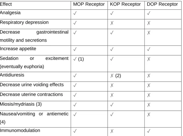
Opioids are chemical compounds, drugs, that promote pharmacological effects like those promoted by opium. However, these drugs are not chemically related. Opioid effects are mediated by opiate receptors. Three major receptors, and several subtypes, have been identified: μ, κ and δ (mu, kappa, and delta or MOP, KOP and DOP). For each receptor there are specific effects (Table 1), but clinically they are more commonly used for their analgesic and sedative proprieties (KuKanich & Papick, 2018).
Opioids can be characterized depending on how they bind to the different receptors. There are full agonists, partial agonists and antagonists.
Table 1 - Effects associated to the three main opiate receptors. Adapted from KuKanich &
Papick, 2018.
Effect MOP Receptor KOP Receptor DOP Receptor
Analgesia ✓ ✓ ✓
Respiratory depression ✓ ✗ ✗
Decrease gastrointestinal motility and secretions
✓ ✓ ✗ Increase appetite ✓ ✓ ✓ Sedation or excitement (eventually euphoria) ✓ (1) ✓ ✗ Antidiuresis ✓ ✗ (2) ✗
Decrease urine voiding effects ✓ ✗ ✗
Decrease uterine contractions ✓ ✗ ✗
Miosis/mydriasis (3) ✓ ✓ ✗
Nausea/vomiting or antiemetic (4)
✓ ✓ ✗
Immunomodulation ✓ ✗ ✓
1 – Dose and species dependent; 2 – Promotes diuresis; 3 – Species dependent; 4 – Drug specific.
Analgesic effect of opioids is primarily related with the binding to the spinal and supraspinal receptor (MOP, KOP and DOP). Despite this, peripheral effects were also studied and local injection in joints revealed some positive results. Binding to the receptors induce activation of
6
cyclase, diminished formation of cyclic adenosine monophosphate (cAMP) activation of potassium linked channels and inhibition of voltage gated calcium channels. This translates into a decrease in the release of neurotransmitters from the pre-synaptic membrane, where the three receptors are located, to the synaptic cleft because of the decreased influx of calcium ions. In addition, there are MOP receptors in the post-synaptic membrane that promote a hyperpolarization of the neuron by increasing the entrance of potassium ions affecting the nociceptive signal conduction. Considering this, full MOP agonists provide more profound analgesia than partial MOP agonists and KOP agonists. Nevertheless, for mild and moderate pain a partial MOP agonist and KOP agonist can be employed (KuKanich & Papick, 2018). Substance P has a role in the modulation of nociception. Some receptors for this substance are located in afferent fibers. Opioid peptides act at the MOP, KOP and DOP which modulate the nociceptive stimuli conduction on the dorsal horn of the spinal cord where the afferent fibers meet the spinal cord (Pascoe & Steffey, 2018). Opioids promote analgesia when systemic administered, the effects are primarily limited to the c-fibers (unmyelinated nociceptor fibers with slow conduction). Epidural administration demonstrated effect on both c-fibers and A δ-fibers (myelinated nociceptor fibers with fast conduction) providing greater analgesia (KuKanich & Papick, 2018).
MOP receptors activation have an important role on respiratory physiology. Depression of the medullary ventilatory control centers lead to a decreased response to increases in carbon dioxide partial pressures, hypoventilation (Pascoe & Steffey, 2018). In animals with previous respiratory disease, increased intracranial pressure (ICP) or neonates care should be taken when pure MOP agonists are administered because they could require respiratory support (KuKanich & Papick, 2018).
Some opioids, such as morphine, can promote excitement or even convulsions in high doses. The same happens when opioids are administered intravenously (IV) rapidly. Different species have different reactions to opioids. This can be explained by the different locations and number of opioid receptors depending on the specie. In dog, opioids receptors are distributed in the brain more than in the horse. Opioids produce dose-dependent sedation used alone or in combination with other sedative drugs. Excitatory and inhibitory neurotransmitters can also have a part on the possible excitement reaction. Increased release of gamma-aminobutyric acid (GABA) or glycine (inhibitory neurotransmitters) or increased release of acetylcholine and histamine were suggested as reasons for the sedation or excitement, respectively, of some species. This indicates that pharmacokinetics is not the major responsible for these differences (KuKanich & Papick, 2018).
Minimal effects on cardiac output can be cause by some opioids, when dealing with recommended doses. Bradycardia may occur in dogs, but in healthy animals there is no need for pharmacological intervention. In horses, the cardiac output may be compensated by the excitement promoted by opioids in this specie. Arterial blood pressure is minimally
affected by opioids administration. As discussed for the respiratory depression, the concurrent use of anesthetic drugs can promote cardiovascular depression.
MOP and KOP agonists have shown antitussive effects, not related with the respiratory depression (KuKanich & Papick, 2018; KuKanich & Wiese, 2015).
Some opioids stimulate the chemoreceptor trigger zone (CTZ) and central dopamine receptors leading to emesis and nausea. Hydromorphone and morphine are examples of opioids that can induce emesis or nausea. Emesis occurs in animals that are not in pain, like subjects undergoing surgery and receiving pure MOP agonists as pre-anesthetic medication. On the opposite, butorphanol, methadone and fentanyl have been reported to have some anti-emetic proprieties. Oral administration can also lead to emesis because CTZ is not protected by the blood-brain barrier and so, small plasma concentrations are enough to interact with this trigger zone. Again, histamine and dopamine receptors play a role on the emesis promoted by opioids. Apomorphine, a commonly used drug to induce emesis, acts as a central acting dopamine agonist (KuKanich & Papick, 2018; KuKanich & Wiese, 2015). Binding MOP, KOP and DOP receptors opioids can decrease gastrointestinal motility. Either from central and peripheral mechanisms or exogenous and endogenous opioids. Opioids slow down the gastric emptying, reduce propulsive motility, increases pyloric sphincter tone, increases fluid absorption and decrease fluid secretion. MOP receptors are present in the ileum (submucosal, muscle, and myenteric plexus). MOP, KOP and DOP receptors promote changes in mobility by inhibiting the calcium channels and decreasing the acetylcholine release. MOP and DOP receptors also hyperpolarize the neuronal membrane reducing impulse conduction and leading to less motility. MOP and DOP receptors have an important role in decreasing the fluid secretion by inhibition of chloride secretion and water movement to the colon lumen. As occurs with emesis, after initial vomiting and/or defecation, motility decreases, and sphincter tone are increased delaying the stomach emptying and the bowel movements, leading to constipation. This takes special importance when dealing with horses due to the often ileus and on patients undergoing gastrointestinal endoscopy (KuKanich & Papick, 2018).
As for gastrointestinal tract sphincters, MOP receptors agonists act in the urinary tract sphincters. Increased tone and urinary retention can be consequences of administration of opioids MOP agonists. Decreased urine production can also be due to anti-diuretic effect of MOP agonists. On the other hand, KOP agonists act as diuretics (KuKanich & Papick, 2018; KuKanich & Wiese, 2015).
Immune system is also target of opioid action. However, further studies are needed to better understand the immunomodulatory effects of this family of drugs and it’s role in some processes such as spreading of metastasis in oncologic subject (KuKanich & Papick, 2018).
8
Butorphanol, MOP partial agonists and KOP agonists, can be used in animals who presented excitement or dysphoria which could be caused or enhanced by a full MOP agonist (KuKanich & Papick, 2018).
2. Butorphanol
Butorphanol is a synthetic opioid, known as opiate (Semjonov et al., 2018). It is an highly lipophilic molecule that binds to the three principal opioid receptors mu, kappa, and delta, it acts as a κ receptor agonist and a μ receptor partial agonist or antagonist, depending on the source (Warne, Beths, Holm, & Bauquier, 2013; World Health Organization, 2006). Butorphanol is used as an analgesic, a sedative and an antitussive drug. Its efficacy as analgesic drug is dose dependent, presenting a plateau (or ceiling) effect, which means its efficacy increases with higher doses. The plateau effect it’s reached at the 0.9 mg/kg in the cat and the duration of effect does not increases with higher doses (KuKanich & Papick, 2018).
Commercially, butorphanol is presented has a preparation of butorphanol tartrate (BSAVA, 2017a; KuKanich & Papick, 2018).
Concerning pharmacokinetics, an important difference between the half-life and clearance of dogs and cats is noticed (Table 2). This difference indicates a much slower rate of drug elimination in cats. Oral administration (PO) and bioavailability is poor (KuKanich & Papick, 2018; KuKanich & Wiese, 2015).
Table 2 - Pharmacokinetic parameters of butorphanol intramuscular (IM) and PO bioavailability in dogs and cats (Adapted from KuKanich & Papick, 2018 and KuKanich & Wiese, 2015). Half-life (h) Clearance (ml/min/kg) Volume of distribution Oral bioavailability IV LD50 (mg/kg) Dog 1.6 57 8.0 < 20% 10
Cat 6.6 12.7 7.7 Data not
available
Data not available
Administered intramuscular, butorphanol peak of action occurs after 20 to 30 minutes of administration, but when administered IV this peak changes to 2 to 5 minutes after administration. Both IM and IV administrations can lead to 60 to 90 minutes period of action (Murrel, 2016).
2.1 Dosage
The recommend doses are: 0.2 to 0.5 mg/kg IV, IM, subcutaneous (SC) or PO for both dogs and cats for analgesia purposes and 0.05 to 0.1 mg/kg IV, IM, SC or 0.5 to 1.0 mg/kg PO for antitussive purposes (BSAVA, 2017a).
2.2 Primary uses/effects
Butorphanol is normally used as a sedative in dogs and cats but is also used as analgesic in patients with mild to moderate pain states. Butorphanol shows to be less effective in severe pain than methadone (Warne et al., 2013). When using butorphanol, it is important to know the unset of sedation occurs at lower dosages than analgesic effects. Therefore, sedation should not be assumed as analgesia also (KuKanich & Papick, 2018).
Butorphanol can be used alone or in combination as pre-anesthesia medication. Butorphanol shown to promote greater sedation degree end used in combination with dexmedetomidine in comparison with methadone-dexmedetomidine. Also demonstrated better reliability, less failed sedations, when compared with methadone (Trimble, Bhalla, & Leece, 2018).
Butorphanol is also an antitussive agent, superior to morphine and codeine. It can be used as an anti-emetic agent in patients undergoing chemotherapy. In contrast, morphine and apomorphine can lead to emesis. Butorphanol has got minimal impact on the cardiovascular and respiratory system, when used at the clinically recommended dosages. Unwanted effects, that characterize the opioids, may also be present in patients administered with butorphanol. These effects include: dysphoria, mydriasis (in cats), decreased motility of the gastro-intestinal tract and in some cases even constipation. Considering the MOP antagonist activity of butorphanol, undesired effects occur with less severity when compared with pure MOP agonists (KuKanich & Papick, 2018).
2.3 Special concern
Some authors have been speculating that, because butorphanol is a P-glycoprotein substrate, the sedation promoted by this drug is more profound and prolonged in multidrug resistance 1 (MDR1) mutation gene carrier dogs. With this possible concern, doses are suggested to be decreased in at least 50%. However, without a well-designed study yet published, this speculation is not scientifically confirmed (Mealey, 2006).
Part B - General Anesthesia 1. Definitions
In greek, the word anaisthaesia means insensibility, a loss of sensation. Nowadays the term anesthesia is used to describe a much wider concept.
The American Society of Anesthesiologists (ASA) published a consensus on the definitions of Sedation and General Anesthesia (American Society of Anesthesiologists, 2014). In this consensus, ASA divided sedation in three stages: minimal sedation or anxiolysis, moderate sedation or conscious sedation and deep sedation.
10
Minimal sedation is defined as “… drug-induced state during which patients respond normally to verbal commands (…) cognitive and physical coordination may be impaired …”. Moderate sedation differs from the minimal sedation, because in this stage there are depression of conscious. Finally, Deep sedation involves the depression of consciousness, diminished response to verbal stimuli but maintained response to repeated physical stimuli or provoked pain. In these three stages, the latter is the only one where the patient may require external help to maintain a patent airway or spontaneous ventilation.
General Anesthesia produce loss of consciousness, “reversible alteration of wakefulness and cognitive function of the brain” (Bonhomme & Hans, 2004), “… during which patients are not arousable, even by painful stimulation …”. These patients often require assistance to maintain spontaneous ventilation and the cardiovascular function may be impaired (American Society of Anesthesiologists, 2014).
On the other hand, another definition for General Anesthesia was proposed “drug-induced, reversible state characterized by amnesia, unconsciousness, analgesia, immobility, and muscle relaxation” (Tranquilli & Grimm, 2015).
Anesthesia plan depends on the patient and their medical condition. Type of procedure is also relevant. Depending on the procedure, the patient may need to unconscious, analgesia or even be in total muscle relaxation (for example for phacoemulsification procedure).
2. General Anesthesia Mechanisms
As written above, general anesthesia is induced by drugs that act like depressant in the central nervous system (CNS) (Otto, 2015).
After several thesis about how the anesthetics agents provoke general anesthesia, some studies open the door to the hypothesis that the action occurs on the proteins (rather than on fat tissue), more specifically on the integral membrane proteins. Therefore, the anesthetics agents promote reversible effects on the ion channels ligand or voltage gated in the central nervous system (Garcia, Kolesky, & Jenkins, 2010; Krasowski & Harrison, 1999). However different anesthetic agents exert actions in different proteins.
The volatile halogenated agents act on several proteins, some of them are inhibited and other are enhanced. For instance, they are reported to activate the potassium channels, acting like enhancers of GABA and glycine receptors (Forman & Chin, 2008). Volatile agents also act as inhibitors of the excitatory glutamate receptors, serotonin subtype 3 channels, also known as 5-HT3, and on ion channels associated with nicotinic acetylcholine (nACh) receptors (Flood & Role, 1998; Stevens, Rüsch, Davies, & Raines, 2005).
Injectable anesthetics seem to act on only one pathway. It is proposed that injectable anesthetics act mainly at inhibitory GABA receptors (Otto, 2015). The GABAa seem to be the most relevant neurotransmitter of hypnosis in the mammals (Lambert, 2004).
However, more sites of action have been discovered to be play a role in the anesthetic mechanisms, including: glycine receptors, 5-HT3 receptors, nACh receptors and inotropic
glutamate receptors as N-methyl-4-isoxazoleprpionic acid (NMDA)(Flohr, Glade, & Motzko, 1998; Yamakura & Harris, 2000), 2-carboxy-3-carboxymethyl-4-isopropenylpyrrolidine (kainate) and alfa-amino-3-hydroxy-5-methyl-4-isoxazoleprprionic acid (AMPA) receptors (Yamakura & Harris, 2000). From all this, the NMDA receptors gain special attention because is where ketamine and some of the volatile agents acts (Hollmann, Liu, Hoenemann, Liu, & Durieux, 2001; Lambert, 2004; Petrenko, Yamakura, Sakimura, & Baba, 2014).
3. Anesthesia Depth and Planes
When maintaining a general anesthesia, a classification of the depth in which the patient is in takes relevance. In this field, Doctor Arthur Guedel had an important role by proposing guidelines in a form of a chart that the anesthesiologists could use to evaluate how deep is the patient, since the induction to possible death. In the Table 3, later named Guedel’s classification, there are four stages and three planes in the third stage.
Table 3 - Guedel's table for monitoring anesthesia depth (Adapted from Cascella, 2015).
Due to the modern technics of anesthesia monitoring, the Guedel’s classification is getting less importance in our days. Besides new and improved monitoring equipment, the use of multi-drug combinations to induce and maintain general anesthesia has also a role in the loss of reliance on this classification. Having this said, today’s monitoring relies on
12
physiological parameters like blood pressure, respiration and neuromuscular tone. The next step of the anesthesia monitoring, that it’s starting to gain application and relevance, seem to be the monitoring of the neuro-electric function of the brain with the electroencephalographic monitoring (Tranquilli & Grimm, 2015).
4. Anesthesia Types
There are types of anesthesia maintenance, anesthetic drug or drug administration. With all these we can classify the anesthesia in several types, however for practical, economical and safety proposes there are three that are more often used: inhalation, injectable and total intravenous anesthesia (TIVA). The inhalation is applied with anesthetic gases or vapors that are inhaled mixed with air and oxygen. Injectable is applied with anesthetics that can be injected intravenously, intramuscularly and subcutaneous, other routes are not commonly used. TIVA is applied when a continuous rate infusion of an anesthetic drug or a combination of drugs are administered intravenously, usually by automated infusion systems (Tranquilli & Grimm, 2015).
5. Anesthesia Risks
When anesthesia is needed, a risk analysis should be performed, and the advantages and disadvantages of this anesthesia should be taken in consideration, to discuss the several options with the owners. In recent studies, the ASA status was associated with the percentage of animal death due to anesthesia. Patients with higher ASA status were associated with an increased risk of death (Bille et al., 2012; Gil & Redondo, 2013; Itami et al., 2017). In another study, poor prognostic factors were observed in dogs and cats with increased ASA score and colloid administration (Smith, Barletta, Young, & Hofmeister, 2017).
It is well known that anesthetic agents can cause cardiopulmonary disturbances, most of the times depression, and if there is a pre-existing pathology associated with the animal, it is most likely to have disturbances caused by the anesthesia. In fact, most of the disturbances or death related to anesthesia are related with the cardiopulmonary system (Itami et al., 2017). For instance, in gastrointestinal surgery, dogs that presented intraoperative hypotension had increased risk of developing septic peritonitis post-surgery (Grimes, Schmiedt, Cornell, & Radlinksy, 2011). For this reason, at least a good physical examination including cardiopulmonary auscultation and blood pressure measurement is advised.
Also, basic blood test including hematologic and liver and kidneys biochemistry and biochemical control is also important. To demonstrate this, it is known that serum glucose values below 77mg/dL and a white cell count above 15.200 cel/µL had greater risk of anesthesia-related death (Itami et al., 2017). Anemia is also important because will predispose to hypoxemia, hypoproteinemia will interfere with the drugs that have high protein-bounds that can predispose to overdose (Brodbelt, Flaherty, & Pettifer, 2015). In
patients with renal pathology, dehydration and uremia increase the changes of acute renal insufficiency because of the least resistance to the anesthetic and non-anesthetic drugs, as non-steroidal anti-inflammatory drugs, and the possible hypotension related with anesthesia that will impair the normal renal perfusion (Bednarski et al., 2011; Brodbelt et al., 2015). Curiously, in one study, animals that received analgesic therapy with nonsteroidal anti-inflammatory drugs (NSAID’s) or opioids had less risk of anesthesia-related death than those who did not receive it (Gil & Redondo, 2013). Also, liver and endocrine disease are able to affect the anesthesia, special concern with diabetes mellitus because of the possible changes on blood glucose intraoperatively (Bednarski et al., 2011; Brodbelt et al., 2015). On a retrospective study with dogs that underwent phacoemulsification surgery, hypotension was observed to be more frequent on diabetic dogs and 44% of the diabetic dogs had at least one episode of severe hyperglycemia during anesthesia. In the same study, the authors suggested that concomitance of other endocrine diseases like hyperadrenocorticism may also have a role on the peri-anesthetic complications (Oliver, Clark, Corletto, & Gould, 2010). For all the risks written above, there are some of them that the clinician is not able to avoid (Brodbelt et al., 2015; Flemming & Scott, 2004).
6. Monitoring
6.1 Cardiovascular system
When monitoring the cardiovascular system, a variety of constants should to be checked every time it is possible.
6.1.1 Heart rate and rhythm
The cardiac output is determined by several factors, but the heart rate gains a special importance. The heart rate is measured in beats per minute (bpm) and for a dog it varies between 60 and 160 bpm, depending on the breed and size and for a cat it is higher between 120 and 220 bpm. On one hand, bradycardia states lead to a decreasing on the cardiac output even so the refiling time on diastole and stroke volume both increase. On the other hand, tachycardia diminishes the refilling time and decrease the stroke volume that will also decrease the cardiac output. When discussing if intra-anesthetic bradycardia and tachycardia should be treated, the agreement standing is to treat every time that these are associated with poor cardiac output, blood pressure or tissue perfusion. There are some causes of bradycardia that does not respond to medical pharmaco-therapy like hypothermia, severe myocardial hypoxemia and malfunction of cardiac electric conduction. The current treatment advised is based on sympathomimetic or anticholinergic drugs (Haskins, 2015). Concerning tachycardia, the major impact can be how the diastolic filling is affected and if there are ventricular arrhythmias. When tachycardia is present, the anesthesiologist should address if it is a compensatory or primary cause of hemodynamic problems. For this reason, treating
14
compensatory tachycardia leads to hemodynamic decompensation of the animal and pharmacological treatment should be administered depending on the primary cause.
6.1.2 Arterial blood pressure
The arterial blood pressure is an important part of the monitoring of the cardiovascular system for the anesthesiologist during the general anesthesia of a patient. The arterial blood pressure can be divided in three measurements: systolic arterial pressure (SAP), diastolic arterial pressure (DAP) and mean arterial pressure (MAP). The SAP is mainly determined by the stroke volume and arterial system compliance. On the other hand, DAP is mainly determined by the systemic vascular resistance and circulating volume. The MAP is not the real arithmetic mean between these two values, but it is used often as so. Normal range values for dog and cat are 100 to 160 mmHg of SAP, 60 to 100 mmHg of DAP and 80 to 120 of MAP. Values of MAP below 50 mmHg will compromise the perfusion pressure and blood flow of the brain and coronary arteries. There are several reasons for hypotension development during anesthesia like hypovolemia, vasodilation or insufficient cardiac output. However, when an acute hypertension, edema or hemorrhage happen, the brain is one of the organs that more suffers with the increase of the arterial blood pressure values. An animal with a MAP value over 140 or 150 mmHg is considered as a hypertensive patient. When dealing with a hypotensive or hypertensive animal, the anesthesiologist as to consider these values as suggestions, because there are a few factors that can alter the blood pressure values without compromising the wellbeing of the animal. For instance, it is frequent altered values when the measurements are no reliable or hypotensive state when anesthesia induction starts. For these reasons the pharmacological treatment should be carefully administered because it can cause more damage than good (Haskins, 2015; Schauvliege, 2016).
There are two methods to measure the arterial blood pressure: indirectly with a sphygmomanometer or directly with an arterial catheter (Haskins, 2015). In a study performed in pigs, the direct measurement technique seems to be more reliable. However, the authors state that indirect measurement technique appears to be acceptable in general anesthesia monitoring (Gladczak, Shires, Stevens, & Clymer, 2013). In a different study, when comparing doppler measurement with direct blood pressure measurement in cats, the results had poor agreement and the doppler values were considered misleading. In this case the authors advise the caution when using and interpreting the doppler values (Da Cunha et al., 2014).
6.2 Respiratory system 6.2.1 Respiratory effort
The breathing pattern allows the anesthesiologist to have a variety of information about the patient status. Altered respiratory patterns can indicate respiratory disease or can be physiological, depending on the medication and plane of anesthesia which the animal is in. A slower frequency of respiratory cycles, bradypnea, may indicate that the depth of anesthesia is increasing or hypothermia. Meanwhile a tachypnea can indicate pain, change on the anesthesia depth or hypotension for example. Just after induction with propofol, apnea is usual (Haskins, 2015).
6.2.2 Partial pressure of carbon dioxide
The arterial partial pressure of carbon dioxide (PaCO2) is an indicator of the effective alveolar
minute ventilation and it should be between 35 to 45 mmHg in healthy animals. When PaCO2
increases over 45 mmHg the animal is in hypercapnia and when it decreases under 35 mmHg the animal is in hypocapnia. Hypercapnia occurs when the anesthesia plane is to deep, when there is an airway obstruction or a neuromuscular or respiratory disease. On the other hand, hypocapnia occurs when the animal is in hyperventilation that can be due to hypoxemia, pulmonary disease, hyperthermia or superficial anesthesia depth (Haskins, 2015).
However, most of the times when performing anesthesia in small animal practice there are no blood gas analysis intra-anesthesia, and for this reason the PaCO2 value is calculated
from the End tidal or Expired concentration of carbon dioxide (FE’CO2) that is given by the
measuring of the carbon dioxide (CO2) exhaled by the animal. There are technologies that
can be used to measure it, but the monitoring equipment is always called capnograph. This said, End tidal CO2 is 5 units higher than PaCO2 and so the normal range of End tidal CO2 is
between 40 to 50 % in dogs and 35 to 45 % in cats (Haskins, 2015; Schauvliege, 2016). 6.3 Temperature
Hypothermia is one of the most common complications during and after anesthesia (Kennedy, Tamburello, & Hardie, 2011). The normal values of temperature, or normothermia, in the dog range from 38,5 to 39,5 Celsius degrees (Redondo et al., 2012).
16
Hypothermia happens due to depression of metabolism, induced by drugs, decrease of muscular activity and possible hypothalamic thermostatic impairment, for example, with the use of opioids. Also, some anesthetic drugs have vasodilation effect which will increase the heat loss as well, enhanced with the time that the animal is under anesthesia, the body surface that is exposed, temperature and fluid therapy volume that the animal receives, exposure of body cavities to the environment and contact with cold surfaces.
Although, it is not a consensus, it is accepted that intra anesthesia, the temperature is usually monitored using esophageal thermometers (Haskins, 2015). The anesthesia recovery is longer for hypothermic patients and presents some detrimental collateral effects (Pottie, Dart, Perkins, & Hodgson, 2007).
7. Anesthetics influence on vascular tone
The vascular tone can also be altered by some anesthetic drugs. In 1974, the vasodilation and cerebral metabolic depression caused by inhalant agents, isoflurane and enflurane, used in dogs, was well demonstrated in two studies (Cucchiara, Theye, & Michenfelder, 1974; Michenfelder & Cucchiara, 1974). When using volatile anesthetics like isoflurane or sevoflurane, the vasodilation effect is dose-dependent, but the CO2 reactivity of the vessels
is not compromised. In humans, some studies proved that sevoflurane acts as the least vasodilator agent when the minimum alveolar concentration is less than 1 (Pang, 2016). For this reason, the sevoflurane appears to be the most indicated volatile agent in general anesthesia maintenance in a patient with intracranial pathology (Pang, 2016). Other agent, that theoretical presents advantages for neurological patients with a cerebral vasoconstriction effect reducing ICP and the cerebral blood volume, and at the same time maintain the vasoreactivity to the cerebrospinal fluid (CSF) pH and the autoregulatory mechanisms, is propofol. However, these advantages were still not well demonstrated in a clinical study (Dagal and Lam, 2009).
8. Pre-medication
A dog that needs sedation to perform an ultrasound will not receive the same anesthetic protocol than one undergoing surgery for fracture repair. At the same extend, a excited or aggressive dog will be approached differently that a calm one. For these reasons, there are no universal anesthesia protocol and the approach should be tailored to each subject. Opioids are important to provide sedation and analgesia, but not general anesthesia (Bednarski, 2015). For these reasons, premedication takes an important role on the anesthesia plan. Premedication is usually used to provide sedation and analgesia, reduce anesthetic drug used for induction and maintenance of general anesthesia and to allow a smooth induction, maintenance and recovery of general anesthesia. With premedication, intravenous catheter placement should be easier by reducing the animal movement and
decreasing its stress level. In case of aggressive or difficult to restrain animals, handling them after premedication administration should also be easier. Intramuscular administration is often used, however for better uptake reliability premedication should be administered intravenously (Brainard & Hofmeister, 2012).
Considering that magnetic resonance imaging is not a painful procedure (the position may be uncomfortable for spinal cord injured patients), butorphanol presents to be indicated for sedation in patients with suspected intracranial pathology. In addition, butorphanol has a maximum duration of effect around 90 minutes, which avoids long recoveries in patients used in our study because the longest general anesthesia time was 95 minutes.
The bradypnea caused by MOP partial agonist like butorphanol is not that relevant when compared with MOP agonists. General anesthesia is also needed to prevent movement besides the respiratory ones.
Part C – Neuroanesthesia 1. Brain physiology
One important concern when dealing with a dog with suspected brain pathology is the increase of the ICP (Leece, 2016; Otto, 2015). So, to better understand how these dogs should be clinically managed and safely submitted to anesthesia a brief overlook of the brain physiology is important.
1.1 Intracranial pressure
Two of the most import investigators for a better knowledge of the brain physiology were Monroe and Kellie. They proposed a thesis that stated that the cranium acts like a closed compartment composed by three major components: brain tissue occupying 80 to 85% of the cranium, cerebrospinal fluid with 7 to 10% and blood (sub divided in arterial and venous) responsible for 5 to 8% of the volume. For this reason, when an increase in the volume in one of these three compartments occurs, the ICP increases. The volume may increase for several reasons as: space-occupying lesion, hematoma, edema, trauma or inflammation (Leece, 2016).
Dog normal value of the ICP range between 0 and 10 mmHg (Leece, 2016) or 5 to 12 mmHg (Madison, Sharma, & Haidekker, 2015) depending on the authors, so above this value, 20 to 30 mmHg, the dog presents an increased ICP. However, there are some physiological mechanisms in the healthy animals that can avoid this increase in the initial stages as illustrated by Graph 1. The most relevant is the fluid displacement with movement of CSF from the cranium the medullary canal, and blood from the brain vessels to the rest of the body. This problem presented by increased ICP turns out to be very relevant, because with the limited space of the cranium an increase in the ICP can lead, in a short period of time, to tentorial and tonsillar herniation from the cranium to the medullary channel or hydrocephalus
18
secondary to blocked or decreased CSF drainage (Leece, 2016). However, there are other important issues for the when ICP increases, namely a vasomotor paralysis, a decrease in the cerebral blood flow (CBF) and insufficient delivery of oxygen occur (Pasternak & Lanier, 2014). All these lead to an impaired irrigation and nutrition of the brain tissue and eventually will promote an ischemia and a neuronal necrosis (Lassen & Christensen, 1976).
Graph 1 - Relation Intracranial volume-pressure (mmHg) and Ischemia
1.2 Cerebral blood flow and cerebral perfusion pressure
For a correct functioning of the brain, two major assumptions must be considered: the cerebral perfusion, and the metabolism. To be assured that these two are not compromised, an adequate blood flow to deliver oxygen and glucose is crucial (Otto, 2015).
As in many other organs, the blood flow depends on the perfusion and vascular resistance. For the brain the formula is: Cerebral blood flow = cerebral perfusion pressure/ cerebral vascular resistance (Leece, 2016).
Healthy animals present several autoregulatory mechanisms, that can maintain a constant CBF independently of systemic blood pressure and cerebral perfusion pressure (CPP) changes. However, under general anesthesia or a pathological condition, some of this autoregulatory mechanisms might present difficult work or even be abolish. A study suggests that volatile agents such as isoflurane turn the CBF “passively dependent upon CPP” which can be traduced by the equation: CPP=MAP – ICP. Some authors also add the central venous pressure value to the formula, however, this pressure values are so low (mean 5 cmH2O) that most of the times are ignored. As it seems to be obvious, when monitoring a
general anesthesia of a confirmed or suspected animal with intracranial pathology the blood pressure, represented by the mean arterial pressure (MAP) should be closely monitored. For this reason, a special concern with the cardiovascular depression and fluid therapy is very important. In the specialized literature it’s advised a CPP over 70 mmHg which correlates with a MAP of 70 to 80 mmHg (Leece, 2016; Otto, 2015).
The major influencer of the blood flood regulation in the brain is the arterial CO2 (PaCO2),
which for each 1 mmHg of change it can increase or decrease in 4% the cerebral blood flow (Lassen & Christensen, 1976). The reactions to the PaCO2 are mediated by the alteration on
the CSF pH that induce near to the arterioles, their dilation or contraction depends on the pH alterations (Lassen & Christensen, 1976).
However, the homeostasis of the CSF pH and PaCO2 will affect the pulmonary ventilation,
when changes in CSF pH occur near the brainstem (Otto, 2015; Leece, 2016). Hyperventilation is one away to induce mild vasoconstriction in the brain arterioles during surgery, because it leads to hypocapnia. This technique works when it is performed for a limited period, and when applied over several days for coma patients an adaptation takes place, and the PaCO2 and the CSF pH will have a limited effect on the cerebral
vasoconstriction. However, PaCO2 values below 25 mmHg start to cause ischemia of the
neuronal cells which will affect their normal function (Lassen & Christensen, 1976; Otto, 2015). For this reason, it is recommended to maintain a PaCO2 between 30 to 33 mmHg (Leece, 2016).
1.4 Oxygen (O2)
On the other hand, oxygen partial pressure (PaO2) can only have an influence on the
cerebral blood flow with a value below 50mmHg (Gupta, Smielewski, Menon, Czosnyka, & Jones, 1997). When this happens, the CBF increases but at the same time a shift from aerobic to anaerobic respiration occurs, leading to an increase of lactic acid production with ultimately unset of brain tissue acidosis and total abolishment of autoregulatory mechanisms (Otto, 2015). This hypoxia in conjunction with hypercapnia will promote a pronounced cerebral vasodilation, increasing therefore the ICP and eventually the herniation of brain tissue into the medullary canal, promoting edema or hemorrhage (Lassen & Christensen, 1976).
All this brain physiology and autoregulatory mechanisms should be considered when dealing with healthy animals, because in trauma, space occupying lesions, inflammation, (…), are capable to modify all these assumption (Lassen & Christensen, 1976).
1.5 MAP
Although, the arterial blood pressure control gains a special importance it is known that in a range of MAP, the blood flow that arrives to the brain stays constant. This interval of 50 to 150 mmHg is possible because of the autoregulation mechanisms present in the healthy animals, as illustrated by the Graph 2 (Leece, 2016).
20
When MAP reaches a value lower than 45mmHg (Duke-Novakovski, 2016) or 60mmHg (Lassen & Christensen, 1976), the blood flow drops and the difference between arterial and venous blood oxygen increases. At this point, and apart from systemic symptoms, as acute renal insufficiency because of low renal perfusion (Duke-Novakovski, 2016), hyperventilation that we can see during general anesthesia or in an awake animal, neurological symptoms start to appear, like dizziness and mental impairment (Lassen & Christensen, 1976). On the other hand, with a MAP above 130 mmHg (Lassen & Christensen, 1976) or 150 mmHg (Leece, 2016), vasodilation starts on leading an ICP increasing because of the increased volume in the cranium, eventually promoting loss of the blood-brain barrier with the rupture of blood vessels inducing hemorrhage or cerebral edema (Lassen & Christensen, 1976). However, small periods of hypertensive states will not cause blood-brain barrier disruption (Lassen & Christensen, 1976). A special concern must be taken when dealing with chronic hypertensive animals. When a hypertensive state is present for a long time, brain vessels adapt along with the progressive state of the hypertension (Lassen & Christensen, 1976). The specific adaptation mechanism is the hypertrophy of the vessels halls. In practical, these patients tolerate much better high MAP values than normotensive animals. However, this means that when dealing with lower MAP values, their limit of autoregulation it’s reached at higher values, 60 to 70 mmHg and not the 50 mmHg of the healthy animal (Lassen & Christensen, 1976; Strandgaard, 1976).
1.6 Management of Neurological Patients
When dealing with animals with confirmed or suspected increased ICP, a head elevation of 30 degrees was proved to promote a good venous drainage of the head, decreasing the ICP, without compromising the MAP or the brain oxygenation (Ng, Lim, & Wong, 2004). An important concern when applying this technic is to avoid jugular compression that would diminish the head blood drainage. For this is not just the head that should be elevate, but the hole animal.
A careful approach to the fluid therapy of patients with suspected or increased ICP must be taken. Recent studies cannot differentiate the usage of hypertonic saline and mannitol (Schreibman et al., 2018; Witherspoon & Ashby, 2017), the only valid consideration is that in
hypovolemic patients the hypertonic seems to be the ideal fluid, but when the patient does not respond to that, mannitol should be administered (Kuo, Bacek, & Taylor, 2018).
2. MRI Anesthesia
The Magnetic Resonance Imaging (MRI) technique is known to be better in diagnosing several neurological pathologies (Beltran et al., 2012; Da Costa, Parent, Dobson, Holmberg, & Partlow, 2006). However, a few concerns about the magnetic field necessary for the MRI performance must be taken in concern.
In summary, the MRI functioning is based on a magnetic field that changes during the scans promoting alteration on the atomic magnetic fields and electrons movement on the body. This imaging procedure does not have harmful biological effects yet documented, however, there are not studies on long term exposition to the MRI magnetic field. Because of this magnetic field, some concerns regarding metallic objects should be taken, because these objects can be turned into flying projectiles. This point is relevant when performing the anesthetic monitoring, because the appliances must be MRI safe. Another concern can be the noise produced by the MRI, and in this case, ear plugs and/or headphones seem to be a good option for personnel safety. Skin burns were also reported in humans because there are electrical currents induced by the magnetic field changes that are present in the conductive materials as electrocardiogram cables. If the scanner is in a separate room there is no need for a protective cage, however most of the times a Faraday cage is needed to separate the scanner from the anesthetic monitors, imaging technician and other appliances such as computers.
For this reason, extension cables and lines for anesthetic monitoring and injection points can be applied for a easier monitoring and administration of drugs, fluids and contrast to the patient (Dempsey & Condon, 2001; Hartwig et al., 2009; Smith, 2016).
Patients (animal or human) submitted to a MRI procedure are positioned on a long or round table, depending on the MRI scanner. The recumbency depends on the coil used and the area of the body subjected to the imaging examination. For a better management of the patient, some technics that can be applied, as for example: covering the animal to avoid hypothermia, sand bags and foam pillows can be placed to help positioning and special care when positioning an animal with intervertebral disc herniation because of the pain related to the injury (Otto, 2015).
An important concern when performing an MRI in a dog is the need of total immobilization, besides the respiratory movements that can also diminish the quality of the images which can imply a scan repetition with more study time and overall more anesthesia time as well. For this reason, heavy sedation or general anesthesia induction is advised animal patients (Smith, 2016).
22
As explained above, special concern should be taken about the monitoring equipment. All the equipment that remains inside de cage as to be MRI-safe to the magnetic field do not suffer any change that may affect the quality of the image and most important to avoid accidents with flying material (Smith, 2016).
Hypothermia is common in MRI patients not well covered or heated, because the room must be climatized to avoid overheating of the magnet and the exam may take some time to perform. Covering the patient with blankets may be enough to maintain normothermia (Smith, 2016).
2.2 Gadolinium Contrast Agent
To allow a better definition and contrast between tissues, a gadolinium-based contrast may be administered to the animal. However, concerning the patient, there may be some adverse reactions to this contrast (Smith, 2016), which can be divided in two groups: 1) depending on the onset time: acute or delayed; 2) by the severity of the symptoms caused: mild, moderate and severe (Scarabelli, Cripps, Rioja, & Alderson, 2016).
There are just a few studies in veterinary medicine about this subject and some of them are contradictory. Some state that no changes on heart rate and MAP were observed (Mair, Woolley, & Martinez, 2010) and others say there are 5% of dogs and 14% of the cats that had hemodynamic alterations (Pollard, Puchalski, & Pascoe, 2008).
Some anaphylactoid reactions have been reported, however this are just suspect reactions and the number of cases is too low to be considered relevant (Girard & Leece, 2010).
In a study performed with 238 dogs with suspected intracranial pathology that underwent brain MRI, there were no significant difference between the percentage of dogs that suffer peri-anesthesia complications, however there was a difference between recovery. The recovery time was longer in dogs with MRI image of potential intracranial pathology (Hicks, Kennedy, & Patterson, 2013).