Ophthalmic and anesthetic evaluation of topical 1% tetracaine and 0.5%
proparacaine in dogs
[Ação anestésica e oftálmica da administração de colírio de tetracaína 1% e proparacaína 0,5% em cães]
H.D. Parchen1, M.L. Izar1, P.S. Branco2, C. Lacowicz2, D.H. Sano2, C.E.P. Belo2, R.G.D’O.C. Vilani3*
1
Aluno de pós-graduação – Universidade Federal do Paraná
2Aluno de graduação – Universidade Federal do Paraná 3
Universidade Federal do Paraná Rua Funcionários, 1540
80035-060 - Paraná
ABSTRACT
The aim of this study was to establish the action of 1% tetracaine eye drops in combination with 0.1% phenylephrine in two different posologies and their effects on the eye compared to the 0.5% proparacaine drops in dogs. 22 animals were divided into two groups: TG (11 animals), received 1% tetracaine associated with 0.1% phenylephrine eye drops, one drop instilled in the left eye and two drops, with one-minute interval between each, instilled in the right eye; PG (11 animals) received 0.5% proparacaine eye drops following the same dosage. The average duration of the observed anesthetic action was 25 minutes for tetracaine and 15 minutes for proparacaine. The instillation of two drops increased anesthetic time in five minutes. No changes in intraocular pressure, pupil diameter and tear production was observed. The drops of tetracaine triggered chemosis in four (36.4%) animals. Topical anesthesia with proparacaine eye drops showed no adverse reactions and is thus recommended preferentially.
Keywords: dog, topical anesthesia, eye, aesthesiometry, Cochet-Bonnet
RESUMO
Estudou-se a ação do colírio de tetracaína 1%, em associação com a fenilefrina 0,1% em duas posologias diferenciadas, bem como seus efeitos oculares, comparando-a com a do colírio de proparacaína 0,5% em cães. Vinte e dois animais foram separados em dois grupos. Os do GT (n=11) receberam colírio de tetracaína 1% associada à fenilefrina 0,1%, sendo uma gota instilada no olho esquerdo e duas gotas, com intervalo de um minuto entre cada, instiladas no olho direito; e os do GP (n=11), receberam colírio de proparacaína 0,5% seguindo a mesma posologia. A média de duração da ação anestésica observada foi de 25 minutos para a tetracaína e 15 minutos para a proparacaína. A instilação de duas gotas aumentou o tempo anestésico em cinco minutos. Não ocorreram alterações na pressão intra-ocular, no diâmetro pupilar e na produção lacrimal. O colírio de tetracaína desencadeou quemose em quatro (36,4%) animais. Na anestesia tópica do olho com proparacaína não ocorreram reações adversas sendo, assim, recomendada preferencialmente.
Palavras-chave: cão, anestesia tópica, olho, estesiometria, Cochet-Bonnet
INTRODUCTION
Currently, in the topical ophthalmic anesthesia routine, proparacaine (also called proximetacaine) is the most widely used drug (Stiles et al., 2001). Its use is largely devoted to
Recebido em 18 de julho de 2010 Aceito em 2 de agosto de 2011
*Autor para correspondência (corresponding author) E-mail: [email protected]
Parchen et al.
2005), cats (Binder and Herring, 2006), rabbits (Seabaugh et al., 1993), horses (Kalf et al., 2008) and men (Ezra and Allan, 2007).
Besides the formulation of proparacaine 0.5%, tetracaine 1% eye drops are commercially available associated with 0.1% phenylephrine, a vasoconstrictor which aims to prolong the anesthetic effect. The tetracaine ophthalmic solution is widely used in veterinary medicine in Brazil, but few studies elucidate its action time and anesthetic efficacy in animals. Its ocular effects have been well established only in humans, which has slightly less anesthetic duration than the proparacaine, and moreover, is being associated with discomfort in the application (Bartfield et al., 1994). The comparison of analgesic and ophthalmic effects, as well as the adverse reactions, of these two commercially available formulations of anesthetic eye drops in dogs will set up the indications for each drug for this species.
The aim of this study was to determine and compare the anesthetic and the ocular effects of the two main commercial formulations, the drops of tetracaine 1%, in combination with phenylephrine 0.1% and 0.5% proparacaine eye drops in two different doses in dogs.
MATERIALS AND METHODS
A total of 22 healthy, male and female dogs of different breeds were randomly divided into two groups: TG animals (n=11) received tetracaine ophthalmic drops 1% associated with phenylephrine 0.1% (Colírio Anestésico®, Allergan Prod. Farm. Ltda, São Paulo, Brazil), being one drop instilled in the left eye (1) and two drops, with one-minute interval between each, instilled in the right eye (2), and PG animals (n=11) received proparacaine 0.5% eye drops (Anestalcon®, Alcon Lab. do Brasil Ltda, São Paulo, Brazil), being instilled a drop in the left eye (1) and two drops, with one-minute interval between each in the right eye (2).
Before application (T0) corneal sensitivity was measured in both eyes, using a Cochet-Bonnet esthesiometer (Luneau Ophtalmologie, Chartres,
France), noting the value of the longest length that triggered responses of retraction of the eyeball, eye movement or blinking, intraocular pressure, using Schiotz tonometer (Miltex, Tuttlingen, Germany), photomotor and consensual reflexes and the pupillary diameter with a digital caliper (Vonder Ferramentas, São Paulo, Brazil). The measurements continued after instillation of the drops, being recorded at 5 minutes intervals until 60 minutes (T5, T10, … T60). The Schirmer tear test (Ophthalmos Farmacêutica, São Paulo, Brazil) was performed at T0 and repeated at T60 in order to detect changes in tear production. Attention has been focused to the occurrence of conjunctiva and cornea reactions as adverse effects to the application of eye drops.
Statistical analysis was performed using the t test for comparisons between doses and between groups, except for data on reflexes and the occurrence of adverse reactions, for which was employed the chi-square test.
RESULTS
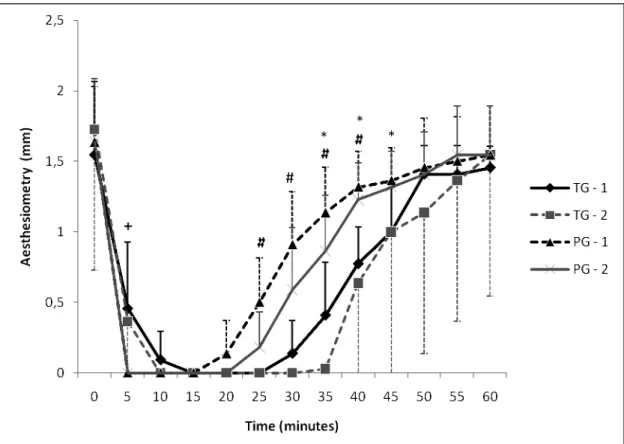
In the TG, six males and five females with a mean weight of 17.63±7.69kg, and in the PG four males and seven females with a mean weight of 11.81±8.62kg, were used. The average duration of anesthetic action observed was 25 minutes for tetracaine and 15 minutes for proparacaine, both times for the dosage using a single drop (Fig. 1).
In TG dogs no significant effect of treatment was observed, and in PG dogs, there was difference between the two doses at 25min (P<0.05), with an increase of 5min in time of action for the instillation of two drops.
Figure 1. Values of corneal sensitivity in dogs measured by Cochet-Bonnet esthesiometer for animals treated with tetracaine 1% drops associated with phenylephrine 0.1% at the dosage of one drop (GT-1) and the dosage of two drops with a 1 min interval between drops (GT-2) and animals treated with proparacaine 0.5% eye drops at the dosage of one drop (GP-1) and the dosage of two drops, with a 1 min interval between instillations (GP-2). Time 0 corresponds to an assessment prior to instillation. + Over time shows significant difference (P<0.05) between GT and GP. # Of times above shows a significant difference (P<0.05) between GT-1 and GP-2. * Over time shows a significant difference (P<0.05) between GT-1 and GP-2.
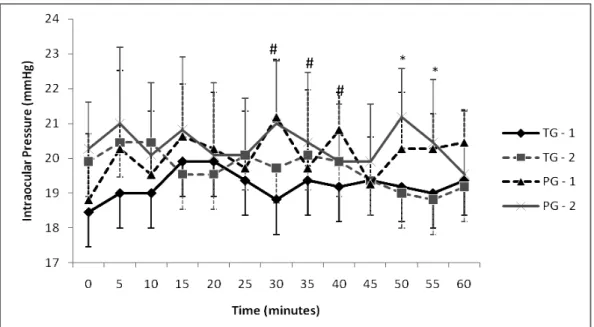
The intraocular pressure showed no differences between the doses in both groups (Fig. 2). When comparing the two eye drops, significant difference was observed with a lower eye pressure in the left eye (from T30 to T40; P<0.05) and in the right eye (T50-T55; P<0.05) for the TG.
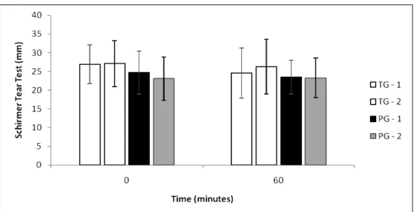
The groups did not present homogeneity in the pupil diameter size before the trial (T0), but differences in eye diameter (Fig. 3) and in the Schirmer tear test (Fig. 4) were not observed between groups or doses.
There was a decrease in fotomotor reflex in TG, three animals in both eyes and one animal only in the right eye, from 5 to 20 min (P<0.05), but no difference between the dosages. No decrease was observed in photomotor reflex on PG.
The chemosis was seen as an adverse reaction in the TG, with three animals presenting bilaterally and one animal only in the right eye, with no difference between the dosages, but with significant difference between the groups (P<0.01). In PG no dog had chemosis.
Parchen et al.
Figure 2. Intra-ocular pressure in dogs measured using Schiotz tonometer for animals treated with tetracaine 1% drops combined with phenylephrine 0.1% at the dosage of one drop (GT-1) and the dosage of two drops, with a 1 min interval between drops (GT-2) and animals treated with proparacaine 0.5% eye drops at the dosage of one drop (GP-1) and the dosage of two drops, with a 1 min interval between instillations (GP -2). Time 0 corresponds to an assessment prior to instillation. # Of times above shows a significant difference (P<0.05) between GT-1 and GP-2. * Over time shows a significant difference (P<0.05) between GT-1 and GP-2.
Figure 3. Pupillary diameter observed in dogs measured by digital caliper in animals treated with tetracaine 1% drops associated with phenylephrine 0.1% at the dosage of one drop (GT-1) and the dosage
#
#
Figure 4. Schirmer tear test in dogs treated with tetracaine 1% drops associated with phenylephrine 0.1% at the dosage of one drop 1) and the dosage of two drops, with a 1 min interval between drops (GT-2) and animals treated with proparacaine 0.5% eye drops at the dosage of one drop (GP-1) and the dosage of two drops, with a 1 min interval between instillations (GP-2). Time 0 corresponds to the evaluation before instillation and at 60 the end of evaluations.
DISCUSSION AND CONCLUSIONS
The cornea is a richly innervated tissue sensitively connected to the trigeminal nerve (Marfurt et al., 2001). Topical anesthetics work by promoting reversible blockade of sodium channels, preventing the spread of noxious stimuli from the cornea, conjunctiva and sclera (Page and Fraunfelder, 2009).
The rapid onset of action and the absence of tissue reaction popularized proparacaine, the most commonly used local anesthetic in ophthalmology. The onset time of proparacaine in dogs is approximately 30-60 seconds, its effects lasting between 10 to 20 minutes (Stiles et al., 2001). The instillation of two drops of 0.5% proparacaine, with a of one minute interval between each drop increased the time of action to 25 minutes (Herring et al., 2005). These results are similar to the ones of this experiment, with a median time of 15 minutes of action with the instillation of a drop and 20 minutes with two drops. The increase in duration of corneal anesthesia with the instillation of an additional drop is due, probably, to the mere increase of drug concentration in the cornea and surrounding tissues.
In studies with other species, the average duration of proparacaine was 10 minutes (Bartfield et al., 1994) and 11 minutes (Weiss end Goren, 1991) in humans, 10 minutes in rabbits (Seabaugh et al., 1993), 25 minutes in cats (Binder and Herring, 2006) and 25 minutes in horses (Kalf et al., 2008).
In a prospective, randomized double-blind clinical study comparing 0.5% proparacaine and tetracaine eye drops in persons, the anesthetic effect of proparacaine lasted 1.3 minutes longer than tetracaine (Bartfield et al., 1994). In this study, conversely, the comparison between two commercial formulations of commonly used anesthetic eye drops showed that tetracaine lasts about five minutes longer than proparacaine. This fact is due to the presence of phenylephrine associated with commercial tetracaine eye drops, a sympathomimetic, which induces local vasoconstriction, increasing the time of action of local anesthetic (Fukuda et al., 1994). Since in Brazil there is only commercial solution containing 1% tetracaine associated with phenylephrine 0.1%, there was no interest in observing the effects of tetracaine alone.
Parchen et al.
eyelid glands reflex secretion, reducing tear production (Hamor et al., 2000).
According to Patton and Robinson (1975), decreased tear production afforded by the action of local anesthetics is responsible for increasing the bioavailability of the drug in the tissues of the cornea. This would be a possible explanation for the increase in the time of anesthetic action found in the dosage of two drops. However, decreased tear production was not detected in either group, although it was compared to Schirmer test before instillation and after 60 minutes, when there was more activity of local anesthetics.
There is a tendency to decrease intraocular pressure promoted by local anesthetics, probably by reducing the tension of the eyelids and auxiliary muscles of the eyeball (Ehongo et al., 2009). Unlike the study by Almubrad and Ogbuehi (2007) in humans, which found a significant reduction in intraocular pressure of 0.9mmHg two minutes after the instillation of proparacaine and 0.8mmHg after instillation of oxybuprocaine, in this study an increase of 0.9mmHg with the use of proparacaine and 0.5mmHg with tetracaine in intraocular pressure of the dogs after 5 minutes was found, with no significant statistic with the baseline. These results also differ from those observed in people with two other local anesthetics, betoxicaine and oxybuprocaine, in which a significant decrease in intraocular pressure was observed (Baudouin and Gastaud, 1994).
The increased thickness of the cornea can lead to a reading greater than the real tonometry (Ko et al., 2005). The instillation of one drop of proparacaine 0.5% is capable of increasing, though transient, corneal thickness (Herse and Siu, 1992; Nam et al., 2006), thus a greater reading of IOP is expected.
The lower intraocular pressure observed with the use of tetracaine, when compared to proparacaine, just after the end of the therapeutic action of drugs may be due to the longer time of action of tetracaine, promoting greater recovery time of the tone of the eyelids and accessory
since its concentration when combined with tetracaine is 0.1%. Phenylephrine is widely used as a mydriatic in hospitalar routine made at 10%, concentration 100 times greater than that present in the anesthetic eye drops. Even at this higher concentration, it cannot promote adequate pupil dilation. The 10% concentration was not sufficient to promote mydriasis in cats (Trinavarat and Pituksung, 2009). Phenylephrine, however, even at concentrations as low as 0.1%, is a drug that can potentially trigger immediate or delayed allergic blepharoconjunctivitis. Furthermore, in humans, tetracaine promotes more pain or discomfort than proparacaine (Bartfield et al., 1994; Shafi and Koay, 1998).
There are minor structural differences between the two drugs. Tetracaine is a para-aminobenzoic ester, while proparacaine is a meta-aminobenzoic ester. The implication of this subtle difference is not well defined, but it is known that the type of ester group may be linked to the development of allergic reactions (Lawrenson et al., 1998). The higher the contrast of anesthetic eye drops pH with 7.4 (physiological pH), the greater the irritation of the ocular tissues (Lawrenson et al., 1998). Proparacaine has an average pH of 4.8 (Garcia-Arumi et al., 2007), while that of tetracaine is 4.54 (Weaver et al., 2003), which creates greater eye pain and adverse reactions. Both the ability of phenylephrine and tetracaine to trigger allergic reactions, since tetracaine has higher acidity, may explain the high incidence of chemosis animals in this group.
There is a tendency to use the commercial solution of tetracaine in veterinary medicine in Brazil. One reason may be the need for refrigerated storage of proparacaine. This research, however, shows that proparacaine eye drop presents less tissue reaction and should be indicated both in diagnostic and surgical procedures.
REFERENCES
ALMUBRAD, T.M.; OGBUEHI, K.C. Clinical investigation of the effect of topical anesthesia on intraocular pressure. Clin. Ophthalmol., v.1, p.305-309, 2007.
BARTFIELD, J.M.; HOLMES, T.J.; RACCIO-ROBAK N.A comparison of proparacaine and tetracaine eye anesthetics. Acad. Emerg. Med., v.1, p.364-367, 1994.
BAUDOUIN, C.; GASTAUD, P. Influence of topical anesthesia on tonometric values of intraocular pressure. Ophthalmologica, v.208, p.309-313, 1994.
BINDER, D.R.; HERRING, I.P. Duration of corneal anesthesia following topical administration of 0,5% proparacaine hydrochloride solution in clinically normal cats. Am. J. Vet. Res., v.67, p.1780-1782, 2006.
CAREL, R.S.; KORCZYN, A.D.; BOYMAN, R. Amethocaine and intraocular pressure. Ophthal. Res., v.11, p.212-215, 1979.
EHONGO, A.; de MAERTELAER, V.; POURJAVAN, S. Effect of topical corneal anaesthesia on ocular response analyzer parameters: pilot study. Int. Ophthalmol., v.29, p.325-328, 2009.
EZRA, D.G.; ALLAN, B.D.S. Topical anaesthesia alone versus topical anaesthesia with intracameral lidocaine for phacoemulsification. Cochrane Database of Systematic Reviews Issue 3.: Art. No.: CD005276. DOI: 10.1002/14651858.CD005276.pub2. 2007.
FUKUDA, T.; DOHI, S.; NAITO, H. Comparisons of tetracaine spinal anesthesia with clonidine or phenylephrine in normotensive and hypertensive humans. Anesth. Analg., v.78, p.106-111, 1994.
GARCIA-ARUMI, J.; FONOLLOSA, A.; SARAROLS, L. et al. Topical anesthesia: Possible risk factor for endophthalmitis after cataract extraction. J. Cataract. Refract. Surg., v.33, p.989-992, 2007.
HAMOR, R.E.; ROBERTS, S.M.; SEVERIN, G.A. et al. Evaluation of results for Schirmer tear tests conducted with and without application of a topical anesthetic in clinically normal dogs of 5 breeds. Am. J. Vet. Res., v.61, p.1422-1425, 2000.
HERRING, I.P.; BOBOFCHAK, M.A.; LANDRY, M.P. et al. Duration of effect and effect of multiple doses of topical ophthalmic 0.5% proparacaine hydrochloride in clinically normal dogs. Am. J. Vet. Res., v.66, p.77-80, 2005.
HERSE, P.; SIU, A. Short-term effects of proparacaine on human corneal thickness. Acta. Ophthalmol., v.70, p.740-744, 1992.
KALF, K.L.; UTTER, M.E.; WOTMAN, K.L. Evaluation of duration of corneal anesthesia induced with ophthalmic 0.5% proparacaine hydrochloride by use of a Cochet-Bonnet aesthesiometer in clinically normal horses. Am. J. Vet. Res., v.69, p.1655-1658, 2008.
KO, Y.C.; LIU, C.J.; HSU, W.M. Varying effects of corneal thickness on intraocular pressure measurements with different tonometers. Eye, v.19, p.327-332, 2005.
LAWRENSON, J.G.; EDGAR, D.F.; TANNA, G.K. et al. Comparison of the tolerability and efficacy of unit-dose, preservative-free topical ocular anaesthetics. Ophthal. Physiol. Optics, v.18, p.393-400, 1998
MARFURT, C.F.; MURPHY, C.J.; FLORCZAK, J.L. Morphology and Neurochemistry of canine corneal innervation. Invest. Ophthalmol. Visual Sci., v.42, p.2242-2251, 2001.
NAM, S.M.; LEE, H.K.; KIM, E.K. et al. Comparison of corneal thickness after the instillation of topical anesthetics: proparacaine versus oxybuprocaine. Cornea, 25, p.51-54, 2006.
PAGE, M.A.; FRAUNFELDER, F.W. Safety, efficacy, and patient acceptability of lidocaine hydrochloride ophthalmic gel as a topical ocular anesthetic for use in ophthalmic procedures. Clin. Ophthalmol., v.3, p.601-609, 2009.
PATTON, T.F.; ROBINSON. J.R. Influence of topical anesthesia on tear dynamics and ocular drug bioavailability in albino rabbits. J. Pharmac. Sci., v.64, p.931-939, 1975.
Parchen et al.
SEABAUGH, V.M.; CHAMBERS, W.A.; GREEN, S. et al. Use of ophthalmic anaesthetics. Food Chem. Toxicol., v.31, p.105-109, 1993.
SHAFI, T.; KOAY, P. Randomised prospective masked study comparing patient comfort following the instillation of topical proxymetacaine and amethocaine. Br. J. Ophthalmol., v.82, p.1285-1287, 1998.
STILES, J.; KROHNE, S.; RANKIN, A. et al. The efficacy of 0.5% proparacaine stored at room temperature. Vet. Ophthalmol., v.4, p.205-207, 2001.
TRINAVARAT, A.; PITUKSUNG, A. Effective pupil dilatation with a mixture of 0.75% tropicamide and 2.5% phenylephrine: A randomized controlled trial. Indian J. Ophthalmol., v.57, p.351-354, 2009.
WEAVER, C.S.; RUSYNIAK, D.E.;
BRIZENDINE, E.J. et al. A prospective, randomized, double-blind comparison of buffered versus plain tetracaine in reducing the pain of topical ophthalmic anesthesia. Ann. Emerg. Med., v.41, p.827-831, 2003.