O
R
I
G
I
N
A
L
A
R
T
I
C
L
E
Potential doxorubicin delivery system based on magnetic
gelatin microspheres crosslinked with sugars
Josefa Souza
1, Manoel Silva
2and Marcos Costa
1*
1
Laboratório de Química de Polímeros, Instituto de Química, Universidade do Estado do Rio de Janeiro –
UERJ, Rio de Janeiro, RJ, Brazil
2
Laboratório de Física, Departamento de Física e Química, Universidade Federal de Itajubá – UNIFEI,
Itajubá, MG, Brazil
*marcos.costa@uerj.br
Abstract
The preparation and characterization of magnetic microspheres based on gelatin for use in drug delivery systems are reported. Sugars were employed as crosslinking agents and type A gelatin and type B gelatin were compared to prepare
microspheres by water-in-oil emulsion. The influence of gelatin and sucrose concentration, temperature and stirring
speed on microbeads’ characteristics was studied. The gelatin concentration and stirring speed were the parameters directly associated with the particle sizes. We found no relevant difference between the use of type A and type B gelatin.
In addition, the gelatin crosslinking study revealed that sucrose is not a crosslinking agent but fructose can crosslink the protein chains when the reaction medium has pH 9. The size of the microspheres varied from 5 to 60 μm as measured
by optical microscopic images. Doxorubicin adsorption and release were successfully performed using the microspheres
crosslinked with fructose under the action of an external magnetic field. It was observed that the microspheres absorbed 69% of the doxorubicin that was in solution. After 24 h, about 45% of the DOX was displaced from microspheres to
saline medium in the free form in the solution.
Keywords: gelatin microspheres, magnetic properties, sugar crosslinking.
1. Introduction
Gelatin is a mixture of water-soluble proteins obtained by
hydrolysis of collagen from the skin, bones and connective
tissues of animals[1]. There are two types of gelatins and they are characterized by their mode of manufacture. The Type A gelatin (pH 3.8-6.0; iso- electric point 6-8) is obtained from acidic hydrolysis of pork skin and the Type B gelatin (pH 5.0-7.4; isoelectric point 4.7-5.3) is obtained from basic hydrolysis of bones and animal skin[1].
Attributable to the excellent biocompatibility and biodegradability[2,3], gelatin has been widely used in biomedical
materials for controlled drug release. In this application,
can be found gelatin in different forms: films[4-6], disks[7], hydrogels[8,9], sponges[10] and frequently microspheres[7,9,11-14]. Microspheres are usually prepared by water-in-oil emulsion.
However, the main preparation parameters vary widely in the literature. When microspheres are produced, these
parameters can influence particle size and the microsphere’s size is very important to define the administration route[15,16] and the liberation rates[16]. Because of this, in this work, we designed experiments to determine the most important parameters that can influence particle size.
Because gelatin is a water-soluble polymer, its must
be modified for application in the human body (where
the medium is aqueous). Thus, gelatin hydrogels can be
prepared as three-dimensional hydrophilic networks that are able to release drugs at the controlled rates. Such networks can be physical as those obtained by gelatin mixed with other polymers such as sodium carboxymethyl cellulose[17], hydroxyethyl cellulose[18] and carboxymethyl
guar gum[19] to form interpenetrating polymer networks (IPNs) or chemical as those obtained by using chemical crosslinking agents[2]. The chemical crosslinking agents are bifunctional or polyfunctional compounds that act by binding to carboxylic or amino groups of adjacent molecules of gelatin. Examples of this type of crosslinker include
formaldehyde, glutaraldehyde, glyceraldehyde, imines, ketones, saccharides, dyes, calcium carbonate, carbodiimides,
genipin and other bifunctional compounds[1].
There are many chemicals that can be used for gelatin
crosslinking, but the crosslinking process in biomedical materials must be done with reagents that, like the polymer, are biocompatible and biodegradable. Most of
these crosslinking agents can cause some cytotoxic effects because of unreacted fractions[7]. To avoid undesirable
reactions, some studies have investigated the use of sugars
as crosslinking agents[7,10,11,20]. Among the studied sugars, calls our attention the fact that the researchs conclude that sucrose is a crosslinking agent able to significantly
reduce the gelatin water solubility. Additionally, sucrose is biocompatible, easy to obtain and inexpensive, making it a
good candidate for use in controlled drug release. For these
reasons, we will use it as a crosslinking agent in this work. Besides the main features of biocompatibility, biodegradability and low water solubility, the device
designed by us should possess the ability to be transported
inside the human body directly to target cells. To this end, in one of the phases of this study, magnetite is incorporated
magnetic properties to the device. Thus, it can be injected into the patient’s circulatory system and, with the aid of an external magnetic field, it is possible to concentrate the
drug/biocomposite complexes at a specific target site in the body where the particles have entered the bloodstream[21].
Once the biocomposite is concentrated at the target, the
drug can be released to act on the target cells by enzymatic
activity or changes in pH, temperature or magnetic field.
These magnetic drug delivery systems have many advantages
over normal, non-targeted methods, such as: ability to target
specific locations in the body; reduction of the drug quantity needed to attain a particular concentration in the vicinity of the target and reduction of the drug’s concentration at
non-target sites, minimizing side effects[22].
The above described characteristics are essential for obtaining optimum system for controlled drug release.
Despite its potential applications, few studies have investigated
the use of sugars as a crosslinking agent to obtain gelatin microspheres and there is no literature about magnetic
gelatin microspheres sugar crosslinked. Thus, in order to obtain a similar device, we decided to evaluate the effect of gelatin type, sucrose concentration, magnetite concentration
and crosslinking time on the physical properties of the
microspheres based on gelatin, sucrose and magnetite.
2. Materials and Methods
2.1 Materials
Type B gelatin (225 bloom), type A gelatin (300 bloom), sucrose, fructose, corn oil and doxorubicin were purchased from Sigma-Aldrich Co. Acetone, and sodium hydroxide
were acquired from B. Herzog Varejo de Produtos Químicos
Ltda. Ferric chloride, sodium chloride and ferrous sulfate
were purchased from Proquimios Comércio e Indústria Ltda. All chemicals were analytical grade and used as received.
2.2 Preparation of gelatin microspheres
Microspheres were produced by thermal gelation.
Briefly, 10 mL of 10% w/v gelatin solution preheated to
60 °C containing 40% w/w of sucrose was added dropwise to 40 mL of corn oil to form an emulsion by stirring with a two-paddle stirrer (1000 rpm). As the emulsion was
obtained, the temperature was kept at 60 °C for different
time periods and then lowered to 5 °C by rapid cooling in an ice bath. The microspheres formed were maintained in
this condition for 30 minutes. Then, to completely solidify the droplets of the dispersed phase, 50 mL of precooled
(5 °C) acetone was added and the mixture was stirred for
another hour. The microspheres were filtered, washed with
cool acetone (5 °C) and rapidly dried.
2.3 Preparation of magnetite
Magnetite nanoparticles were synthesized using an adaptation of a previously described co-precipitation method[23]. This involved adding 100 mL of an aqueous solution of sodium hydroxide (concentration of 10 mols/L) dropwise to a mixture of iron salts with Fe2+/Fe3+ molar ratio
of 1/2, forming an immediate dark brown/black solution.
The solution was stirred for 1 h at room temperature and
then was heated at 90 °C for another 1 h, which resulted
in the formation of a black colloidal magnetite solution.
Subsequently, the dispersion was cooled to room temperature
and was washed several times with distilled water until neutral pH. The magnetite formed was separated by magnetic decantation/separation and was dried in an oven at 60 °C for 24 h. The Fe3O4 nanoparticles’ precipitation happened according to the Equation 1 below:
2 3
3 4 2
Fe++2Fe++ 8OH−→Fe O +4H O (1)
2.4 Preparation of the magnetic gelatin microspheres
Magnetic microspheres were produced by the same method described before. The magnetite was added in the gelatin solution and this mixture was then added dropwise to corn oil in order to form an emulsion.
2.5 Size particle distribution and average diameter
These analyses were performed using a method described by Allen[24]. In this method, optical microscopic images were used to measure the diameter of 625 microspheres of each sample.
2.6 Magnetic properties
The magnetic properties (saturation magnetization, residual
magnetization and coercivity) were analyzed by using a Lake Shore series 7400 vibrating sample magnetometer (VSM).
2.7 DSC and FTIR analysis
The thermal properties of the gelatin microspheres were analyzed by using a Perkin-Elmer Pyris 1 differential scanning calorimeter. The melting temperature (Tm) of the microspheres was determined under nitrogen atmosphere.
Samples were scanned in aluminum pans, under static air atmosphere, at a heating rate of 20 °C/min in the temperature
range of 50-200 °C.
FTIR spectra of microspheres were measured by the
KBr pellet method using a Perkin Elmer Spectrum One
spectrophotometer.
2.8 Morphological analysis
The gelatin microspheres’ morphology was determined by observation of the samples with a FEI Inspect 550 scanning electron microscope. The samples were coated with gold in an argon atmosphere for 120 s and the images were captured using acceleration voltages of 5 kV and 20 kV.
2.9 Swelling ratio
Gelatin microspheres in the dry state were put on filter paper and weighed. Then the microspheres were immersed
in distilled water at room temperature. Subsequently, the
weight of the swollen microspheres was determined after 60 minutes. The swelling ratio (Rsw) of each test sample was calculated as follows (Equation 2):
(
)
(
)
sw s d d
R = W – W / W ×100 (2)
2.10 Atomic absorption spectroscopy
The iron concentration of the gelatin microspheres was determined by atomic absorption spectroscopy. About 10 mg of each sample was heated in a flat-bottomed flask with 20 mL of aqua regia at reflux temperature for 24 h. Then the
solution was cooled to room temperature, filtered into a
100 mL volumetric flask and the volume was completed with distilled water. The solution was analyzed by a Perkin Elmer Analyst 300 spectrometer.
2.11 Doxorubicin loading and in vitro doxorubicin release
The doxorubicin (DOX) loading and in vitro DOX release
were determined using gelatin magnetic microspheres made with 50% magnetite and fructose crosslinked at pH = 9.
The loading of DOX was performed by allowing the magnetic
gelatin microspheres (50 mg) to contact a freshly prepared
DOX solution (200 ppm) for 1 hour. Then, the amount of free DOX in the solution was quantified by UV-Vis spectroscopy (Fentom 600S) at 480 nm. The DOX loading efficiency
(DL)(%) was calculated using the following Equation 3:
( ) DOX DOX
DL % 100
DOX
Total amount of Free amount of Total amount of
−
= × (3)
The in vitro DOX release experiments were carried out
in phosphate buffer saline (PBS) (pH 7.4, 1.2 mM KH2PO4, 1.15 mM Na2HPO4, 2.7 mM KCl, 1.38 mM NaCl) in presence of constant magnetic field (6000 gauss) using a magnet.
In order to determine the released amount of the DOX, 0.1 g of DOX-loaded magnetic gelatin microspheres was added to 8 mL of PBS (release medium, pH 7.4). The resulting
suspension was gently shaken under a constant magnetic field
of 6000 Gauss for predetermined time period. After shaking,
3 mL of supernatant was withdrawn and assayed for
DOX spectrophotometrically (Fentom 600S at 480 nm).
Each experiment was carried out in triplicate.
3. Results and Discussions
3.1 Influence of gelatin and sucrose concentration, temperature and stirring speed on particle size of materials obtained
A full factorial design at two levels, 23, was applied to evaluate the main effects. The variables considered and the levels studied are shown in Table 1. The experiments
involved fixing the oil phase (corn oil), aqueous phase/oil phase ratio (1/4), heating time (10 minutes), cooling time
(30 minutes) and acetone cooling time (1 hour). In order
to replace cytotoxic crosslinkers, sucrose was chosen as
crosslinking agent because it is a well-known biocompatible reagent.
The particle size distribution showed in Figure 1 revealed that the diameter of the microspheres produced in
all experiments ranged from 5 to 60 µm. However, there was
predominance in the range from 11 to 30 µm. For application
in drug delivery systems, gelatin microspheres should have
sizes below 5 µm for intravenous administration and should be smaller than 125 µm for arterial administration[15]. Thus, the particles obtained in all experiments were adequate for use in drug delivery by the arterial route.
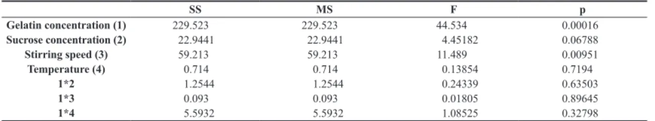
The analysis of variance (ANOVA) was used to analyze the effect of gelatin concentration, sucrose concentration, stirring
speed and temperature on gelatin microspheres’ particles size
Table 1. Experimental conditions in the 23 factorial design to evaluate their effects on particle size of gelatin microspheres.
Sample Gelatin concentration (%) Sucr
ose
concentration (%) Temperatur
e
(°C) Stirring speed (rpm)
P1 10 0 40 500
P2 20 0 60 500
P3 10 40 60 500
P4 20 40 40 500
P5 10 0 60 1000
P6 20 0 40 1000
P7 10 40 40 1000
P8 20 40 60 1000
Figure 1. Particles size distribution of gelatin microspheres obtained according to Table 1.
Table 2. Analysis of the effects of variables on particles size by ANOVA.
SS MS F p
Gelatin concentration (1) 229.523 229.523 44.534 0.00016
Sucrose concentration (2) 22.9441 22.9441 4.45182 0.06788
Stirring speed (3) 59.213 59.213 11.489 0.00951
Temperature (4) 0.714 0.714 0.13854 0.7194
1*2 1.2544 1.2544 0.24339 0.63503
1*3 0.093 0.093 0.01805 0.89645
1*4 5.5932 5.5932 1.08525 0.32798
As can be seen in Table 2, with a 95% confidence level, the parameters that are directly associated with the particle sizes are the gelatin concentration and stirring speed. The data evaluation shows that smaller diameters are obtained with solutions of low gelatin concentration and higher stirring
speeds. Because of this, the microspheres made afterward
were prepared with a 10% gelatin solution and 1000 rpm stirring speed.
3.2 Gelatin type influence
Manufacturers offer a wide variety of gelatins that are
simple combinations of type A and type B gelatins. Thus, it
is important to know if the gelatin type will influence the
properties of the final particles. In this study, microspheres
were prepared with both gelatin types and their magnetic and solubility properties were compared.
The magnetic properties are extremely important for application in the device proposed in this paper. The saturation magnetization of the particles should be known in order to calculate the magnetic field strength that must be applied externally. Another very important characteristic is
superparamagnetism. For application in the human body, this property is essential to prevent the particles’ agglomeration, which can lead to clogging of blood vessels. For this reason,
we assessed the effects of the magnetite concentration on the magnetic properties of the microspheres.
Table 3 shows the magnetic properties obtained. As might
be expected, the saturation magnetization increased with
rising magnetite concentration. Higher magnetization was observed for the microspheres obtained with type A gelatin
when 50% was added during preparation, but this behavior
was not observed for the other magnetite concentrations. All microspheres prepared in this experimental series had superparamagnetic behavior because remnant magnetization close to zero was observed.
The data show that the experimental values are all larger than the theoretical values. The most probable hypothesis for these experimental results is that magnetite in the pure state forms clusters but is evenly dispersed when placed in a gelatin matrix. The formation of clusters tends to decrease the saturation magnetization of the particles while homogeneous dispersion has the opposite effect[25].
FTIR experiments were performed to find evidence of sugar-mediated crosslinking. Figure 2a shows the spectra of raw materials used to produce gelatin microspheres
(type A gelatin, type B gelatin and sucrose). According to
Cortesi et al.[7], the absorption band located at 1450 cm-1
is characteristic of an aldimine stretching vibration, which provides evidence of the crosslinking of gelatin. However, this
band is already present in the microspheres’ raw material. Figure 2b shows the FTIR spectra of type A gelatin and type B gelatin microspheres with no sucrose and with 40% (w/w) of sucrose. All microspheres showed the same peaks with similar intensities in the infrared region and no difference between type A and Type B gelatin was noted.
Table 3. Influence of gelatin type and magnetite concentration on magnetic properties of materials.
Sample Gelatin type Magnetite
concentration1 (%)
Iron concentration2 (%)
Magnetite concentration3 (%)
Theoretical Ms (emu/g)
Experimental Ms (emu/g)
Magnetite - - - 42.33
GAM10 A 10 4.34 5.99 2.54 3.27
GAM20 A 20 6.32 8.73 3.7 4.42
GAM50 A 50 12.2 16.86 7.14 9.24
GBM10 B 10 3.65 5.05 2.14 3.64
GBM20 B 20 5.57 7.71 3.26 4.80
GBM50 B 50 10.4 14.38 6.09 8.18
1Magnetite concentration usen on microsphere fabrication; 2Iron concentration analysed by atomic absorption analysis; 3Magnetite concentration
calculated by atomic absorption analysis.
Therefore, these spectra present no evidence of sucrose
crosslinked gelatin.
In addition, water solubility tests were conducted to
verify how the solubility was affected by addition of sugar. All the samples dissolved completely in under 3 hours. These results indicate that the microspheres have a low level of crosslinking.
Type A and type B gelatin had similar properties with respect to solubility in water and minor differences in relation
to magnetic properties. Thus, we decided to use only type B
gelatin to produce microspheres from this point on.
3.3 Gelatin microsphere crosslink study
In order to optimize the efficiency of gelatin microspheres
as drug carriers, their number of crosslinks in the polymer matrix should be evaluated. Because of this, we decided to
observe how the heating time influences protein crosslinking in a series of experiments using type B gelatin. The obtained microspheres were characterized by differential scanning
calorimetry (DSC), swelling analysis and scanning electronic micrography. The microspheres’ composition, as well as
swelling and DSC results are in Table 4.
As can be seen in Table 4, the melting point of gelatin microspheres increased when the heating time increased. The only difference between sample GB and GBt10 is that
in the last one, sucrose was added during the preparation.
The melting points of samples GB (no sucrose) and GBt10 (40% sucrose) did not differ greatly and the sample without sucrose showed a slightly higher melting point than that in the presence of sugar.
We carried out swelling tests because they are a relatively easy way to measure the ability of gelatin microspheres to retain water. The results showed a significant decrease in the water retention with longer heating time. The values of
Tm and swelling ratio corroborate each other, confirming
that longer heating time increases the number of crosslinks
in the polymer matrix. In contrast, the presence or absence
of sucrose in the microspheres had little influence on the
data analyzed, which leads us to believe that sugar had little
influence on the crosslinks formed.
Figure 3 shows scanning electron micrographs of microspheres of gelatin obtained at different heating times. As can be seen in Figure 3 (left side), no significant differences
were observed. The particles present spherical morphology, but there are many agglomerates. Probably, these agglomerates
are formed because it was not added a surfactant agent during emulsion preparation. Figure 3 (right side) shows the
difference on particles’ surface according the heating times. It can observed that the surfaces became smoother when the
heating time increased. With a higher magnification, this
difference can clearly be seen when comparing the heating times of 10 and 2880 min (Figure 4).
Based on these results, there are two possible explanations
for these experimental observations. The first one is based on Russo’s[26] paper. According to him, crosslinks can occur
by intermolecular bonds (interstrand), which occur between arginine-lysine or arginine-arginine within the same strand,
while amino acid residues from two neighboring strands can also interact and form intramolecular (intrastrand) crosslinked
strands, providing strength to the gelatin. The second theoretical
explanation is based on carbohydrate chemistry. Sugars commonly exist as cyclic molecules because alcohols react reversibly with aldehydes and ketones to give hemiacetals
and hemicetals, respectively. However, in the equilibrium state, there is a mixture of carbohydrate isomers and a small fraction of aldehyde or ketone source. Although small, the
fractions of aldehyde and ketone allow the occurrence of common reactions of these organic functions[27]. Sucrose is a disaccharide composed of one glucose and one fructose
molecule, both reducing sugars. The link between the two
monosaccharides (glycosidic bond) forming disaccharide prevents the opening of the cyclic-form portions of fructose
and glucose, resulting in the absence of aldehydic and ketonic forms in equilibrium, so the common reactions of
these functions do not occur[27]. Sucrose is liable only if there is a hydrolysis reaction of the molecule to form the
start of monosaccharides, which can only happen with a
strongly acidic medium or under the influence of catalysts or enzymes. Since the reaction medium for preparation of gelatin microspheres here did not provide the main conditions
for hydrolysis of sucrose, the gelatin remained in its original form, i.e., unable to form crosslinking reactions. Considering
the theoretical foundations presented and the results of
thermal analysis and swelling, we assume that sucrose does not react with the gelatin chains, so the increase in melting
point of the microspheres was only due to the crosslinks
formed by intermolecular and intramolecular bonds, which
were favored by increasing the heating time.
Although some authors[7,11,20] have indicated the use of sucrose as a biocompatible and biodegradable alternative to
crosslink gelatin, we found no evidence of chemical reaction between the gelatin amino groups and sucrose. Thus, we
decided to test fructose as crosslinking agent because the ketone functional group of fructose is more reactive than the aldehyde functional group of glucose. In order
Table 4. Melting temperature and swelling ratio of the gelatin microspheres as a function of heating time and sucrose concentration.
Sample Heating time (min) Sucrose concentration (%) Tm (°C) Rsw
Sucrose - - 191
-Type B gelatin - - 161
-GB 10 0 166 570
GBt10 10 40 165 443
GBt30 30 40 - 441
GBt1440 1440 40 175 365
GBt2880 2880 40 197 225
Figure 3. Scanning electronic micrographs of the samples GB (a), GBt10 (b) and GBt2880 (c) with 1,000X (1) and 15,000X (2).
to compare this substance with a traditional crosslinking
agent, microspheres crosslinked with glutaraldehyde were
also prepared.
Preliminary solubility tests were performed comparing gelatin microspheres crosslinked with fructose (GBF sample) with gelatin microspheres crosslinked with glutaraldehyde
(GBG sample). In these tests, a few milligrams of sample
were left in contact with water for 24 h. The GBG sample was insoluble while the GBF was soluble. This result showed that the crosslinking of the protein chains’ gelatin using sugar as crosslinking agent does not occur as easily under
normal conditions, so more factors should be investigated. Therefore, we decided to modify the pH in order to change
the equilibrium between cyclic and open fructose forms and thus provide more ketone available for the formation of
crosslinks. According to the literature, the kinetics of bond
formation in chemical crosslinking of gelatin solutions is strongly affected by the solution’s pH[28], but at pH values higher than 9 and lower than 5 the denaturation enthalpy
decreases, indicating that the triple helix amount is reduced[29].
Thus, gelatin microspheres were produced by varying the pH.
Preliminary solubility tests were performed and since
the aim was to reduce solubility, the gelatin microspheres
made with pH 9 solution (GBF9) were chosen.
The thermogravimetric analysis revealed an initial degradation temperature (Tonset) of 288 °C for the GBG sample and 294 °C for the GBF9 sample. This analysis also showed that the GBF sample had a residual 10 percentage points higher than the GBG sample. Higher degradation temperatures indicate higher crosslinking degree because
more energy is required to break chemical bonds. Likewise,
a larger amount of residue confirms that the particle has more strongly linked protein chains. These results show that the fructose crosslinking was successful.
3.4 Preliminary drug release tests
We observed that the microspheres absorbed 69% of the doxorubicin that was in solution. If this value is compared in the literature for drug absorption by gelatin microspheres[13,14,30], one can considerer that a satisfactory amount of the drug was incorporated into the gelatinous matrix. Figure 5 shows the results of in vitro DOX release tests.
The saline solution mimics the biological environment because it has similar pH and osmotic pressure. In these
conditions, the gelatinous support gradually increased DOX
Figure 5. Release of doxorubicin from gelatin microspheres crosslinked by fructose.
release over the time. After 24 h, about 45% of the drug was
displaced from microspheres to saline medium in the free
form in the solution. In this way, these preliminary release
tests show that the method described in this study can be successfully used for the magnetic gelatin microspheres obtainment to incorporation and controlled release of doxorubicin.
4. Conclusions
With the aim of obtaining gelatin microspheres with
suitable properties for use in drug delivery systems, we
evaluated the experimental parameters using a set of experiments. The statistical results showed that smaller particles can be prepared when low gelatin concentration and high stirring speed are used. By applying these parameters we obtained microspheres with appropriate size to use in arterial drug delivery systems. Because of the large variety
of types available in the market, we decided to investigate
whether there are significant differences between the use of type A gelatin and type B gelatin. The analyses showed no difference between the two types regarding crosslinking or adsorption of magnetic material in the gelatinous matrix.
Furthermore, superparamagnetic samples were obtained
with both gelatin types.
With respect to crosslinking of the protein chains, we
analyzed whether use of sucrose is effective to make the beads more biocompatible. The microspheres obtained
remained very soluble in aqueous media and, so sucrose is not a suitable sugar to crosslink gelatin. Nevertheless, the
extent of crosslinking increased as a function of heating time
periods. Because of this, we analyzed the use fructose in place
of sucrose. Taken together the results obtained indicate that crosslinked gelatin microspheres can be prepared using fructose when the reaction pH is 9. The microspheres crosslinked with fructose were successfully used in preliminary tests of adsorption and release of doxorubicin (a drug that is
widely used in the treatment of cancer patients). Thus, the
material prepared in this paper has great potential for use in drug delivery systems.
5. Acknowledgements
We gratefully acknowledge FAPERJ for a scholarship to
J.V.S. Souza, and FAPERJ and CNPq for financial support.
6. References
1. Singh, S., Rama Rao, K. V., Venugopal, K., & Manikandan, R. (2002). Alteration in dissolution characteristics of
gelatin-containing formulations: a review of the problem, test methods, and solutions.Pharmaceutical Technology, 26, 36-58. Retrieved in 2016, March 11, from http://www.pharmtech. com/pharmaceutical-technology-04-01-2002
2. Sahoo, N., Sahoo, R. K., Biswas, N., Guha, A., & Kuotsu, K. (2015). Recent advancement of gelatin nanoparticles in drug and vaccine delivery. International Journal of Biological Macromolecules, 81, 317-331. PMid:26277745. http://dx.doi. org/10.1016/j.ijbiomac.2015.08.006.
(1-3), 256-274. PMid:16266768. http://dx.doi.org/10.1016/j. jconrel.2005.09.023.
4. Coimbra, P., Gil, M. H., & Figueiredo, M. (2014). Tailoring the properties of gelatin films for drug delivery applications: Influence of the chemical cross-linking method. International Journal of Biological Macromolecules, 70, 10-19. PMid:24971558. http://dx.doi.org/10.1016/j.ijbiomac.2014.06.021. 5. Matsuda, S., Se, N., Iwata, H., & Ikada, Y. (2002). Evaluation
of the antiadhesion potential of UV cross-linked gelatin films
in a rat abdominal model. Biomaterials, 23(14), 2901-2908. PMid:12069331. http://dx.doi.org/10.1016/S0142-9612(01)00418-5.
6. Mutalik, V., Manjeshwar, L. S., Wali, A., Sairam, M., Sreedhar, B., Raju, K. V. S. N., & Aminabhavi, T. M. (2007). Aqueous-solution
and solid-film properties of poly(vinyl alcohol), poly(vinyl pyrrolidone), gelatin, starch, and carboxymethylcellulose
polymers. Journal of Applied Polymer Science, 106(2), 765-774. http://dx.doi.org/10.1002/app.25427.
7. Cortesi, R., Nastruzzi, C., & Davis, S. S. (1998). Sugar cross-linked gelatin for controlled release: microspheres and disks.
Biomaterials, 19(18), 1641-1649. PMid:9839999. http://dx.doi. org/10.1016/S0142-9612(98)00034-9.
8. Rathna, G. V. N. (2008). Gelatin hydrogels: enhanced
biocompatibility, drug release and cell viability.Journal of Materials Science: Materials in Medicine, 19(6), 2351-2358. PMid:18157687. http://dx.doi.org/10.1007/s10856-007-3334-9. 9. Lau, T. T., Lee, L. Q. P., Leong, W., & Wang, D. (2012). Formation of model hepatocellular aggregates in a hydrogel scaffold using degradable genipin crosslinked gelatin microspheres as cell carriers. Biomedical Materials, 7(6), 065003-065011. PMid:23117748. http://dx.doi.org/10.1088/1748-6041/7/6/065003. 10. Ulubayram, K., Aksu, E., Gurhan, S. I. D., Serbetci, K., & Hasirci, N. (2002). Cytotoxicity evaluation of gelatin sponges prepared with different cross-linking agents. Journal of Biomaterials Science: Polymer Edition, 13(11), 1203-1219. PMid:12518800. http://dx.doi.org/10.1163/156856202320892966.
11. Samad, A., Sultana, Y., Khar, R. K., Chuttani, K., & Mishra, A. K. (2009). Gelatin microspheres of rifampicin cross-linked with sucrose using thermal gelation method for the treatment of tuberculosis. Journal of Microencapsulation, 26(1), 83-89. PMid:18608799. http://dx.doi.org/10.1080/02652040802172638. 12. Saravanan, M., Anbu, J., Maharajan, G., & Pillai, K. S. (2008). Targeted delivery of diclofenac sodium via gelatin magnetic microspheres formulated for intra-arterial administration.
Journal of Drug Targeting, 16(5), 366-378. PMid:18569281. http://dx.doi.org/10.1080/10611860802046224.
13. Phadke, K. V., Manjeshwar, L. S., & Aminabhavi, T. M. (2014). Biodegradable polymeric microspheres of gelatin and carboxymethyl guar gum for controlled release of theophylline.
Polymer Bulletin, 71(7), 1625-1643. http://dx.doi.org/10.1007/ s00289-014-1145-y.
14. Kajjari, P. B., Manjeshwar, L. S., & Aminabhavi, T. M. (2011). Semi-interpenetrating polymer network hydrogel blend microspheres of gelatin and hydroxyethyl cellulose for controlled release of theophylline. Industrial & Engineering Chemistry Research, 50(13), 7833-7840. http://dx.doi. org/10.1021/ie200516k.
15. Saravanan, M., Anbu, J., Maharajan, G., & Pillai, K. S. (2008). Targeted delivery of diclofenac sodium via gelatin magnetic microspheres formulated for intra-arterial administration.
Journal of Drug Targeting, 16(5), 366-378. PMid:18569281. http://dx.doi.org/10.1080/10611860802046224.
16. Narayani, R., & Panduranga Rao, K. (1996). Gelatin microsphere cocktails of different sizes controlled release of anticancer drugs. International Journal of Pharmaceutics, 143(2), 255-258. http://dx.doi.org/10.1016/S0378-5173(96)04685-6.
17. Rokhade, A. P., Agnihotri, S. A., Patil, S. A., Mallikarjuna, N. N., Kulkarni, P. V., & Aminabhavi, T. M. (2006). Semi-interpenetrating polymer network microspheres of gelatin and sodium carboxymethyl cellulose for controlled release of ketorolac tromethamine. Carbohydrate Polymers, 65(3), 243-252. http://dx.doi.org/10.1016/j.carbpol.2006.01.013. 18. Kajjari, P. B., Manjeshwar, L. S., & Aminabhavi, T. M.
(2011). Semi-interpenetrating polymer network hydrogel blend microspheres of gelatin and hydroxyethyl cellulose for controlled release of Theophylline. Industrial & Engineering Chemistry Research, 50(13), 7833-7840. http://dx.doi. org/10.1021/ie200516k.
19. Phadke, K. V., Manjeshwar, L. S., & Aminabhavi, T. M. (2014). Biodegradable polymeric microspheres of gelatin and carboxymethyl guar gum for controlled release of theophylline.
Polymer Bulletin, 71(7), 1625-1643. http://dx.doi.org/10.1007/ s00289-014-1145-y.
20. Schuler, B. J. (2004). Evaluation of novel cross-linking agents for gelatin/collagen matrices (Doctoral thesis). School of
Pharmacy, West Virginia University, West Virginia, USA. 21. Pankhurst, Q. A., Connolly, J., Jones, S. K., & Dobson, J.
(2003). Applications of magnetic nanoparticles in biomedicine.
Journal of Physics D: Applied Physics, 36(13), R167-R181. http://dx.doi.org/10.1088/0022-3727/36/13/201.
22. Arruebo, M., Fernández-Pacheco, R., Ibarra, M. R., & Santamaría, J. (2007). Magnetic nanoparticles for drug delivery. Nano Today, 2(3), 22-32. http://dx.doi.org/10.1016/ S1748-0132(07)70084-1.
23. Amali, A. J., & Rana, R. K. (2009). Stabilisation of Pd(0) on
surface functionalised Fe3O4 nanoparticles: magnetically
recoverable and stable recyclable catalyst for hydrogenation and Suzuki–Miyaura reactions. Green Chemistry, 11(11), 1781-1786. http://dx.doi.org/10.1039/b916261p.
24. Allen, T. (1997). Particle size measurement: powder sampling and particle size measurement. 5th ed. London: Chapman & Hall.
25. Santa-Maria, L. C., Costa, M. A. S., Hui, W. S., Santos, F. A. M., & Silva, M. R. (2006). Preparation and characterization of polymer metal composite microspheres. Materials Letters, 60(2), 270-273. http://dx.doi.org/10.1016/j.matlet.2005.08.033. 26. Russo, P. S. (1987). A perspective on reversible gels and related systems (ACS Symposium Series, Vol. 350, pp. 1-21). Washington: American Chemistry Society Symposium. 27. Allinger, N. L., Cava, M. P., Jongh, D. C., Johnson, C. R.,
Lebel, N. A., & Stevens, C. L. (1976). Organic chemistry. 2nd ed. New York: Worth Publishers.
28. Abete, T., Del Gado, E., Arcangelis, L., Serughetti, D. H.,
& Djabourov, M. (2008). Re-entrant phase diagram and pH effects in cross-linked gelatin gels. The Journal of Chemical Physics, 129(13), 134902. PMid:19045122. http://dx.doi. org/10.1063/1.2985655.
29. Gioffrè, M., Torricelli, P., Panzavolta, S., Rubini, K., & Bigi, A. (2012). Role of pH on stability and mechanical properties of gelatin films. Journal of Bioactive and Compatible Polymers,
27(1), 67-77. http://dx.doi.org/10.1177/0883911511431484. 30. Gaihre, B., Khil, M. S., Lee, D. R., & Kim, H. Y. (2009). Gelatin-coated magnetic iron oxide nanoparticles as carrier system: drug loading and in vitro drug release study. International Journal of Pharmaceutics, 365(1-2), 180-189. PMid:18790029. http://dx.doi.org/10.1016/j.ijpharm.2008.08.020.