*Correspondence: A. R. Baby. Departamento de Farmácia. Faculdade de Ciências Farmacêuticas. Universidade de São Paulo. Av. Prof. Lineu Prestes, 580, Bloco 15, 05508-900 – São Paulo - SP, Brasil. E-mail: [email protected]. Phone number: +55 11 3091-2358
A
vol. 52, n. 4, oct./dec., 2016 http://dx.doi.org/10.1590/S1984-82502016000400004
Gelatin-based microspheres crosslinked with glutaraldehyde and
rutin oriented to cosmetics
Fabiana Graziola
1, Thalita Marcílio Candido
1, Camila Areias de Oliveira
1, Daniela D’Almeida
Peres
1, Michele Georges Issa
1, Joana Mota
2, Catarina Rosado
2, Vladi Olga Consiglieri
1, Telma Mary
Kaneko
1, Maria Valéria Robles Velasco
1, André Rolim Baby
1*1 Department of Pharmacy, School of Pharmaceutical Sciences, University of São Paulo, São Paulo, SP, Brazil, 2CBios - Centre for Research in Biosciences & Health Technologies, Universidade Lusófona, Lisboa, Portugal
Glutaraldehyde (GTA) has been extensively used as a gelatin crosslinking agent, however, new natural
ones have been suggested as more biocompatible. Polyphenols are possible candidates and the lavonols,
such as rutin (RUT), also exhibit potential synergism with sunscreens and antioxidant agents used in cosmetics. In this work, gelatin microspheres (M0) were obtained and crosslinked with GTA 10 mM (MG) or RUT 10 mM (MR), dissolved in acetone:NaOH 0,01M (70:30 v/v). MG exhibited crosslinking extent of 54.4%. Gelatin, M0, MG and MR did not elicit any signs of skin damage, regarding the formation of erythema, the barrier function disruption and negative interference in the stratum corneum
hydration. Oily dispersions containing M0, MG or MR, isolated or combined with benzophenone-3 or octyl methoxycinnamate, suggested that the microspheres, at a 5.0% w/w, had no additional chemical or
physical photoprotective efect in vitro. Crosslinking with RUT had occurred, but in a lower degree than GTA. Microspheres had not improved sun protection parameters, although, non-treated gelatin interfered
positively with the SPF for both UV ilters. The in vivo studies demonstrated that these materials had very good skin compatibility.
Uniterms: Protection solar/tests. Gelatin/microspheres/safety testing. Glutaraldehyde/safety testing. Flavonoids/Rutin/use in cosmetics. Natural products/use in cosmetics.
INTRODUCTION
Microspheres are microparticles in which a substance of interest is homogeneously dispersed within a polymer or a wax matrix, forming a monolithic system in which it is not possible to identify a distinct nucleus (Bresolin, Filho, 2003). Gelatin is a biodegradable natural polymer that can be used to produce microparticles. However, due to the aqueous solubility and limited mechanical and thermal properties of gelatin microparticles, improvements, such as crosslinking reactions, are necessary in order to provide the use in long term applications (Bigi et al., 2002; Hayashi, Tabata, 2011).
Crosslink reactions can be performed by chemical or physical medium and they cause structural changes that improve the gelatin mechanical strength and water
resistance (Ratanavaraporn et al., 2010). Glutaraldehyde (GTA) is the most used aldehyde as chemical crosslinking,
but its toxicity concerns and laws in materials like heart
valves, that triggers the search for new crosslinking substances (Catalina et al., 2013). Flavonoids are polyphenolic and aromatic substances constituted by 15 carbon atoms. They have a diphenylpropane skeleton (C6C3C6) with two benzene rings bonded to a pyran ring. Due to such structure, flavonoids are characterized as intrinsic agents of protection against ultraviolet radiation (UV) by acting as chemical filters that absorb in this wavelength range (Bruneton, 1991; Baby et al., 2009). Flavonoids are further divided into 13 classes according to the chemical structure, including flavonols (rutin, quercetin and mericitin). Because of its strong antioxidant activity, rutin is used in the prevention or treatment of
venous or lymphatic insuiciency or capillary fragility and
as a new alternative to chemical crosslinking agent for gelatin (Bigi et al., 2002; Strauss, Gibson 2004).
Chemical ilters are substances that may be included
in cosmetic formulations to protect the skin from damage caused by solar radiation. However, they can generate free radicals when exposed to sunlight and present problems related to photoallergenicity and skin irritation, as well as, photoinstability (Hanson, Gratton, Bardeen, 2006). Such scenario has driven the search for alternatives to minimize
or eliminate these efects and there is a tendency to develop
photoprotective cosmetic formulations with reduced
concentration of chemical ilters, but with high protection
against UVA and UVB radiation, preferentially, with
multifunctional beneits. The research of new photostable
molecules for use in sunscreens are still performed extensively and, currently, we notice growing interest for the development of sunscreens based on natural products (Schlumpf et al., 2004; Tanaka, Setsuko, Utsumi, 1963; Oliveira et al., 2016).
In this research work, we investigated the use of the
lavonoid rutin as an alternative to GTA for crosslinking
gelatin microparticles. The good cutaneous compatibility of our samples was established by non-invasive methods and the physicochemical and functional properties of microparticles were determined to evaluate their applications as adjuvants for performance improvement of photoprotective dermocosmetic products.
MATERIAL AND METHODS
Obtainment of gelatin microspheres
Gelatin microspheres were obtained by emulsion polymerization method, using surfactant and solvent extraction (Tanaka, Setsuko, Utsumi, 1963; Ugwoke, Kinget, 1998). Gelatin (bovine, food grade, bloom 180 and mesh 30) was purchased from NP® (São Paulo, Brazil).
An aqueous gelatin solution at 10.0% w/w (240.0 g) was transferred by dripping to a mixture of mineral oil (560.0
mL) and sorbitan monooleate (inal concentration of 1.0%
w/v) involving heating (55.0 °C) in a ceramic hot plate and mechanical agitation at 2400 rpm. The resulting emulsion was maintained under the conditions described previously for 15 min and then it was cooled to room temperature (25.0 °C) using water bath and 2400 rpm agitation. Then, to enable the gelation of the sample, the emulsion was cooled between 10.0 and 15.0 °C with an ice bath while stirring. After 30 min, acetone (400.0 mL), at of 5.0 °C, was added dropwise with a burette to the emulsion and the mixture was stirred for another 30 min, keeping the temperature at 10.0-15.0 °C, and then it was fractionated
on 50.0 mL in plastic tubes to be centrifuged. After centrifugation (2 min, 3400 rpm, room temperature), the supernatant was discarded. To the amount of solid residue was added in an equal volume of acetone at 5.0 °C. The tubes were centrifuged again at 3400 rpm for 2 min. This procedure was repeated three times. After the last acetone washing, the solid residues were combined and dried at room temperature for at least 12 hours and the dry residue was sieved in 80 mesh sieve (0.177 mm opening).
Crosslinking of gelatin microspheres
The crosslinking medium was consisted of acetone: NaOH 0.01 M (70:30 v/v), in the proportion of 5.0 g of microspheres for each 100.0 mL of medium. The
crosslinking agents were GTA (inal concentration: 0.1% w/v, equivalent to 10.0 mM) and RUT (inal concentration:
0.6% w/v, equivalent to 10.0 mM) and we accomplished the crosslinking process for 4 hours with magnetic stirring at maximum speed at room temperature. After, the crosslinking medium was removed and the crosslinked microspheres were washed 5 times with acetone and agitated for 15 min with magnetic stirring at maximum speed. Subsequently, the crosslinked microspheres were kept in an open beaker to allow air drying for, at least, 12 hours. The microspheres were sieved at 80 mesh sieve.
Appearance by scanning electron microscopy (SEM)
Morphology of gelatin, M0, MG and MR was assessed by SEM. Dry and aqueous samples of gelatin used for obtaining the microparticles were observed and the microparticles were observed in triplicate. The observation of the samples was performed in scanning electron microscope Quanta 600 FEG, FEI® Company, (Hillsboro, OR, U.S.A.) in microscopic magniication of
150 to 50,000 times.
Determination of extent of crosslinking
There were added to 15.0 mL plastic tubes about 15.0 mg of each sample. Then, to each tube was added 1.0 mL of NaHCO3 (4% w/v) and 1.0 mL of TNBS (0.5% w/v).
The tubes were transferred to a bath and kept for 2 hours in this condition. After, the TNBS reaction was stopped by adding 3.0 mL of HCl (6.0 N) and the tubes were transferred to an ultrasonic bath, where they remained at 60.0°C for 90 min, to ensure complete dissolution of samples. Subsequently, there were added 5.0 mL of
puriied water to each tube. The resulting solution was
analysis, in triplicate, at 345.0 nm, using UV-VIS Spectrophotometer 600 Evolution®, Thermo Scientiic
(San Jose, CA, U.S.A.) (Sheu et al., 2001).
A standard curve of L-leucine with concentration range from 0.0 to 3.0 mM was performed to measure free ɛ-amino groups in samples, which is equivalent to L-leucine groups (Gan, Cheng, Easa, 2009).The extent of crosslinking was calculated as shown in Equation 1.
Determination of surface area
Measurements of surface area and porosity were performed in gas adsorption analyzer NOVA® 2200E,
Quantachrome Corporation (Boynton Beach, FL, U.S.A.). Samples were subjected to freezing in liquid nitrogen for 20 min and then analyzed with ultra-pure nitrogen. For each sample, the isotherms were constructed utilizing multipoint method by collecting 30 points of adsorption and 20 of desorption. Results were generated by NOVA®
P -Win 9.0 program.
Determination of true density
The determination of the true density of the samples was performed in a helium gas pycnometer, Ultrapycnometer® 1000, Quantachrome Corporation.
Approximately, 1.5 to 4.0 g of each sample was subjected to three successive measurements of volume and density and, ultimately, the average density was determined.
Determination of particle size distribution
The determination of particle size distribution was
done in laser difraction particle size analyzer CILAS® 1090L (Orléans, France) by wet method. Suicient amount
of sample to obtain obscuration around 15% was dispersed in 50 mL of isopropyl alcohol and submitted to reading. Each sample was analyzed in triplicate in a range from 0.04 to 500 µM per 100 classes.
In vivo cutaneous compatibility assays
Cutaneous compatibility was performed with 12 male and female volunteers, after informed oral and written consent. The procedures were in accordance with the ethical standards of the local responsible committee on human
experimentation (Comissão de Ética da ULHT, Lisbon, Portugal) and with the Helsinki Declaration. Epicutaneous patches (Finn Chambers®, Epitest) were applied in the
volar forearms of the volunteers for 24 h. Each patch had 5 chambers, containing each of the samples (gelatin,
M0, MG and MR) at 10% w/v in puriied water, as well
as, purified water as the negative control. Non-invasive measurements were conducted in each tested site: stratum corneum (SC) hydration was assessed with a Corneometer®
CM825 (CK Electronics GmbH); skin barrier was probed with a Tewameter® TM 300 (CK Electronics GmbH). A
Chroma Meter CR-300 (Minolta Camera, Japan) was used to quantify a possible increase in erythema by the parameter
a* that relects the red chromaticity. All measurements were
performed in triplicate and using the CIE Lab system. The basal values were determined before patch application, and further measurements were made at 24 hours, 2 hours
after its removal. To minimize the efect of inter-individual
variability, the results were analyzed as the ratio between the values obtained after patch application and the basal values (Oliveira et al., 2015; Peres et al., 2016).
In vitro efficacy determination
Fifteen dispersions (formulations) were prepared with mineral oil, and mineral oil (and) polyethylene (Crodabase® SQ). There were added to dispersions:
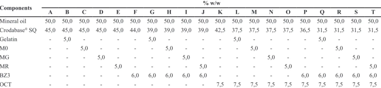
gelatin, M0, MG, MR, benzophenone-3 (BZ3) and octyl methoxycinnamate (OCT), alone or in combination, in order to obtain the sunscreen activity in vitro. Formulations followed the proportions shown in Table I.
For functional in vitro characterization of the
samples, it was used difuse relectance spectrophotometer,
equipped with integrating sphere, UV Transmittance Analyzer, Labsphere® UV-2000S (North Sutton). In order
to mimic the surface properties of human epidermis, it was used a quartz plate substrate coated with Transpore®
tape (Springsteen et al., 1999; Velasco et al., 2008)where human volunteers are subjected to potentially damaging and carcinogenic doses of ultraviolet radiation, has been the method of choice by regulatory agencies for
determining the eicacy of sunscreens to protect humans
from both sunburn (solar erythema.
Aliquots of the samples, accurately 0.75 mg/cm²,
were applied uniformly in the form of ilm, in a circular
motion on the surface of the substrate. The records of spectrophotometric absorbance values were performed in the wavelength range between 290.0 and 400.0 nm, rate of progression of 1.0 nm. The analysis was performed in triplicates for obtaining in vitro SPF (Springsteen et al.,
Analysis of results
Statistical analysis was performed using a Minitab
(Version 16) software, with a signiicance level of 5.0% (α = 0.05). Data were treated using one-way ANOVA
followed by Tukey test for multiple comparisons.
RESULTS AND DISCUSSION
Aspect of microspheres and yield of the processes of preparation and crosslinking
Protein-based microspheres were developed with
gelatin, a raw material that ofers low cost, biocompatibility,
biodegradability, low antigenicity and potential application in several dermocosmetic products (Nahar et al., 2008). The gelatin microspheres were slightly yellowish fine powder, unlike the coarse yellowish powder aspect of the gelatin, as raw material. After the crosslinking process, the powder aspect changed to slightly beige, when the GTA was the crosslinking agent and, to greenish yellow, when the RUT was the crosslinking agent.
The loss of, approximately, 25% in the process of obtaining microspheres can be mainly explained by the separation and removal of the low molecular weight gelatin in the supernatant of the emulsion when adding acetone, which is an agent that precipitates high molecular weight gelatin (Nahar et al., 2008). The loss observed between 15 and 18% in the crosslinking process of the microspheres can be explained mainly by the process of separation, by sieving and washes with acetone (Table II).
Scanning electron microscopy
It was observed that the gelatin used to obtain microspheres showed amorphous granules larger than
100 micron size (Figure 1). In increase of 5,000 times, it was possible to observe in detail the texture of gelatin suggesting compact and dense structure.
The gelatin microspheres obtained in this research work showed heterogeneity spherical shape and size (Figure 2). This observation confirmed the variation description of the particle size obtained by emulsion polymerization method using a surfactant and solvent extraction (Rabanel et al., 2009).
With a magnification of 50,000 times, it was possible to observe that the surface of all microspheres had a slight roughness, suggesting that crosslinking had
no impact on surface morphology. This inding is contrary
to that described by Choy et al. (2008), who observed expressive wrinkles on the surface of MG and described by Patel et al. (2006), who obtained microspheres with smooth surface (Choy et al., 2008). The use of NaOH and acetone, as a medium of crosslinking, may have led
to diferences in relation to the literature. The presence
of agglomerates was greater in MG and MR than in M0.
Extent of crosslinking
The extent of crosslinking was determined by measuring the absorbance of the chromophore formed in the reaction of the free ɛ-amino groups of M0, MG and MR with 2,4,6-trinitrobenzenesulfonic acid (TNBS) with modifications (Sheu et al., 2001). The extent of
TABLE I - Composition of the dispersions (%w/w)
Components % w/w
A B C D E F G H I J K L M N O P Q R S T
Mineral oil 50,0 50,0 50,0 50,0 50,0 50,0 50,0 50,0 50,0 50,0 50,0 50,0 50,0 50,0 50,0 50,0 50,0 50,0 50,0 50,0 Crodabase® SQ 45,0 45,0 45,0 45,0 45,0 44,0 39,0 39,0 39,0 39,0 42,5 37,5 37,5 37,5 37,5 36,5 31,5 31,5 31,5 31,5
Gelatin - 5,0 - - - - 5,0 - - - - 5,0 - - - - 5,0 - -
-M0 - - 5,0 - - - - 5,0 - - - - 5,0 - - - - 5,0 -
-MG - - - 5,0 - - - - 5,0 - - - - 5,0 - - - - 5,0
-MR - - - - 5,0 - - - - 5,0 - - - - 5,0 - - - - 5,0
BZ3 - - - 6,0 6,0 6,0 6,0 6,0 - - - 6,0 6,0 6,0 6,0 6,0
OCT - - - 7,5 7,5 7,5 7,5 7,5 7,5 7,5 7,5 7,5 7,5
A to T: dispersions; (-): Raw material is not added; M0: Microspheres uncrosslinked; MG: microspheres crosslinked with glutaraldehyde; MR: microspheres crosslinked with rutin. BZ3: benzophenone-3; OCT: octyl methoxycinnamate
TABLE II - Microspheres process yield
Process Process yield (%)
Obtaining 74.8 ± 8.5
Crosslinking with glutaraldehyde 82.0 ± 13.3
FIGURE 1 - SEM photomicrographs of gelatin (raw material) at a magniication of 150x (a), 5,000 x (b) and 650x (c).
crosslinking was calculated in relation to non-crosslinked samples.
Reaction extent for gelatin and M0 were equal, demonstrating that the obtaining process of the microspheres had no impact on crosslinking of the free amino groups on the gelatin. Comparing to M0, the process of MG crosslinking resulted in the reduction of free amino groups with extent of 54.4%. This value indicated that the free amino groups were successfully crosslinked when treated for 4h, under agitation in room temperature in a NaOH 0.01M and acetone solution containing GTA 10 mM (0.1%). As exposed, this crosslinking occurred faster than the reaction described by Bigi et al. (2001) that registered an extent of crosslinking of 60.0% in gelatin
ilms treated with 7.4 phosphate bufer containing GTA
0.05% for 24 hours at room temperature (samples were remained in rest). Regarding the values obtained with RUT reaction, it was observed that MR extent of crosslinking occurred quite discreetly (14.4%).
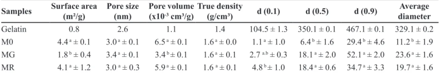
Surface area, Porosity and True Density
Table III presents the results of surface area, pore volume, pore size and true density for gelatin, M0, MG and MR. Surface area and pore volume of MG were
signiicantly lower than the values obtained for M0 and
MR. However, this occurrence brought no changes to
surface morphology of microspheres that can be identiied
by the SEM photomicrographs. The amount of pore sizes for all samples was in the range of mesoporosity
classiication (20-500 nm) (Teixeira, Coutinho, Gomes, 2001). No signiicant diferences in this parameter were
observed amongst M0, MG and MR. Values of true density
for all samples showed no signiicant variations. This fact indicated that crosslinking with GTA inluenced the results of surface area and pore volume, but it had no efect on
true density of the powder.
Particle size distribution and particle diameters
The particle size distribution is presented in Figure 3. MR presented the smaller particle size distribution amplitude than M0 and MG. Diameter values at 10, 50, 90% and average diameter of gelatin, M0, MG and MR are presented in Table II.
Results presented that average diameters were higher in groups subjected to crosslinking (MG and MR), although, this value was only consistent with that one observed for surface area regarding the MG, which showed that the increase in particle diameter implied in its surface area reduction. Also, it has been reported that GTA is an
efective crosslinker agent that provides stability and shape
to gelatin microspheres by forming a network structure from the GTA aldehyde groups and the gelatin amino
groups undergoing Schif base reaction (Karthikeyan et al., 2013).
These results may indicate that, during the microspheres obtaining process, the washes with acetone, aiming at removing any oil residual, promoted deagglomeration of the particles, although, the crosslinking agents and some oily remained residual contributed to the highest average diameter for MG and MR.
In vivo cutaneous compatibility assays
According to the in vivo cutaneous biophysical properties established before (basal values) and 24 hours subsequently to the patch test, aqueous dispersions at 10.0% w/v of gelatin (non-treated raw material), M0, MG and MR did not elicit any signs of skin damage, regarding the formation of erythema (skin redness), disruption of the barrier function (increase in transepidermal water loss) and negative interference in the SC hydration, in
comparison with the negative control (puriied water).
For all the variables, the ratio between the values after
TABLE III - Surface area, volume and pore sizes, true density, diameters of 10%, 50%, 90% and average diameter by laser difraction of gelatin and gelatin microspheres
Samples Surface area (m²/g)
Pore size (nm)
Pore volume (x10-3 cm³/g)
True density
(g/cm³) d (0.1) d (0.5) d (0.9)
Average diameter
Gelatin 0.8 2.6 1.1 1.4 104.5 ± 1.3 350.1 ± 0.1 467.1 ± 0.1 329.1 ± 0.2
M0 4.4 a ± 0.1 3.0 a ± 0.1 6.5 a ± 0.1 1.6 a ± 0.0 1.1 a ± 1.0 6.4 b ± 1.6 29.4 b ± 4.6 11.2 b ± 1.9 MG 1.8 b ± 0.4 3.4 a ± 0.1 3.4 b ± 0.1 1.6 a ± 0.1 2.7 a.b ± 0.3 18.1 a ± 2.0 52.1 a ± 2.0 23.6 a ± 1.6 MR 4.1 a ± 1.2 3.0 a ± 0.3 5.9 a ± 0.1 1.6 a ± 0.1 4.8 b ± 1.0 18.4 a ± 0.6 34.7 a ± 3.3 19.7 a ± 1.6
Diferent letters in the same column represent statistically diferent results according to the ANOVA followed by Tukey test. M0:
the patch application and the basal values was, hence, close to 1 (Figure 4). The ratio was used to decrease the inter-individual variability impact (Oliveira et al., 2015).
Photoprotective activity in vitro: SPF and protection against UVA
Table IV presents the results of photoprotective
eicacy in vitro for dispersions containing gelatin, M0,
MG, MR, and/or UV ilters, alone or in combination.
SPF values for dispersions containing gelatin (dispersion B), M0 (dispersion C), MG (dispersion D) or MR (dispersion E) were equal to base dispersion (dispersion A), that presented value close to 1.0 (no
anti-UVB eicacy). All dispersions containing OCT and gelatin
or microspheres (dispersions L, M, N and O) presented SPF values equal to or greater than 4.0. However, regarding to dispersions containing BZ3 and gelatin or microspheres, only the one containing gelatin (dispersion G) presented SPF values equal to or greater than 4.0.
The dispersions containing gelatin and BZ3 or OCT (dispersions G and L) had higher SPF values than
the dispersions containing only one of these UV ilters
(dispersions F and K), suggesting that the gelatin, as
raw material, when combined with ilters, showed good
ilm formation and/or absorption in the UVB region that contributed with the SPF improvement (dispersions G and L) and, interestingly, M0 did not achieve an enhanced
FIGURE 3 - Particle size distribution by laser difraction of gelatin (a), M0 (b), MG (c) and MR (d).
performance as the gelatin, even though, there was not the use of crosslinking agent.
According to our results, it may be suggested that
M0, MG and MR performed discreetly ilm formation and with similar proile among them. It was supposed that the
microspheres could develop a booster SPF activity by dispersing the UV radiation, considering the more rigid spherical structures achieved by the reaction with GTA and RUT, or by absorbing the UVB and UVA radiation,
regarding the presence of the lavonoid. It is also allowed
to suppose that the polymorph aspect of the gelatin, as
raw-material, could have inluenced the ilm formation more eiciently considering the arrangement of the irregular shape gelatin particles, that structured a thicker ilm, since
the gelatin is not soluble at the oily vehicle adopted in this
research. Still considering this property, the efect of MR
could have been inhibited by two mechanisms: (i) rutin was inaccessible to absorb the UV radiation since it was bound to the gelatin through the crosslinking reaction; and
(ii) rutin was not suicient soluble at the oily vehicle, than
it was not adequately free for exerting its UV absorbing property. Rutin is a natural compound that does not possess the tendency to permeate across the skin and it is a notable antioxidant that has been studied as sunscreen adjuvant with promising results. For example, our research group observed that an O/W emulsion containing 0.1% rutin in association with 3.5% octyl methoxycinnamate and 1.0%
benzophenone-3 developed a signiicant improvement
of the in vitro sun protective properties in comparison
with a similar preparation without the lavonoid (Baby et al., 2008; de Oliveira et al., 2015). Oliveira et al. (2015) additionally described the positive interaction of 0.1% rutin and 6.0% BZ3 in an O/W emulsion. Authors observed
that the lavonoid contributed with the elevation of the in vitro SPF of 24 to 33. It is noteworthy to mention that the gelatin, M0, MG and MR concentrations were not suitable enough to interfere more positively with the efficacy parameters of sun protection.
Regarding λC (critical wavelength, nm), only
dispersions without ilters (dispersions A, B, C, D and E)
presented values of λC greater than 370 nm. However, the average SPF values were approximately 1.0, indicating low protection against UVB radiation. The UVA/UVB ratio of dispersions containing OCT (dispersions K, L, M, N and O) was the lowest of all dispersions and it can be explained by UVB action, that is assigned to this
chemical UV ilter. Dispersions without ilters showed no signiicant absorbance, indicating that gelatin and isolated
microparticles did not develop substantial photoprotective action. In mixtures containing BZ3 and/or OCT, it was
observed that there were no changes in the peak proiles
due to the presence of gelatin or M0, MG or MR.
CONCLUSION
Physicochemical properties of MG and MR indicated that crosslinking method was more effective when the crosslinking agent was GTA and suggested that crosslinking occurred with RUT, however, to a lesser extent that would reinforce further researches to establish optimal conditions for the use of rutin as a potential substituent for GTA. The SPF analysis of dispersions containing M0, MG or MR, isolated or combined with chemical UV filters (benzophenone-3 and octyl methoxycinnamate) suggested that the microspheres (M0, MG or MR), at a 5.0% w/w, had
no additional chemical or physical photoprotective efect
TABLE IV - Photoprotective eicacy in vitro for dispersions
containing gelatin and gelatin microspheres, and UV ilters alone
or in combination. Values were expressed as mean ± standard
deviation (n = 3)
Dispersions
Sun protection
fator (in vitro SPF)
λC (nm)
UVA / UVB ratio
A 1.0 a ± 0.1 378.6 a ± 9.6 0.8 a ± 0.2 B 1.1 a ± 0.2 383.0 a ± 10.0 0.8 a ± 0.3 C 1.0 a ± 0.0 377.5 a ± 20.0 0.8 a ± 0.4 D 1.0 a ± 0.0 386.0 a ± 6.6 0.8 a ± 0.3 E 1.0 a ± 0.0 374.6 a ± 12.4 0.6 a ± 0.2 F 2.8 b ± 0.3 354.7 b ± 1.2 0.5 b ± 0.0 G 4.1 c ± 0.5 354.5 b ± 1.9 0.5 b ± 0.0 H 3.7 b.c ± 0.3 356.1 b ± 1.7 0.5 b ± 0.0 I 3.3 b.c ± 0.3 354.9 b ± 0.5 0.5 b ± 0.0 J 3.8 b.c ± 0.5 356.3 b ± 0.7 0.5 b ± 0.0 K 6.4 d ± 0.4 328.3 d ± 2.2 0.1 d ± 0.0 L 8.4 e ± 1.0 327.3 d ± 2.9 0.1 d ± 0.0 M 6.8 d.e ± 0.6 328.7 d ± 3.4 0.1 d ± 0.0 N 7.7 d.e ± 0.3 327.6 d ± 2.1 0.1 d ± 0.0 O 7.3 d.e ± 0.8 328.0 d ± 3.7 0.1 d ± 0.0 P 10.1 f ± 4.1 351.3 f ± 1.7 0.3 f ± 0.0 Q 16.2 f ± 3.4 350.0 f ± 1.1 0.3 f ± 0.0 R 10.7 f ± 1.1 350.7 f ± 0.2 0.3 f ± 0.0 S 11.0 f ± 2.1 351.2 f ± 1.2 0.3 f ± 0.0 T 12.4 f ± 1.4 350.9 f ± 0.9 0.4 f ± 0.0 Different letters in the same column represent statistically
diferent results according to the ANOVA followed by Tukey
in vitro, and showed no inluence on the estimated eicacy
when combined with UV ilters, that implies in formulation
adjustments such as increasing of the gelatin microsphere concentrations and, as well as, use of new classes of UV
ilters. The in vivo studies demonstrated that these materials had very good skin compatibility.
ACKNOWLEDGMENT
Authors are greatly thankful to The State of São Paulo Foundation - FAPESP - (2012/04435-9 and 2012/19972-0). Authors thank Prof. Humberto Gomes Ferraz for his valuable support regarding the analysis of particle size distribution, surface area, pore volume, pore size and true density. Authors also thank Prof. Carlota de Oliveira Rangel-Yagui and Prof. Tânia Santos de Almeida for their kind supports.
REFERENCES
BABY, A.R.; HAROUTIOUNIAN-FILHO, C.A.; SARRUF, F.D.; TAVANTE-JÚNIOR, C.R.; PINTO, C.A.S.O.; ZAGUE, V.; ARÊAS, E.P.G.; KANEKO, T.M.; VELASCO, M.V.R. Estabilidade e estudo de penetração cutânea in vitro da rutina veiculada em uma emulsão cosmética através de um modelo de biomembrana alternativo. Rev.Bras.Ciênc. Farm.,v.44, p.233-248, 2008.
BABY, A.R.; HAROUTIOUNIAN-FILHO, C.A.; SARRUF, F.D.; PINTO, C.A.S.O.; KANEKO, T.M.; VELASCO,
M.V.R. Inluence of urea, isopropanol and propylene glycol
on rutin in vitro release from cosmetic semisolid systems estimated by factorial design. Drug Dev. Ind. Pharm., v.35, p.272-282, 2009.
BIGI, A.; COJAZZI, G; PANZAVOLTA, S.; RUBINI, K.; ROVERI, N. Mechanical and thermal properties of gelatin
ilms at diferent degrees of glutaraldehyde crosslinking. Biomaterials, v.22, n.8, p.763-68, 2001.
BIGI, A.; COJAZZI, G; PANZAVOLTA, S.; RUBINI, K.;
ROVERI, N. Stabilization of gelatin ilms by crosslinking
with genipin. Biomaterials, v.23, n.24, p.4827-32, 2002.
BRESOLIN, T.M.B.; CECHINEL FILHO, V. Ciências
farmacêuticas: contribuição ao desenvolvimento de novos fármacos e medicamentos. Itajaí:Editora da Universidade do Vale do Itajaí, 2003.
BRUNETON, J. Elementos de Fitoquímica y de Farmacognosia. Zaragoza: Acribia, 1991.
C ATA L I N A , M . ; AT T E N B U R R O W, G . E . ; C O T, J . ; COVINGTON, A.D.; ANTUNES, A.P.M. Influence of crosslinkers and crosslinking method on the properties of
gelatin ilms extracted from leather solid waste. J. Appl.
Polym. Sci., v.119, n.4, p.2105-2111, 2011.
CHOY, Y.B.; CHENG, F.; CHOI, H.; KIM, K.K. Monodisperse gelatin microspheres as a drug delivery vehicle: release
proile and efect of crosslinking density. Macromol. Biosci.,
v.8, n.8, p.758-765, 2008.
GAN, C.Y.; CHENG, L.H.; EASA, A.M. Assessment of cross-linking in combined cross-linked soy protein isolate gels by microbial transglutaminase and maillard reaction. J. Food Sci., v.74, n.2, p.141-146, 2009.
HANSON, K.M.; GRATTON, E.; BARDEEN, C.J. Sunscreen enhancement of uv-induced reactive oxygen species in the skin. Free Radical Biol. Med., v.41, n.8, p.1205-1212, 2006.
HAYASHI, K.; TABATA, Y. Preparation of stem cell aggregates with gelatin microspheres to enhance biological functions.
Acta Biomater., v.7, n.7, 2797-2803, 2011.
K A RT H I K E YA N , S . ; R A J E N D R A P R A S A D , N . ; GANAMANI, A.; BALAMURUGAN, E. Anticancer activity of resveratrol-loaded gelatin nanoparticles on NCI-H460 non-small cell lung cancer cells. Biomed. Prev. Nutr., v.3, n.1, p.64-73, 2013.
NAHAR, M.; MISHRA, D.; DUBEY, V.; JAIN, N.K. Development, characterization, and toxicity evaluation of amphotericin b-loaded gelatin nanoparticles. Nanomed-Nanotechnol., v.4, n.3, p.252-261, 2008.
OLIVEIRA, C.A.; PERES, D.D.; GRAZIOLA, F.; BOU-CHACRA, N.A.; ARAUJO, G.L.B.; FLORIDO, A.C.; M O TA , J . P. ; R O S A D O , C . ; V E L A S C O , M . V. R . ; RODRIGUES, L.M.; FERNANDES, A.S.; BABY, A.R. Cutaneous biocompatible rutin-loaded gelatin-based nanoparticles increase SPF of the association of UVA and
UVB ilters. Eur. J. Pharm. Sci., v.81, p.1-9, 2016.
PATEL, M.; JAIN, S.K.; YADAV, A.K.; GOGNA, D.; AGRAWAL, G.P., Preparation and characterization of oxybenzone-loaded gelatin microspheres for enhancement
of sunscreening eicacy. Drug Deliv., v.13, n.5, p.323-330,
2006.
PERES, D.D.; OLIVEIRA, C.A.; COSTA, M.S.; TOKUNAGA, V.K.; MOTA, J.P.; ROSADO, C.; CONSIGLIERI, V.O.; KANEKO, T.M.; VELASCO, M.V.R.; BABY, A.R. Rutin increases critical wavelength of systems containing a
single UV ilter and with good skin compatibility. Skin Res.
Technol., v.22, p.325-333, 2016.
R A B A N E L , J . M . ; B A N Q U Y, X . ; Z O U A O U I , H . ; MOKHTAR, M.; HILDGEN, P. Progress technology in microencapsulation methods for cell therapy. Biotechnol. Progr., v.25, n.4, p.946-963, 2009.
R AT A N AVA R A P O R N , J . ; R A N G K U PA N , R . ; J E E R ATAWAT C H A I , H . ; K A N O K PA N O N T, S . ; DAMRONGSAKKUL, S. Influences of physical and chemical crosslinking techniques on electrospun type a
and b gelatin iber mats. Int. J. Biol. Macromol., v.47, n.4,
p.431-438, 2010.
SCHLUMPF, M.; SCHMID, P.; DURRER, S.; CONSCIENCE, M.; MAERKEL, K.; HENSELER, M.; GRUETTER, M . ; H E R Z O G , I . ; R E O L O N , S . ; C E C C AT E L L I , R.; FAASS, O.; STUTZ, E.; JARRY, H.; WUTTKE, W.; LICHTENSTEIGER, W. Endocrine activity and
developmental toxicity of cosmetic uv ilters - an update. Toxicology, v.205, n.1-2, p.113-122, 2004.
S H E U , M . T. ; H U A N G , J . C . ; Y E H , G . C . ; H O , H . O . Characterization of collagen gel solutions and collagen matrices for cell culture. Biomaterials, v.22, n.13, p.1713-1719, 2001.
SILVA, D.G.; SARRUF, F.D.; OLIVEIRA, L.C.D.; ARÊAS, E.P.G.; KANEKO, T.M.; CONSIGLIERI, V.O.; VELASCO, M.V.R.; BABY, A.R. Influence of particle size on appearance and in vitro efficacy of sunscreens. Braz. J. Pharm. Sci., v.49, n.2, p.251-261, 2013.
SPRINGSTEEN, A.; YUREK, R.; FRAZIER, M.; CARR, K.F. In vitro measurement of sun protection factor of sunscreens
by difuse transmittance. Anal. Chim. Acta, v.380, n.2-3,
p.155-164, 1999.
STRAUSS, G.; GIBSON, S.M. Plant phenolics as cross-linkers of gelatin gels and gelatin-based coacervates for use as food ingredients. Food Hydrocolloid, v.18, n.1, p.81-89, 2004.
TANAKA, N.; SETSUKO, T.; UTSUMI, I. A new oral gelatinized sustained-release dosage form. J. Pharm. Sci., v.52, n.7, p.664-667, 1963.
TEIXEIRA, V.G.; COUTINHO, F.M.B., GOMES, A.S. Principais métodos de caracterização da porosidade de resinas à base de divinilbenzeno. Quim. Nova, v.24, n.6, p.808-818, 2001.
UGWOKE, M.I.; KINGET, R. Influence of processing variables on the properties of gelatin microspheres prepared by the emulsification solvent extraction technique. J. Microencapsul., v.15, n.3, p.273-281, 1998.
VELASCO, M.V.R.; SARRUF, F.D.; SALGADO-SANTOS, I.M.; HAROUTIOUNIAM-FILHO, C.A.; KANEKO, T.M.; BABY, A.R. Broad spectrum bioactive sunscreens. Int. J. Pharm., v.1/2, p.50-57, 2008.