Fisioter. Mov., Curitiba, v. 31, e003119, 2018 DOI: http://dx.doi.org/10.1590/1980-5918.031.AO19 Licensed under a Creative Commons attribution
The effects of functional training and stationary
cycling on respiratory function of elderly
with Parkinson disease: a pilot study
Os efeitos do treino funcional e da bicicleta
estacionária na função respiratória de idosos com
Doença de Parkinson: um estudo piloto
Raíssa Almeida da Silveira, Karen Valadares Trippo, Gabriel Pereira Duarte, Mansueto Gomes Neto, Jamary Oliveira Filho, Daniel Dominguez Ferraz*
Universidade Federal da Bahia (UFBA), Salvador, BA, Brazil
[R]
Abstract
Introduction: Respiratory function changes in patients with Parkinson’s disease (PD) contribute to aspiration pneumonia, the main cause of mortality in PD population. Objective: To compare the effects of functional training and cycle exercise in respiratory function of elderly with PD. Methods: Twenty participants were randomised into two groups. Experimental group (EG) (n = 10) performed cycle exercise and control group (CG) (n = 10) was trained with functional training. A blinded researcher evaluated maximal inspiratory pressure (MIP), maximal expiratory pressure (MEP), thoracoabdominal amplitude, forced vital capacity (FVC) and forced expiratory volume in the first second (FEV1) before
and after interventions. Results: Both groups did not improve significantly respiratory function. Only CG improved significantly FVC (p = 0.01). No differences were found between groups. Conclusion:
8-week exercise programs are not able to improve the respiratory function of elderly in 2 to 3 PD
* RAS: BS, e-mail: [email protected]
KVT: Doctoral student, e-mail: [email protected] GPD: BS, e-mail: [email protected]
2
Hoehn & Yahr stage. Both functional training and cycle exercise do not to improve respiratory function in elderly with PD.
Keywords: Aged. Exercise. Respiratory Function Tests.
Resumo
Introdução: Mudanças na função respiratória em pacientes com Doença de Parkinson (DP) contribuem para a pneumonia aspirativa, principal causa de mortalidade nessa população. Objetivo: Comparar os efeitos do treino funcional e do exercício em bicicleta estacionária na função respiratória de idosos com Doença de Parkinson (DP). Métodos: 20 participantes foram randomizados em 2 grupos. O grupo experimental (GE) (n = 10) realizou exercício em bicicleta estacionária e o grupo controle (GC) (n = 10) realizou o treino funcional. Um pesquisador cegado avaliou a pressão inspiratória máxima (PIM), a pressão expiratória máxima (PEM), a amplitude toracoabdominal, a capacidade vital forçada (CVF) e o volume expiratório forçado no primeiro segundo (VEF1) antes e após as intervenções. Resultados: Ambos os grupos não melhoraram significativamente a função respiratória. Apenas o GC melhorou de forma significante a CVF (p = 0.01). Nenhuma diferença foi encontrada entre os grupos. Conclusão: Programas de exercício com duração de 8 semanas não são capazes de melhorar a função respiratória de idosos com DP nos estágios 2 a 3 de Hoehn & Yahr. Ambos grupos que realizaram o treino funcional e exercício em bicicleta estacionária não melhora a função respiratória de idosos com DP.
Palavras-chave: Idoso. Exercício. Testes de Função Respiratória.
Introduction
Parkinson’s disease (PD) patients present neurological symptoms as resting tremor, rigidity, bradykinesia and postural instability which intensify with disease progression [1]. These patients also have respiratory dysfunction, a major cause of mortality in this population. They tend to reduce their daily activities and do not notice these breathing dysfunctions in early disease stages [2].
Chest expansion decrease, and lung volumes reduction may induce respiratory changes in patients with PD. Trunk and spine mobility restriction, osteoarticular degeneration and trunk
flexion posture can also compromise the inspiration
and expiration movements inducing respiratory dysfunctions [3]. Patients with PD presented reduction in cough effectiveness and decreasing in peak electromyography amplitude of abdominal muscles during voluntary and induced cough4. These changes hinder the removal of airway secretions by the respiratory system, contributing to aspiration pneumonia, the main cause of mortality in PD population [4].
Although PD is an irreversible and neurodegenerative disease, pharmacological and non-pharmacological treatment can help to minimize and control its signs and symptoms. Neurorehabilitation is a complement to drug treatment and works primarily to prevent or reduce disease complications [5]. The conventional therapeutic exercises aim to maintain the physical conditioning, preserve the activity and musculoskeletal flexibility and improve postural balance [6].
3
Even if breathing dysfunction is a major cause of death in PD, assessment and treatment of respiratory function is not common in clinical practice. The aim of this study was to compare the effects of functional training and cycle exercise in respiratory function of elderly with PD.
Methods
We performed a pilot randomised controlled clinical trial with blinded assessment. The study was conducted at the Centro de Referência Estadual de Atenção à Saúde do Idoso (CREASI) (State Reference Health Care Centre for Elderly), a public reference outpatient clinic for the elderly in Salvador, Bahia, Brazil. Data were collected in the period between June/2015 and June/2016. The trial design followed the recommendations of the Consolidated Standards of Reporting Trials (CONSORT) [10]. This is an exploratory analysis of clinical trial registered
under ClinicalTrials.gov (clinical trial identifier:
NCT02622737). The study was carried out in accordance with the Declaration of Helsinki and approved by the Ethics Human Research Committee of the Institute of Health Sciences — Federal University of Bahia (ref n° 1.016.971). All participants provided voluntary written informed consent.
Population and sample
The sample consisted of 20 elderly patients with PD divided randomly into two equal groups. The method of sequence generation was determined with a computerized random number generator. Participants of CG underwent functional training based on conventional therapeutic exercises and the EG performed an aerobic training on a stationary bike.
Elderly candidates with idiopathic PD according to the London Brain Bank Criteria were recruited and included in the study according to the following
inclusion criteria: ≥ 60 years-old, regular use of
medication for PD and stage 2, 2.5 or 3 according to
modified Hoehn and Yahr scale. The exclusion criteria were: regular use of medication for PD modified
Hoehn and Yahr stages 2, 2.5 or 3 (the interventions were developed for patients with bilateral disease involvement without severe disability). The exclusion criteria were: visual or hearing impairment;
parkinsonian syndromes other than PD; bone, joint or muscle diseases that limit the practice of physical activity; chronic uncontrolled diseases (hypertension, diabetes mellitus, chronic pain); unstable cardiovascular disease (acute heart failure, recent myocardial infarction, unstable angina and arrhythmias uncontrolled); current alcohol and other toxic substance use; contraindications for performing physical exercise according to the American College of Sport Medicine; practicing any physical exercise program in the last 6 months or participating in
regular resistance training (≥ 2 times per week) in
the previous 12 months.
Evaluation procedures
Unified Scale Evaluation of Parkinson's disease
(UPDRS) was used to evaluate the clinical and functional status of all participants [11].
We evaluated the thoracoabdominal amplitude, respiratory muscle strength, forced vital capacity
(FVC) and forced expiratory volume in the first
second (FEV1). All participants were evaluated before
and after treatment by a single blinded researcher. The thoracoabdominal amplitude measure was obtained in centimetres by cirtometry. The measurement was performed horizontally into three levels: axillary, xiphoid and abdominal. In the axillary region the tape was placed below axillary fold. We used the lower edge of the xiphoid to measure xiphoid amplitude, and to check abdominal amplitude, we used average distance between xiphoid process and umbilicus [12]. The participants were examined in standing position. In each benchmark were performed three maximum inspiration and expirations under the
examiner's voice command. Three values were noted
and the differences obtained between inhalation and
exhalation (amplitude coefficient). For data analysis
4
Spirometry was performed to evaluate FVC and FEV1, respecting technical procedures, acceptability
and reproducibility criteria, according to the standards of American Thoracic Society and European Respiratory Society [15].
Intervention procedures
The sessions were performed in all groups lasted 8 weeks with a frequency of three 50-minutes sessions per week. Each session consisted in 10 minutes of stretching (hamstrings, quadriceps, triceps sural, paraspinal, pectoralis minor and major, biceps,
deltoid, trapezius, rhomboids, flexors and extensors
of the wrist); 5 minutes of calisthenics exercises, 30 minutes of intervention and 5 minutes of breathing exercises (diaphragmatic breathing exercises and costal expansions) to slowdown. The heart rate, oxygen saturation and blood pressure were measured before training, after 15 minutes and at the end of each session. The participants with more than 1 missing a week or more than 4 faults were considered losses. and consisted of an exercise circuit consisting of 10 activities lasting 3 minutes each one. Among the functional activities the following stations were: 1) Gait with obstacles; 2) Going up and down stairs and ramp; 3) Sitting and standing exercises; 4) Side gears; 5) Balance exercise in proprioceptive platform; 6) Activities with balls; 7) Step exercises; 8) Foot tip exercises; 9) Graded reaching activities; 10) Gait training. The intensity evolution was performed progressively with barbells and manual resistance, achieving the individual perception of 15 points on Borg scale [17].
Participants of EG performed an aerobic training on stationary bike lasting 30 minutes. The intensity
of the training in the first week was 50% of maximum
heart rate and gradually increased to reach 75% in the eighth week. The Karvonen formula [18] was used to determine heart rate training goals for each session. Heart rate training was calculated using the equation: (MHR – HRR) x intensity + HRR. The maximum heart rate (MHR) was estimated by the equation (220 – age) and resting heart rate (HRR) was checked before starting the workout after 10 minutes of rest, with the patient in seated.
In addition, patients should be in a period “on” of PD and taking antihypertensive medications, in case of hypertension.
Statistical analysis
Variables were expressed as median and interquartile range. To compare independent and paired variables was used nonparametric tests as
Mann-Whitney and Wilcoxon test. The significance
level used was 5 % (p < 0.05).
Results
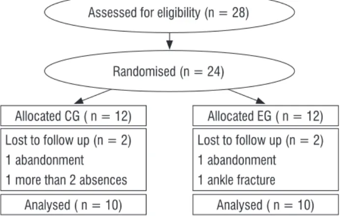
The study began with 24 participants, but only 20
patients finished the treatment protocol (Figure 1).
The participants were evaluated before and after the therapeutic programs. One participant was excluded because it had more than two absences per week, two subjects abandoned the study and one fractured his ankle at home.
Analysed ( n = 10) Allocated CG ( n = 12)
Lost to follow up (n = 2) 1 abandonment 1 more than 2 absences
Analysed ( n = 10) Allocated EG ( n = 12)
Lost to follow up (n = 2) 1 abandonment 1 ankle fracture Randomised (n = 24)
Assessed for eligibility (n = 28)
Figure 1 - Flow diagram of the progress through the phases of a parallel randomised trial of two intervention groups. Salvador, Brazil, 2016.
Table 1 shows no difference between demographic and clinical characteristics of the two groups.
Table 1 - Baseline demographic and clinical characteristics (p-values refer to Mann-Whitney test). Salvador, Bahia, Brazil, 2016.
Variable CG (n=10) EG (n=10) p
Median (IR) Median (IR)
Age (years) 67 (65-73) 68 (63-75) 0.90
BMI (Kg/m2) 26.24
(23.76-30.57)
25.55
(22.70-29.10) 0.59 Hoehn & Yahr 2.75 (2.50-3.00) 2.50 (2.00-3.00) 0.31
UPDRS 23.50
(18.25-27.25)
24.00
(17.50-45.25) 0.58 Illness time (years) 5 (2-12) 6 (6-11) 0.32
5
Table 2 and 3 show the CG and EG results of respiratory function before and after 8 weeks
of intervention. The CG showed significant
improvements in FVC (p = 0.01). There were no
significant differences for the other variables in both
groups (Table 2).
Table 2 - Summary results for each study group (p-values refer to Wilcoxon test). Salvador, Bahia, Brazil, 2016.
Variable CG (n=10) Baseline median (IR) 8 months median (IR) p EG (n=10) Baseline median (IR) 8 months median (IR) p FVC (%) 91.50 (66.50-109.00) 122.00 (72.75-159.00) 0.01 75.00 (72.00-92.50) 92.00 (79.00-112.50) 0.50 FEV1 (%) 71.50 (63.75-99.25) 71.00 (62.50-97.75) 0.95 93.00 (81.50-103.50) 85.00 (77.00-95.50) 0.06
MIP (cmH2O)
-64.00 (-48.25-104.88) -71.50 (-58.75-88.25) 0.72 -68.00 (-76.00-62.00) -70.00 (-85.00-42.00) 0.68
MEP (cmH2O)
60.00 (40.75-91.60) 70.00 (51.50-112.75) 0.13 56.00 (42.00-60.00) 60.00 (44.00-80.00) 0.34 AAC (cm) 6.00 (3.75-7.00) 6.00 (6.00-7.00) 0.14 6.00 (4.25-9.25) 7.00 (5.00-9.00) 0.68 XAC (cm) 5.00 (2.12-7.00) 5.00 (4.00-6.25) 0.43 5.00 (3.75-8.00) 6.00 (4.50-9.00) 0.49 UAC (cm) 3.50 (0.75-5.00) 4.50 (-0.50-7.00) 0.73 -1.00 (-4.50-3.00) 4.00 (-0.50-7.00) 0.14
Note: Number of participants; cm: centimetres; IR: interquartile range; FVC: forced vital capacity; FEV1: expiratory volume in the first second; MIP: maximal inspiratory pressure; MEP: maximal expiratory pressure; AAC: axillar amplitude coefficient; XAC: xiphoid amplitude coefficient; UAC: umbilical amplitude coefficient.
Comparing before and after training values, EG
(Table 3) increased not significantly FVC, MEP, MIP,
abdominal and xiphoid amplitude.
Table 3 - Comparison of respiratory parameters between groups, before and after interventions (p-values refer to Man-Whitney test). Salvador, Bahia, Brazil, 2016.
Variable CG (n=10) Difference (IR)
EG (n=10) Difference (IR) p
FVC (%) 14.50 (3.25-61.50) 7.00 (-8.25-39.50) 0.290
FEV1 (%) -3.00(-9.50-10.00) (-7.25-21.75)-5.50 0.940
MIP (cmH2O) 12.50 (-28.87-25.25)
2.50
(-29.50-17.25) 0.520
MEP (cmH2O) 11.00 (-5.25-32.50)
19.00
(-6.25-30.00) 0.705
AAC (cm) 0.00 (-0.12-2.25) 1.00 (-0.62-2.00) 0.760
XAC (cm) 0.5 (-1.00-1.87) 1.00 (1.00-2.12) 0.314
UAC (cm) 0.00 (-2.00-4.12) 0.00 (-2.25-5.12) 0.878
Note: Number of participants; cm: centimetres; IR: interquartile range; FVC: forced vital capacity; FEV1: expiratory volume in the first second; MIP: maximal inspiratory pressure; MEP: maximal expiratory pressure; AAC: axillar amplitude coefficient; XAC: xiphoid amplitude coefficient; UAC: umbilical amplitude coefficient.
No difference between groups was observed (Table 3).
Discussion
According to our results functional exercise programme and cycle training improved respiratory function of elderly with PD. Although only functional
training group increased significantly the lung
volume capacity, no difference was observed between the two groups.
Studies suggest that physiologic use of exercise can be an important component of neuroplastic changes in human PD brain and support the central hypothesis that self-produced activity is important in slowing, halting or reversing human PD [19].
However, physical activity seems to be insufficient
to improve respiratory function. A multicentre randomised controlled clinical trial has evaluated the effect of physical activity on respiratory outcomes in community-dwelling elders with mobility limitations [20]. Their physical activity programme was like our functional training, and included walking
and strength, flexibility and balance training. In
6
activity was insufficient to improve dyspnoea severity,
FEV1 or MIP. In our study we have found similar results in respiratory parameters of elderly with PD trained with cycling exercise and functional training.
However, the combination of aerobic exercise and resistance training improved respiratory parameters in patients with moderate to severe chronic obstructive pulmonary disease. Twenty-four sessions, three times a week involving 40 minutes on treadmill with a load of 80% of maximal incremental test, 30 minutes of arms exercise with two diagonal movements and 20 minutes of stretching or relaxation improved respiratory muscles strength in patients with chronic obstructive pulmonary disease [21]. Therefore, the combined-training (unsupported training for upper limbs and endurance training by treadmill for lower limbs) decreased dyspnoea of this population. Possibly the high intensity workload
(80%) may explain this significant improvement
of respiratory muscles. Even if physiopathology of muscle strength loss is different between PD and chronic pulmonary disease, both patients presented muscle endurance reduction. Lower training intensities used in cycling exercise and functional
training may have influenced our results.
Maybe respiratory exercises should be necessary to improve respiratory parameters in patients with PD. Respiratory exercises include diaphragmatic and costal breathing, mobilization and stretching of the trunk, cervical region and external intercostal, deep inspiration and incentive spirometry. By performing
five months of respiratory exercises, Alves et al. [22]
found improvement in respiratory symptoms in a 47-year-old male patient in stage IV Hoehn & Yahr scale. Their results showed improvements in FVC,
FEV1, MIP and MEP, peak expiratory flow (PEF) and
Maximum Voluntary Ventilation (VVM). Our patients also have done trunk and cervical stretching and diaphragmatic and costal breathing, performed 3 times a week during 8 weeks. However, it was not
sufficient to improve respiratory function. Koseoglu
et al. [23] also used respiratory exercises in patients with PD I and II Hoehn & Yahr stages and found similar results. They performed diaphragmatic, deep-breath exercises, voluntary hyperpnoea, and strengthening of upper limbs and found similar improvements in spirometry, dyspnoea index and exercise capacity.
Respiratory kinesiotherapy is another intervention procedure consisting of an exercise program
combined with diaphragmatic re-education, costal breathing, thoracic, cervical and shoulder girdle mobilization, stretching of the accessory respiratory muscles, deep-breathing exercises and incentive spirometry training. The study of Pereira et al. [24]
verified the effect of respiratory kinesiotherapy in
three men, aged 65 years, with idiopathic PD, in stages 2.5 and 3 Hoehn & Yahr scale. At two months, after performing 16 sessions of 30 minutes, it was observed good improvements of lung volume and
compliance. These findings also support the necessity
of respiratory exercises to improve pulmonary parameters of patients with PD.
Another complementary physiotherapeutic treatment is the Isotretching method. This therapy acts on the imbalances that affect posture, contributing to the improvement of range of motion, muscular strength, body awareness, respiratory capacity and diaphragmatic mobility. Palacio et al. [25] compared the effects of 20 sessions of hydrotherapy and Isotretching method on the pulmonary capacity of patients with PD aged 45 - 65 years old. Although
they did not perform a specific respiratory training, they observed statistically significant improvement of
the MIP, MEP, FVC and FEV1 in both training groups, which was not found in our study.
Another way to improve respiratory function is associating respiratory muscle training with motor therapy. The study conducted by Moreira et al. [26] have submitted PD patients to respiratory muscle training with threshold, 40% of MIP and MEP protocol, associated with whole body vibration therapy in 24 sessions for two months. The participants, aged 45 to 80 years old, presented stage 3 of the Hoehn & Yahr
classification. They observed an increase in MIP and
MEP values, that is, an improvement on respiratory
muscle resistance after intervention. These findings
demonstrate that the combination of physical and respiratory training may also be able to provide positive results on respiratory functions. A clinical trial conducted by Inzelberg et al. [27] also investigated the effect of inspiratory muscle training on lung function, inspiratory muscle performance, dyspnoea and quality of life of 20 patients with PD in stage 2 or 3 of Hoehn & Yahr scale. The training performance was conducted using 15% of MIP, which increased progressively each session until reaching
60% at the end of the first month. The CG was trained with a very low fixed load of 7 cmH2O. At the end
7 inspiratory muscle performance and a significant
decrease in dyspnoea perception.
The practice of physical activity on a regular basis increase aerobic capacity and cardiac output [28] Pelosin et al. [29] reported that individuals with PD spend approximately 20% more energy than healthy controls when exercising on treadmill or
cycle ergometer. Beneficial changes in hemodynamic,
hormonal, metabolic and respiratory functions may also occur with increased ability to perform physical exercises.
Hydrotherapy, Halliwick method, is another modality of training used to treat patients with PD. Espindola et al. [30] have evaluated the effects
of hydrotherapy on respiratory function of five
elderly patients with PD. The treatment consisted of ten points: mental adjustment, detachment, control of longitudinal rotation, control of sagittal rotation, control of horizontal rotation, control of combined rotation, thrust, balance, turbulence/ slip, simple progressions and basic swimming. They have evaluated thoracic expansibility by cirtometry,
cardiopulmonary fitness using 6-minute walk test, and pulmonary flow and volumes with flowmetry
and ventilometry. The study results demonstrated
beneficial effects of Halliwick method on respiratory
function of participants in all variables studied
without performing a specific respiratory training.
The main limitation of the study is represented by the small sample size.
Further research is needed to determine the optimal exercise training to improve the respiratory function of patients with PD.
Conclusion
The functional training based on conventional therapeutic exercises, the most used treatment for patients with PD, as well as aerobic exercise cycling
training seem to modify, but not significantly the
respiratory parameters of elderly with PD. It is noteworthy that elderlies of the two groups did not obtain losses in respiratory capacity after 8 weeks of
intervention period. Studies indicate that the specific
training for the respiratory function seems to be essential for changes in the respiratory parameters of this population.
References
1. Gelb DJ, Oliver E, Gilman S. Diagnostic criteria for Parkinson disease. Arch Neurol. 1999;56(1):33-9.
2. Haas BM, Trew M, Castle PC. Effects of respiratory muscle weakness on daily living function, quality of life, activity levels and exercise capacity in mild to moderate Parkinson´s disease. Am J Phys Med Rehabil. 2004;83(8):601-7.
3. Cardoso SRX, Pereira JS. Análise da função respiratória na doença de Parkinson. Arq Neuro-Psiquiatr. 2002;60(1):91-5.
4. Pitts T, Bolser D, Rosenbek J, Troche M, Okun MS, Sapienza C. Impact of expiratory muscle strength training on voluntary cough and swallow function in Parkinson disease. Chest. 2009;135(5):1301-8.
5. Vieira FDM. Plataforma de Apoio à Terapia de Reabilitação e Manutenção de Doentes de Parkinson [master´s thesis]. Porto, PT: Universidade de Porto; 2013. 48 p.
6. Loureiro APC, Ribas CG, Zotz TGG, Chen R, Ribas F. Viabilidade da terapia virtual na reabilitação de pacientes com doença de Parkinson: estudo-piloto. Fisioter Mov. 2012;25(3):659-66.
7. Panizzi EA, Nunes AC, Borba C, Kerkoski E. Mobilidade torácica em estudantes na faixa etária de 8 a 14 anos de ambos os sexos: uma análise descritiva. VIII Encontro Latino-Americano de Iniciação Científica e IV Encontro Latino-Americano de Pós-Graduação; 2004 Oct 21-22; São José dos Campos. São José dos Campos: Universidade do Vale do Paraíba; 2004.
8. Lopes RB, Brito RR, Parreira VF. Padrão respiratório durante o exercício – revisão literária. R Bras Cienc e Mov. 2005;13(2):153-60.
9. Tomlinson CL, Herd CP, Clarke CE, Meek C, Patel S, Stowe R, et al. Physiotherapy for Parkinson’s disease: a comparison of techniques. Cochrane Database Syst Rev. 2014;(6):CD002815.
8
11. Goetz CG, Fahn S, Martinez-Martin P, Poewe W, Sampaio C, Stebbins GT, et al. Movement Disorder
Society-sponsored revision of the Unified Parkinson's Disease
Rating Scale (MDS-UPDRS): Process, format, and clinimetric testing plan. Mov Disord. 2007;22(1):41-7.
12. Rocha PA, McClelland J, Morris ME. Complementary physical therapies for movement disorders in
Parkinson's disease: a systematic review. Eur J Phys
Rehabil Med. 2015;51(6):693-704.
13. Abbruzzese G, Marchese R, Avanzino L, Pelosin E.
Rehabilitation for Parkinson's disease: Current
outlook and future challenges. Parkinsonism Relat Disord. 2016;22(Suppl 1):S60-4.
14. Black LF, Hyatt RE. Maximal respiratory pressures: normal values and relationship to age and sex. Am Rev Respir Dis. 1969;99(5):696-702.
15. Miller MR, Hankinson J, Brusasco V, Burgos F, Casaburi R, Coates A, et al. Standardisation of spirometry. Eur Respir J. 2005;26(2):319-38.
16. Canning CG, Sherrington C, Lord SR, Close JC, Heritier S, Heller GZ, et al. Exercise for falls prevention in Parkinson disease: a randomized controlled trial. Neurology. 2015;84(3):304-12.
17. Wilson RC, Jones PW. A comparison of the visual
analogue scale and modified Borg scale for the
measurement of dyspnoea during exercise. Clin Sci (Lond). 1989;76(3):277-82.
18. Whaley MH, Brubaker PH, Otto RM, Armstrong
LE. ACSM's guidelines for exercise testing and
prescription. 7th ed. Philadelphia: Lippincott Williams & Wilkins; 2006.
19. Hirsch MA, Iyer SS, Sanjak M. Exercise-induced
neuroplasticity in human Parkinson's disease: What
is the evidence telling us? Parkinsonism Relat Disord. 2016;22 (Suppl 1):S78-81.
20. Fragoso CAV, Beavers DP, Anton SD, Liu CK, McDermott MM, Newman AB, et al. Effect of Structured Physical Activity on Respiratory Outcomes in Sedentary Elderly Adults with Mobility Limitations. J Am Geriatr Soc. 2016;64(3):501-9.
21. Basso-Vanelli RP, Di Lorenzo VA, Labadessa IG, Regueiro EM, Jamami M, Gomes EL, et al. Effects of Inspiratory Muscle Training and Calisthenics-and-Breathing Exercises in COPD With and Without Respiratory Muscle Weakness. Respir Care. 2016;61(1):50-60.
22. Alves LA, Coelho AC, Brunetto AF. Fisioterapia respiratória na Doença de Parkinson idiopática. Fisioter Pesqui. 2005;12(3):46-9.
23. Köseoglu F, Inan L, Özel S, Deviren SD, Karabiyikoglu G, Yorgancioglu R, et al. The effects of a pulmonary rehabilitation program on pulmonary function tests and exercise tolerance in patients with Parkinson’s disease. Funct Neurol. 1997;12(6):319-25.
24. Pereira FF, Assumpção FF, Rosa JS, Lima MIL, Gonçalves
MCP, Faria L. Efeito da fisioterapia respiratória em
portadores da Doença de Parkinson. Rev Fisioter Ser. 2010;5(4):235-40.
25. Palácio SG, Barroca JB, Toldo KF, Ramalho JBL, Vanzela AL, Facci LM. Estudo comparativo entre a hidroterapia e a cinesioterapia na Doença de Parkinson. Rev Saúde Pesqui. 2011;4(2):191-9.
26. Moreira AM, Galvão MLC, Araújo HAG, Silva AT, Reis LM, Silva AM, et al. Treino muscular respiratório e terapia vibratória em pacientes com doença de Parkinson. Rev Neurocienc. 2015;23(3):479-85.
27. Inzelberg R, Peleg N, Nisipeanu P, Magadle R, Carasso RL, Weiner P. Inspiratory Muscle Training and the Perception of Dyspnea in Parkinson’s Disease. Can J Neurol Sci. 2005;32(2):213-7.
28. Cavalca C, Soldi F. Avaliação de Aptidão Física em Pacientes com Doença de Parkinson submetidos a tratamento hidroterápico através do Método Halliwick [undergraduate thesis]. Florianópolis (SC): Universidade do Sul de Santa Catarina; 2004. 29 p.
29. Pelosin E, Faelli E, Lofrano F, Avanzino L, Marinelli L, Bove M, et al. Effects of treadmill training on walking economy in Parkinson’s disease: a pilot study. Neurol Sci. 2009;30(6):499-504.
30. Espindola AR. Avaliação respiratória em pacientes com doença de Parkinson submetidos ao método Halliwick [undergraduate thesis]. Tubarão, SC: Universidade do Sul de Santa Catarina; 2005. 86 p.
Received in 08/01/2017
Recebido em 01/08/2017
Approved in 10/16/2017