Evaluation of the Rabbit as a Model for Chagas Disease - II.
Histopathologic Studies of the Heart, Digestive Tract and
Skeletal Muscle
Arnaldo Moreira da Silva
+, Luiz Eduardo Ramirez, Marlene Vargas*,
Edmundo Chapadeiro/
++, Zigman Brener**
Curso de Pós-graduação em Patologia Humana, Faculdade de Medicina do Triângulo Mineiro, 38025-440 Uberaba, MG, Brasil * Departamento de Patologia, Faculdade de Medicina Veterinária de Viçosa, MG, Brasil
** Centro de Pesquisas René Rachou-FIOCRUZ, Belo Horizonte, MG, Brasil
In order to investigate the value of the rabbit as an experimental model for Chagas’ disease, seventy one animals were inoculated with different Trypanosoma cruzi strains and routes. The rabbits were submitted to necropsy in acute (earlier than three months of infection), recent chronic (three to six months) and late chronic (later than six months) phases.
Myocarditis, generally focal and endomysial, occurred in 94.1%, 66.7% and 70.8% of the infected rabbits respectively in the acute, recent chronic and late chronic phases. The myocardial inflammatory exudate was composed by mononuclear cells, and also polymorphonuclear cells in the acute phase. In most cases of the late chronic phase, the myocarditis was similar to that described in the indeterminate form of human chagasic patients. Initial fibrosis occurred in the three phases but was more severe and frequent in the early chronic. Advanced fibrosis occurred only in the late chronic phase. Tissue para-sites occurred only in the acute phase. The digestive tract and skeletal muscles showed mild and occa-sional lesions.
Our data indicate that experimentally infected chagasic rabbits repeat some lesions similar to that of humans chagasic patients, specially that of the indeterminate form. So, it may be a useful, however not an ideal, model.
Key words: Trypanosoma cruzi - Chagas’ disease - experimental infection - rabbit - histopathology
+Present address: Serviço de Anatomia Patológica,
Hos-pital de Clínicas da Universidade Federal de Uberlândia, Campus Umuarama, Av. Pará nº 1720, 38405-382 Uberlândia, MG, Brasil
++Corresponding author. Fax: 55-034-312.6640
Received 3 March 1995 Accepted 22 December 1995
Many aspects of Chagas’ disease remain poorly understood, in spite of large number of researches in this subject. One example is the pathogenesis of the cardiac lesions in the chronic phase of the dis-ease. So, it is indispensable to select adequate ex-perimental models that can reproduce all or the majority of the pathologic events observed in hu-man cases, as recommended by WHO (1984). According to Brener and Ramirez (1985), other conditions are necessary, such as the development of the lesions in relatively short time, easy manipu-lation and accessible cost and maintenance.
Regarding to rabbits, there are many contro-versies about its usefulness as an experimental model to the study of Chagas’ disease. Different researchers have obtained discordant results, that were promising for those who utilized strains as Ernestina and Albuquerque of Trypanosoma cruzi
(Teixeira et al. 1975, 1983a, b, Figueiredo et al. 1979, Rezende Filho et al. 1979), and discourag-ing for others who employed strains as Y, CL, MR and 12SF (Andrade & Andrade 1979, Chiari et al. 1980). With the Colombian strain, Figueiredo (1984) and Figueiredo et al. (1985) observed sig-nificative lesions, which was not confirmed by Andrade and Andrade (1979).
The aim of this research is to study the histo-logic lesions of the heart, digestive tract and skel-etal muscle of rabbits infected with different strains of T. cruzi, by different numbers of parasites and routes, during the acute and chronic phases of the infection.
MATERIALS AND METHODS
Eighty three outbred male 2-4 month old rab-bits, weighing from 800 to 1,300 g were utilized. Among them, 71 were infected by intraperitoneal (ip) or conjunctival (conj) routes, with Y, CL or Ernane (Er) strains of T. cruzi. and 12, non-infected, were used as controls.
trypo-200 Histopathologic Studies in Chagasic Rabbits AM da Silva et al.
mastigotes (TcTry) of the Y strain; (c) metacyclic trypomastigotes (MTry) of the three strains, ob-tained from Dipetalogaster maximus.
Forty eight (48) rabbits were infected by ip route with 107 BTry (14 with Y, 16 with CL and 12 with Er) or TcTry (6 with Y) and 23 by conj route with 2 - 4 x 10³ MTry (8 with Y, 7 with CL and 8 with Er).
Seventy six rabbits were sacrificed and seven died spontaneously between 22 days and 24 months post-infection and were submitted to necropsy. Fragments of heart (from the four chambers), di-gestive tract and skeletal muscle (diaphragm, in-tercostal and psoas) were obtained for histopathological examination. Routine histologi-cal sections were stained by hematoxylin-eosin (H&E) and Gomori or Masson trichrome. Immuno-histochemistry reaction with the peroxidase-antiperoxidase method (Taylor 1986) was used in heart sections of 31 rabbits, to amplify the search of amastigote forms.
The histological study of the heart, digestive tract and skeletal muscle samples was systemati-cally done, specially concerning the inflammatory process when present (topography, characteristics of the exudate, fibrosis) and the presence of para-sites. Myocarditis, diagnosed in accordance to the Dallas’ criteria (Aretz 1987), was evaluated quali-tatively (inflammatory exudate, fibrosis), semiquantitatively, topographically (endomysial or perimysial inflammation) and about its extent: fo-cal (F), isolated little injured areas; zonal (Z), many confluent foci; and diffuse (D), large confluent ar-eas. To apprise the evolution of the myocarditis, three aspects were considered: (a) leukocytic in-filtration, characterizing active inflammation; (b) fibroblastic proliferation with minimal young col-lagen deposition, called here “initial fibrosis”; (c) abundant dense collagen deposition, designated “advanced fibrosis”. For semiquantitative evalua-tion, the inflammatory exudate, the initial fibrosis and the advanced fibrosis were characterized as F, Z or D. When F, the number of foci per 100 high power field (HPF) was counted, with a 40x objec-tive. Comparing the infected with the controls ani-mals, it seemed suitable to consider: (a) absence of lesion, less than one focus per 100 HPF (f/100 HPF); (b) very mild focal lesion (VmF), one to two f/100 HPF; (c) mild focal lesion (MF), three to ten f/100 HPF; (d) moderate focal lesion (MoF), 11 to 25 f/100 HPF; (e) severe focal lesion (SF), more than 25 f/100 HPF. Thus, the crescent severity de-gree were: VmF, MF, MoF, SF, Z and D.
For description and analysis of the results, it was considered three phases according to the time lapse from the infection to the necropsy: (a) acute phase, earlier than three months of infection; (b)
recent chronic phase, three to six months of infec-tion; (c) late chronic phase, later than six months of infection.
The chi-square test was utilized to compare the frequencies of the lesions observed in the diverse groups (p value ≤ 0.05 was considered statistically significant).
RESULTS
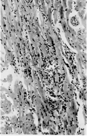
Acute phase - In 16 out of 17 animals submit-ted to necropsy during the acute phase (94.1%), it was observed myocarditis varying from VmF to Z (Table), characterized by myocardial foci of mono-nuclear infiltrate bound to the sarcolemma of myo-cardial cells, and occasional neutrophils and eosi-nophils granulocytes, with endomysial distribution and variable extension (Fig. 1). The exudate was associated with degeneration and destruction of myocardial cells. Nests of T. cruzi amastigotes were seen in six rabbits (35.3%): four infected with Y; one with CL and one with Er.
Initial fibrosis (VmF) occurred in the myocar-dium in only one rabbit, infected with Er. None
2
0
1
M
e
m
I
n
st
O
sw
a
ld
o
C
ru
z
,
R
io
d
e
Ja
n
e
ir
o
, V
o
l.
9
1
(2
),
M
ar
./A
p
r.
1
9
9
6
Inflammatory lesions of the heart, digestive tract and skeletal muscles in the different groups of experimentally chagasic and in the control rabbits
Heart DT SM
Groups Myocarditis
Epi-Phase Strain To Inflammatory exudate Initial fibrosis Advanced fibrosis car
tal Vmf MF MoF SF Z D Total Vmf MF MoF SF Z D Total Vmf MF MoF SF Z D Total ditis
Y 6 - 3 3 - - - 6 - - - 4 - 1
Acute CL 6 1 5 - - - - 6 - - - 1 - 1
Er 5 - - 2 1 1 - 4 1 - - - 1 - - - 3 2 1
Total 17 1 8 5 1 1 - 16 1 - - - 1 - - - 8 2 3
Recent Y 5 1 1 - - 1 - 3 - - - - 1 - 1 - - - 1 1
-chronic Er 1 - - - - 1 - 1 - - - - 1 - 1 - - -
-Total 6 1 1 - - 2 - 4 - - - - 2 - 2 - - - 1 1
-Y 17 1 9 2 - 2 - 14 3 - 1 - 1 - 5 2 2 1 - 2 - 7 3 3 2
Late CL 17 4 10 - - - - 14 - - - 4 - - - 4 5 3 3
chronic Er 14 2 4 - - - - 6 - - - 3 1 - - - - 4 1 3
-Total 48 7 23 2 - 2 - 34 3 - 1 - 1 - 5 9 3 1 - 2 - 15 9 9 5
Total 71 9 32 7 1 5 - 54 4 - 1 - 3 - 8 9 3 1 - 2 - 15 18 12 8
Control 12 2 - - - 2 - - - 1 1
202 Histopathologic Studies in Chagasic Rabbits AM da Silva et al.
had advanced fibrosis.
Epicarditis, distinguished by mononuclear in-filtrate with or without neutrophils and eosinophils, appeared in eight rabbits (47.1%), four infected with Y, one with CL and three with Er strains. Only one animal showed a focus of mononuclear infil-tration in the endocardium .
In the digestive tract it was evidenced myositis, pointed out by mild focal infiltration of mono-nuclear cells on the muscularis propria, in two ani-mals infected with Er. Signs of ganglionitis of the myoenteric plexus were not observed.
Focal myositis in the skeletal muscle was seen, characterized by little foci of mononuclear infil-trate and destruction of muscle cells, in three rab-bits infected with Y, CL and Er strains.
Parasites were seen only in the myocardium and only in the acute phase.
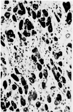
Recent chronic phase - Among the six animals investigated in the recent chronic phase, four (66.7%) showed myocarditis with leukocytic in-filtrate qualitatively similar to that seen in the acute phase varying from VmF to Z (Table) In two ani-mals, besides the mononuclear exudate and the extensive myocardial destruction, there was initial fibrosis of Z extension (Fig. 2). None of them pre-sented advanced fibrosis.
Epicarditis, pointed out by focal mononuclear infiltration, occurred in one rabbit (16.7%) inocu-lated with Y strain. No endocardial lesions were seen.
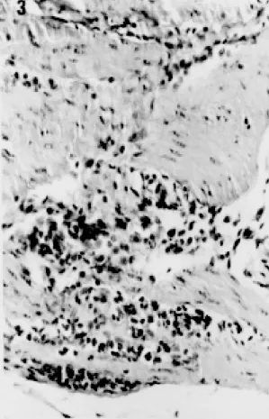
The digestive tract presented exclusively mild focal myositis involving the esophagus and stom-ach of one animal infected with Y. In the stomstom-ach, the process also reached a small area of the myoentheric plexus (Fig. 3).
The skeletal muscles did not show any histo-logic alteration.
Late chronic phase - Among 48 animals sub-mitted to necropsy during the late chronic phase, 34 (70.8%) presented myocarditis characterized by mononuclear, mainly endomisial, exudate and de-generative and necrotic phenomena, similar to those observed in the former groups, varying from VmF to Z (Table). Nevertheless in 14 animals (41.2%), the myocardial inflammation acquired a fibrous feature (Fig. 4). The intensity of the cellu-lar infiltration and fibrosis had great variability, as well as the degree of association between these two phenomena. In three others rabbits, one infected with Y and two infected with Er strains, only mild focal fibrosis without infiltrate was observed. The fibrosis was endomisial and perimisial, being of the advanced type in the great majority of cases and varying from VmF to Z (Table). Initial fibro-sis, in addition to advanced ones, was present in five animals (10.4%), varying from VmF to Z (Table).
Fig. 2: rabbit myocardium in the recent chronic phase (four months of infection by Y strain): multifocal confluent (zonal) myocarditis with initial fibrosis; proliferation of fusiform cells, probably fibroblasts, with young collagen deposition, replac-ing the dead myocardial cells. Gomori’ trichrome x400.
The most severe case of myocarditis was found in a rabbit that died spontaneously in the 6th month of infection by Y strain. In this case, the myocar-dium showed intense multifocal, confluent (Z), mononuclear infiltrate, with severe myocytolysis and confluent (Z) foci of fibrosis of the initial and advanced type. Another rabbit, with 12 months of infection by Y strain, also showed significant Z fibrosis of the advanced type, but with less severe inflammatory exudate and no initial fibrosis. It is noteworthy that one rabbit died suddenly due to right ventricle tip rupture and hemopericardium. In that area, the heart showed more severe myocarditis.
Chronic epicarditis occurred in nine animals (18.8%): three were infected with Y, five with CL and one with Er strains. The epicardial exudate was very mild and represented by one or few groups of mononuclear cells. Subepicardiac nervous ganglia were normal.
Fig. 4: rabbit myocardium in the late chronic phase (six months of infection by Y strain): multifocal confluent (zonal) myocarditis with perimysial and endomysial fibrosis, with heavy deposition of dense collagen (advanced fibrosis). Gomori’ trichrome x200.
Fig. 3: rabbit gastric muscularis propria in the recent chronic phase (three months of infection by Y strain): focal myositis characterized by mononuclear and neutrophil infiltration. H&E x400.
propria, was found in the esophagus of five ani-mals, three infected with Y strain and two other with CL strain; in the stomach of three rabbits, one infected with CL strain and two other with Er strain; in the cecum of one animal inoculated with Er strain; and in the colon of one, infected with CL strain. Muscular fibrosis occurred in the esopha-gus of one animal infected with Y strain and in the cecum of two others, being one infected with CL and the other with Y strains. In the last animal, the fibrosis involved large area of the outer coat of the muscularis propria and the myoenteric plexus. Excepting this case, the intramural nervous sys-tem did not present lesions.
Focal skeletal myositis, pointed out by mild mononuclear exudate and injury of the muscular cells, was found in five rabbits, two infected with Y and three with CL strains.
The Table summarizes the microscopic lesions observed in heart, digestive tract and skeletal muscle of all groups in different phases of the in-fection.
DISCUSSION
Acute phase - The histological lesions observed in heart, digestive tract and skeletal muscle of the acute chagasic rabbits were qualitatively similar to that found in humans (Lopes et al. 1987) and in other experimental models (Andrade & Andrade 1979, Beraldo 1987, Chapadeiro et al. 1988, Bonecini-Almeida et al 1990, Ramirez et al. 1991), although generally milder and scarcer mainly in the digestive tract and in the skeletal muscles.
The acute chagasic myocarditis showed vari-able severity depending on the strain of the para-site: it was more severe with Er strain infection, mild to moderate in the inoculated with Y strain and milder with CL strain. The intensity of the myocardial inflammation did not present relation-ship with cardiac parasitism. In this aspect the rab-bit model is not similar to the human acute cases submitted to necropsy, which usually present in-tense tissue parasitism (Lopes et al. 1987).
204 Histopathologic Studies in Chagasic Rabbits AM da Silva et al.
in rabbits infected by the three strains, was observed only in that inoculated by ip route, that evidenced the highest levels of parasitemia by direct exami-nation (Ramirez 1984, Ramirez & Brener 1987). Nevertheless, tissue parasitism did not show cor-relation with the parasitemia since animals inocu-lated with CL strain presented higher peaks of parasitemia than those infected with Y. The more frequent tissue parasitism in the animals inoculated with Y strain would be due to the virulence behav-ior of this strain (Andrade 1973).
In our material, the acute chagasic myocarditis seemed to be qualitatively similar to that described by others (Andrade & Andrade 1979, Figueiredo et al. 1979, Teixeira et al. 1983a, Figueiredo 1984). The unique case of initial myocardial fibrosis among the acute chagasic rabbits perhaps should be correlated to the acute chagasic myocarditis because the morphological characteristics of this finding are similar to that observed in the recent chronic phase.
The epicarditis, although mild and found in some control animals, may be, at least in part, due to T. cruzi. According to Figueiredo (1984) and our data, it does not seem to occur in the acute chagasic rabbits so severely as in humans (Lopes et al. 1987).
Recent chronic phase - Although researchers who studied previously the chagasic rabbit had considered the chronic phase as a whole, and did not separate events that can occur between three and six months of the infection, we believe such separation to be helpful in order to detect possible lesions interposed between the acute and the late chronic (after six months) phases, and in this way to try to establish the morphogenesis of the patho-logical process.
In spite of the negativity of tissue parasitism in the animals studied in the recent chronic phase, Ramirez (1984) demonstrated the presence of para-sites by the xenodiagnosis and the presence of lytic antibodies, which indicate the infection activity .
The myocarditis was less frequent in this group as compared to the acute phase, albeit the differ-ence was not statistically significant, perhaps due to the low number of animals.
The initial fibrosis presented clear tendency to be more frequent in the recent chronic phase, but the difference in relation to the other phases did not show statistical significance. Being more se-vere in the recent chronic phase, and in two ani-mals allocated in the late chronic phase but stud-ied with six months of infection (at the boundary between the recent and later chronic phases), it is possible that the initial fibrosis represents repara-tive phenomenon following the myocardial de-struction in the acute phase, in the cases of more
severe myocarditis.
The scarcity of the epicarditis and its similarity with the control lesions does not permit compari-sons with the human chagasics. The same occurs with the digestive tract lesions. In skeletal muscle, no lesion was observed in the recent chronic phase.
Late chronic phase - The absence of tissue para-sitism in the late chronic phase in our material is coincident with the majority of published obser-vations in rabbits (Teixeira et al. 1975, Andrade & Andrade 1979, Figueiredo et al. 1979, Figueiredo 1984, Figueiredo et al. 1985, Teixeira et al. 1983a, b). Only Gonzáles Cappa et al. (1977) reported tis-sue parasites in this phase. In this aspect, the in-fected rabbits would look like the human chronic cases (Lopes et al 1987), and other animal models (Lopes et al. 1979, Bolomo et al. 1980, Lana 1982, Bonecini-Almeida et al. 1990).
The myocarditis in the late chronic phase was significantly less frequent than in the acute one, but didn’t show significant difference with the re-cent chronic phase. It was significatively more fre-quent with Y and CL strains infection and more severe in two animals inoculated with Y strain. It is important to stress that the Er strain, that was involved with the more severe acute myocarditis, produced only mild myocardial lesions in the late chronic phase. Such fact seems to indicate that the intensity of the chronic chagasic myocarditis may be at least in part due to the strain behavior with regard to a host, and thus it would not be so depen-dent on the acute lesions intensity.
The advanced myocardial fibrosis, with endomysial and perimysial localization, just emerged in the late chronic phase, and was a little more frequent and intense (but without statistical significance) in the infected with Y, in compari-son to CL and Er inoculated animals. Between the latter, practically there was no difference. Taking in account only the animals that presented myocarditis, the frequency of advanced myocar-dial fibrosis was similar in the infected with Y and Er strains.
(1973) in mice: fibroblastic proliferation (called initial fibrosis in the present investigation) was seen at 90 to 120 days of infection and fibrosis (ad-vanced fibrosis in our designation) was present only after the 6th month.
Although there are no reports on epicarditis in chronic chagasic rabbits, we observed this lesion also in the chronic phase, that was significantly less frequent than in the acute phase.
In our experimental condition, the rabbit did not reproduce the severe digestive tract lesions fre-quently observed in human chronic chagasic pa-tients as well in the same animal by other investi-gators (Teixeira et al. 1975, Rezende Filho et al. 1979, Figueiredo 1984). Again, it is probable that the differences between their finds and ours was dependent on the type and/or the source or T. cruzi
strains and/or the host.
Regarding the skeletal muscles of the chronic chagasic rabbits, our findings agree with those of other investigators (Teixeira et al. 1983a, b, Figueiredo 1984).
It was not detected histopathological differ-ences among rabbits infected by MTry, BTry and TcTry.
In conclusion, in the experimental conditions established in our research, the rabbit model was able to reproduce a chronic focal, endomysial and perimysial myocarditis, with fibrous feature, quali-tatively similar to that observed in human cases but with low potential to develop the more severe forms of chronic myocarditis, and showed only mild histological lesions in digestive tract and skel-etal muscles. In the majority of the animals, the myocarditis was mild and similar to that described in the indeterminate form of the human chagasic patients. Thus, the rabbit did not behave as an ideal model for study of the chagasic infection. Never-theless, as it reproduced some of the lesions de-scribed in the heart of humans naturally infected with T. cruzi, mainly in the indeterminate form, this animal may be useful for the study of some aspects of the Chagas’ disease.
ACKNOWLEDGMENTS
To Prof. Edison Reis Lopes (Faculdade de Medicina do Triângulo Mineiro) and Prof. Ademir Rocha (Universidade Federal de Uberlândia) for critical revi-sions and suggestions, and to Prof. Gisele Pianetti (Faculdade de Medicina do Triângulo Mineiro) for col-laboration with the English version.
REFERENCES
Andrade SG 1973. Caracterização de cepas do
Trypanosoma cruzi isoladas no Recôncavo Baiano: contribuição ao estudo da patologia geral da doença de Chagas em nosso meio. MSc Thesis. Universidade Federal da Bahia, Salvador, 123pp.
Andrade ZA, Andrade SG 1979. Patologia, p. 199-248. In Z Brener, Z Andrade (eds). Trypanosoma cruzi e
doença de Chagas. Guanabara Koogan, Rio de Janeiro.
Aretz HT 1987. Myocarditis: the Dallas criteria. Hu-man Pathology 18: 619-624.
Beraldo PSS 1987. Sobre a infecção chagásica experi-mental no rato; estudo eletrocardiográfico seriado e funcional autonômico do coração, correlacionado à histopatologia. MSc Thesis. Universidade de Brasília, Brasília, 178 pp.
Bolomo N, Milei J, Cossio PM, Segura EL, Languens RP, Fernandez LM, Arana RM 1980. Experimental Chagas’ disease in South American primate (Cebus
sp.). Buenos Aires, Medicina 40: 667-672. Bonecini-Almeida M, Galvão-Castro B, Pessoa MHR,
Pirmez C, Laranja F 1990. Experimental Chagas’ disease in rhesus monkey - I. clinical, parasitologi-cal, hematological and anatomo-pathological stud-ies in the acute and indeterminate phase of the dis-ease. Mem Inst Oswaldo Cruz 85: 163-171. Brener Z, Ramirez LE 1985. Modelo crônico da doença
de Chagas experimental, p. 23-28. In JR Cançado, M Chuster (eds). Cardiopatia chagásica. Fundação Carlos Chagas, Belo Horizonte.
Chapadeiro E, Beraldo PSS, Jesus PC, Oliveira Jr WP, Junqueira Jr LF 1988. Lesões cardíacas em ratos Wistar inoculados com diferentes cepas de
Trypanosoma cruzi. Rev Soc Bras Med Trop 21:
95-103.
Chiari E, Tafuri WL, Bambirra EA, Rezende MM, Ribeiro TO, Castro LP, Salgado JA, Amaral de Padua RA 1980. The rabbit as a laboratory animal for studies on Chagas’ disease; research note. Rev Inst Med Trop São Paulo 22: 207-208.
Figueiredo F 1984. Infecção experimental de coelhos com Trypanosoma cruzi; aspectos de parasitologia, imunologia e patologia. MSc Thesis. Faculdade de Medicina de Ribeirão Preto da Universidade de São Paulo, Ribeirão Preto, 119 pp.
Figueiredo F, Macedo V, Teixeira ARL 1979. Experi-mental model for chronic Chagas’ disease, p. 18. In International congress on Chagas’ disease, Anais, Rio de Janeiro, Instituto Oswaldo Cruz.
Figueiredo F, Rossi MA, Ribeiro dos Santos R 1985. Evolução da cardiopatia experimentalmente induzida em coelhos infectados com Trypanosoma cruzi. Rev Soc Bras Med Trop 18: 133-141.
González Cappa S, Lazzari J, Segal A, Katzin A Mistchenko A, Langues RP, Elizari M 1977. Infección experimental del conejo com Trypanosoma cruzi - II. Correlación entre el electrocardiograma y la histopatología. Buenos Aires, Medicina 37: 508. Lana M 1982. O cão como modelo experimental para o estudo da doença de Chagas. PhD Thesis. Instituto de Ciências Biológicas da Universidade Federal de Minas Gerais, Belo Horizonte, 207 pp.
206 Histopathologic Studies in Chagasic Rabbits AM da Silva et al.
Lopes ER, Pires LL, Chapadeiro E, Tafuri WL, Prata AR 1979. Estudo anatomopatológico das vísceras de cães artificial e naturalmente infectados com o T. cruzi. In 15º Congresso da Sociedade Brasileira de Medicina Tropical, Campinas.
Ramirez LE 1984. O coelho como modelo experimen-tal no estudo da doença de Chagas crônica. PhD Thesis. Instituto de Ciências Biológicas da Universidade Federal de Minas Gerais, Belo Horizonte, 186 pp.
Ramirez LE, Brener Z 1987. Evaluation of the rabbit as a model for Chagas’ disease - I. Parasitological stud-ies. Mem Inst Oswaldo Cruz 82: 531-536. Ramirez LE, Lages-Silva E, Chapadeiro E 1991.
Infecção do hamster pelo Trypanosoma cruzi. Rev Soc Bras Med Trop 24: 119-120.
Rezende Filho J, Figueiredo F, Teixeira ARL 1979. Megacolon syndrome in rabbits with chronic Chagas’ disease. p. 20. In International congress on Chagas’ disease, Anais, Rio de Janeiro, Instituto Oswaldo Cruz.
Taylor CR 1986. Staining protocols and practical pro-cedures, p. 365-367. In CR Taylor. Immunomicros-copy: a diagnostic tool for the surgical pathologist.
Saunders, Philadelphia.
Teixeira ARL, Teixeira ML, Santos-Buch CA 1975. The immunology of experimental Chagas’ disease - IV. Production of lesions in rabbits similar to those of chronic Chagas’ disease in man. Am J Path 80: 163-180.
Teixeira ARL, Figueiredo F, Rezende Filho J, Macedo V 1983a. Chagas’ disease; a clinical, parasitologi-cal, immunologiparasitologi-cal, and pathological study in rab-bits. Am J Trop Med Hyg 32: 258-272.
Teixeira ARL, Junqueira Jr LF, Solórzano E, Zappalá M 1983b. Doença de Chagas experimental em coelhos isogênicos III/J; I. Fisiologia das arritmias e da morte súbita do chagásico. Rev Assoc Méd Bras 29: 77-83.