Abdominal hernia repair with mesh surrounded by fibrous tissue –
Abdominal hernia repair with mesh surrounded by fibrous tissue –
Abdominal hernia repair with mesh surrounded by fibrous tissue –
Abdominal hernia repair with mesh surrounded by fibrous tissue –
Abdominal hernia repair with mesh surrounded by fibrous tissue –
experimental study in Wistar rats
experimental study in Wistar rats
experimental study in Wistar rats
experimental study in Wistar rats
experimental study in Wistar rats
Correção de hérnia abdominal com tela envolta por tecido fibroso – estudo em
Correção de hérnia abdominal com tela envolta por tecido fibroso – estudo em
Correção de hérnia abdominal com tela envolta por tecido fibroso – estudo em
Correção de hérnia abdominal com tela envolta por tecido fibroso – estudo em
Correção de hérnia abdominal com tela envolta por tecido fibroso – estudo em
ratos Wistar
ratos Wistar
ratos Wistar
ratos Wistar
ratos Wistar
BRUNO FILIPPI RICCIARDI1; LUIZ HENRIQUE CHEQUIM2; RICARDO RIBEIRO GAMA3; LEONARDO HASSEGAWA4
A B S T R A C T A B S T R A C T A B S T R A C T A B S T R A C T A B S T R A C T
Objective Objective Objective Objective
Objective: To assess the efficacy and complications of surgical repair of incisional hernias by grafting of polypropylene mesh surrounded by fibrous tissue, compared to polypropylene mesh. MethodsMethodsMethodsMethodsMethods: twenty-five male and female Wistar rats were divided into two groups: control and experimental. The 15 animals of the experimental group underwent implantation of the mesh in the subcutaneous tissue. After 21 days, the screen was removed and served to correct an induced abdominal hernia. The animals were euthanized after 21 days and submitted to macroscopic, microscopic and tension analises. The control group had undergone usual abdominal hernia repair with polypropylene mesh. ResultsResultsResultsResultsResults: There was a significant difference in macroscopic adhesions. The control group had a higher degree of adhesions when compared to the experiment ones (p = 0.003). We could not determine whether there were significant differences in tensile strength and microscopy. ConclusionConclusionConclusionConclusionConclusion: The polypropylene mesh surrounded by fibrous tissue was effective in the correction of induced abdominal hernias, with a lower degree of macroscopic adhesions when compared to polypropylene mesh.
Key words: Key words: Key words: Key words:
Key words: Hernia. Herniorrhaphy. Prostheses and implants. Surgical mesh. Polypropylenes.
Work performed in the Laboratory of Operative Technique and Experimental Surgery of the Medical School of the Evangelical School of Paraná, Curitiba, Paraná State, Brazil.
1. Resident, General Surgery, Holy House of Mercy, Ponta Grossa-PR-BR; 2. Resident, Otolaryngology, Evangelical Hospital of Curitiba-PR-BR; 3. Assistant Professor, Operative technique, Evangelical School of Paraná-PR-BR; 4. Instructor, Discipline of Operative Technique, Evangelical School of Paraná.
INTRODUCTION
INTRODUCTION
INTRODUCTION
INTRODUCTION
INTRODUCTION
H
ernioplasties fall among the most commonly performed operations in the various continents of the globe, and in the United States alone approximately 600,000 such procedures are performed annually1. Despite their frequency, complications and recurrences still make them a great challenge of modern surgery 1.The term derives from the Latin hernia and means rupture. It is defined as an abnormal protrusion of an organ or tissue through a defect in the adjacent abdominal wall. They are found in the abdominal region and in areas where the aponeuroses and fasciae are not covered by striated muscle fibers, characterizing anatomical weaknesses of the muscle wall1
Wall defects are usually the consequences of trau-ma, burns, debridement of necrotizing infections, treatment of compartment syndrome, removal of infected prostheses, resection of tumors, among others2.
Besides these factors, about 11% to 20% of patients undergoing laparotomy develop incisional hernias3,4.
The displacement of the tendinous insertion of the large abdominal supporting muscles aggravates the midline incisional hernias, since there is muscle atrophy, fatty degeneration and fibrosis of the lateral muscles, hampering the reinsertion of the supporting muscle’s tendon 5.
The treatment of these conditions includes the attempt of anatomical reconstruction of the abdominal wall, closing the parietal defect and restoring intra-abdominal pressure. Most small incisional hernias are handled with simple closure of the defect. The challenges, however, are the hernias with rings above 10 cm of diameter, which display more tendency to recur 5.
A great variety of synthetic materials has been used in the abdominal wall, with discrepant results. Complications arising from these types of materials have been largely related to their inherent tendency to promote inflammation8.
Theoretically, the synthetic material replacing the abdominal wall must be strong, biologically inert, non-carcinogenic and stable in the presence of infection. Currently the mesh that comes closest to these features is of expanded PTFE. After review of clinical trials on this mesh, it became apparent that it still has disadvantages such as foreign body reaction, high cost and some degree of adhesion formation8.
Since its advent, the polypropylene mesh is the one that has been used in the repair of hernias. However, clinical experience has shown a diverse range of complications such as sepsis, erosion of intra-abdominal organs and fistulas8.
The reconstruction of abdominal wall is, in fact, one of the few areas in which the surgical practice with synthetic prosthesis precedes the use of autologous tissue8. Despite description in the literature that the use of autografts causes invasion of fibroblasts in the receptor layer, it was found that the grafted tissue contains young fibroblasts. This leads to the inference that the autograft, at a stage later than infiltration, followed by neovascularization, produces young cells, a fact that characterizes the continuing vitality of the grafted tissue. There is therefore active participation of this tissue in the production of fibroplasia 9.
Based on the principles mentioned above, there is the possibility of association between synthetic materials and autologous tissue to maximize induced fibroplasia, responsible for enhancing the desired correction of hernias9. Hence, it is necessary to focus on the development of a new substitute for the repair of abdominal wall defects that could be in contact with the viscera, in addition to the PTFE mesh.
The involvement of the polypropylene mesh with fibroblasts aims to improve the effectiveness of hernioplasties and to reduce recurrences, as well as to enable the development of a new technique for repair of large abdo-minal hernias with this type of mesh.
The present study aimed to verify the effectiveness and complications of surgical repair of incisional hernias by grafting a polypropylene mesh surrounded by fibrous tissue.
METHODS
METHODS
METHODS
METHODS
METHODS
This research was conducted on the premises of the Laboratory of Experimental Surgery and Operative Technique, with support from the Central Vivarium, Histotecnique Laboratory and Laboratory of Pathology of the Evangelical School of Paraná, Number 03/09.
The study was prospective, experimental and comparative in nature, with 25 Wistar rats (Rattus norvegicus Albinus, Rodentia, Mammalia), of both genders, adults, approximately 90 days old and weighing between 250 and 350 grams.
The animals were divided into two groups, an “experiment” group, with 15 animals, and a “control” group, with 10 animals. The study consisted of two surgeries, followed by euthanasia and macroscopic, tensional and pathological analysis of specimens.
In the first surgery, the experimental group underwent standard surgical preparation: anesthesia with 0.1 ml / 100g of ketamine (Ketalar ®) and 0.05 ml / 100g of xylazine (Xilazin ®), intraperitoneally. After anesthesia, the animals were placed in the prone position and fixed to the surgical board. The operation was initiated only after anesthesia had set in, tested by eyelid reflex and paw withdrawal to painful stimuli.
Trichotomy was performed on the dorsal paravertebral region, followed by antisepsis with topic iodine solution and placement of sterile drapes. The animals then underwent a skin incision in the dorsal region, dissection of subcutaneous tissue and implantation of a 3x1 cm elliptical polypropylene mesh in the subcutaneous tissue (Figure 1). After verifying hemostasis, the skin was sutured with 3-0 nylon. After the procedure, analgesia was performed with an intramuscular injection of morphine in the post-operative by the vivarium staff.
After 21 days the rats were submitted to surgery with standard surgical preparation. In this second intervention, the experimental group underwent resection of the mesh surrounded by fibrous tissue adjacent to the dorsal region. The dorsal skin was then sutured with 3-0 nylon.
A new incision was made in the midline of the abdomen, creating a defect in the abdominal wall of the same size of the fibrous tissue, the elliptical 3x1 cm, and dissected the plans to the peritoneal cavity, simulating an abdominal hernia. The mesh surrounded by fibrosis tissue was sutured to the aponeurosis with 4-0 polypropylene single running suture, reconstituting the local anatomy (Figure 2) and the skin was closed with 3-0 nylon sutures.
After 21 days of the second surgery, the animals were euthanized by asphyxiation in a carbon dioxide chamber, as approved by the Ethics Committee of the Evangelical School of Paraná. Dissection was performed by laparotomy, the abdominal cavity was macroscopically inspected and the presence of suture dehiscence, the occurrence and quality of adhesions, fistulas and intra-ab-dominal complications verifying.
abdo-minal wall; grade 3 – firm adhesions, resistant to manipulation, between the abdominal wall and an organ or structure; grade 4 – firm adhesions, resistant to manipulation, between the abdominal wall and over an organ or structure; grade 5 – firm adhesions, resistant to manipulation, between loops and between loops and the abdominal wall, with an enteric fistula. Adhesions were also quantified as mild (thickness 1), moderate (thickness 2), coarse (thickness 3) or too coarse (thickness 4).
Once rated the degree of macroscopic adhesions, we removed a piece of 1x3 cm perpendicular to the main axis of the polypropylene mesh, including muscle/mesh/muscle. This fragment was assessed for tensile strength.
Each fragment was clamped at both muscle ends. The upper end was fixed in a support, with the water tap. The lower extremity was attached to a metal container of known weight. This container was gradually filled with water in a constant flow, and immediately stopped upon rupture of the fragment. We calculated the force required to break each segment by the weight of water added to the container and lower clamp.
Then the material was fixed in 10% formalin and subjected to histological preparation, with dehydration in alcohol and xylene, and embedded in paraffin blocks. Histological cuts were made on microtome and slides prepared with standard hematoxylin/eosin. These slides were submitted to pathological examination to verify the type and degree of inflammation, inflammatory cells, fibroblasts, collagen, and neovascularization in the region. The analysis was quantified in relation to the quantity of each item displayed, ranging from absent, present in small amounts (+), present in moderate quantity (++) and present in large quantities (+++). This analysis was conducted at of Pathology Service of the Paraná Evangelical University.
The control group underwent only the second surgical procedure, but instead bettye repair with the mesh surrounded by fibrosis, we used the polypropylene mesh directly in the induced abdomi-nal defect. These animals were euthanized in the 21st postoperative day and submitted to the same macroscopic, tensional and pathological analysis as the experimental animals.
The results were statistically analyzed using the log-rank test for the analysis of tensile strength, the Wilcoxon test for macroscopic and microscopic analyzes, and results with p <0.05 were considered statistically significant.
RESULTS
RESULTS
RESULTS
RESULTS
RESULTS
The 15 rats used in the experimental group progressed without postoperative complications, with good surgical recovery and availability for food. One animal in the experimental group had extrusion and partial dislocation of the implanted mesh after the first surgery, being excluded from the study. Other complications, such as skin or mus-cular-aponeurotic layer dehiscences, signs of local or systemic infection, intra-abdominal abscess or peritonitis were not seen in any animal. There was satisfactory adherence in both the first intervention, where the polypropylene mesh was placed in the subcutaneous tissue of the dorsal region of animals, and in the second, with the mesh surrounded by tissue fibrous implanted in the peritoneal cavity correcting the induced hernia.
The 10 rats of the control group progressed without postoperative complications, with good surgical recovery and availability for food. No animal showed skin or muscular-aponeurotic layer dehiscences, signs of local or systemic infection, intra-abdominal abscess or peritonitis and extrusion or displacement of the implanted material. The polypropylene mesh satisfactorily adhered to the peritoneal cavity, correcting the induced incisional hernia.
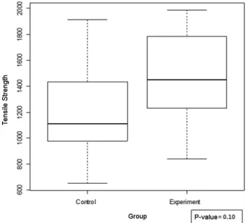
The rupture strength of the control group ranged from 650g and 1,913g, mean 1,204g. The standard deviation was 369g. The experimental group, on its turn, had tensile strength of at least 840g and a maximum of
Figure Figure Figure Figure
Figure 1 - Implantation of polypropylene mesh in the subcutaneous tissue.
Figure 2 Figure 2 Figure 2 Figure 2
1,990g, mean 1,479g and standard deviation 359 g. (Figu-re 3)
Histopathology Histopathology Histopathology Histopathology Histopathology
The animals were evaluated for vascularization, level of inflammation and the presence of fibroblasts. They were classified into four levels, ranging from absent, present in small quantities (+), present in moderate amounts (+ +), present in large amounts (+ + +) and present in very large amounts (+ + + +).
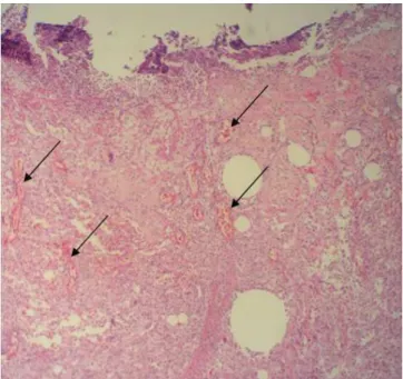
Inflammation was observed in all animals, varying in intensity. In some animals we observed giant cell inflammatory response, characteristic of foreign body (Fi-gure 4), and granulation tissue, angiogenesis and fibrosis (Figure 5).
In the control group, there was inflammation in small quantities in eight animals, comprising 80% of this group. There was also moderate inflammation in one ani-mal and intensive inflammation in another.
The experimental group had six animals with mild inflammation, five moderate, two with inflammation in large quantities and one with inflammatory reaction in very large quantities.
Using the Wilcoxon test, with p-value = 0.09, it was concluded that it was not possible to say that there was significant difference between groups in relation to inflammatory reaction.
The vasculature was identified as one aspect present in both groups. We observed the formation of blood vessels of a typical healthy granulation tissue, characterized by the presence of thin endothelium, with red blood cells in its interior, and little connective tissue support. (Figure 6) Of the control group, two individuals had little formation of neovessels, while two other had them moderate in intensity. The rest of the group, or six animals, presented it in great intensity.
Only one animal in the experimental group had little neovascularization, six moderate, five intense and two great intensity.
Using the Wilcoxon test, with p-value of 0.82, it was not possible to say that there was significant difference between groups regarding vascularization.
Slides analyzed showed varying degrees of formation of fibroblasts, mostly in great intensity. The control group had two animals with a small amount of fibroblasts, one with moderate presence and seven with a large number of this cell type. In the experimental group one animal had small amounts of fibroblasts, five have moderate amounts and eight animals presented with large amount of fibroblasts. Using the Wilcoxon test, with p-value = 0.78l it was not possible to say that there was significant difference between groups regarding the presence of fibroblasts.
All animals were evaluated for gross appearance of the viscera and abdominal adhesions after post-mortem laparotomy. The criteria used for the analysis followed the classification used by Diogo-Filho et al10.
Figure 4 Figure 4Figure 4
Figure 4Figure 4 - Photomicrographs of inflammation, fibrosis and neovascularization triggered by the presence of the polypropylene mesh. A (HE 400x) – giant cell-type reaction to the polypropylene suture (arrow). B – (HE 100x) Presence of foreign body giant cell (arrow). Figure 3
Figure 3Figure 3
Figure 3Figure 3 - Variation in grams (vertical axis) of the rupture strength between the control and experimental groups.
From the 10 animals in the control group, six had adhesions between the wall and an organ, i.e., grade 3, and four had adhesions between the wall and two or more organs, characterizing grade 4.
Figure 5 Figure 5Figure 5
The experimental group had two animals with grade 1 adhesions, six with grade 2 adhesions, another six with grade 3 adhesions and only one with grade 4 adhesions (Figure 7).
Using the Wilcoxon test, with p-value = 0.003, it was possible to affirm that there were significant differences between groups as for macroscopic findings.
DISCUSSION
DISCUSSION
DISCUSSION
DISCUSSION
DISCUSSION
The Wistar rat (Rattus norvegicus Albinus, Rodentia, Mammalia) was the animal of choice because of its ability to adapt to a wide variety of environmental conditions. These animals are also isogenic, meaning that
all are genetically similar individuals. The use of such animals’ lineage allows the development of experiments with a reduced number of animals because of the similarity of response, while not compromising results11.
The weight of the animals was assessed in the preoperative period aiming at using samples similar in both groups and permitting the calculation of the anesthetic dose. They were also subjected to the same environmental conditions throughout the experiment, without any factor that could lead to significant changes in their metabolism. In this way the study was standardized, so that there was not external influence among the animals studied 10.
The surgery was performed in a systematic way, in small groups, and with no more than five animals per day, to avoid changes resulting from execution of the procedure. The interval between the first and second operations, as well as of the end of the experiment with euthanasia of animals and analysis, was 21 days, 11.
The polypropylene mesh was chosen for this study because of its wide use in medical practice, as it has a relatively affordable cost. It approximates the criteria of an ideal material and its surgical technique for the correction of abdominal hernias is widespread due to its advantages such as less tissue reaction, easy sterilization and handling 4. We found no prior studies using polypropylene mesh surrounded by fibrous tissue. There is data in the literature about studies comparing polypropylene with expanded polytetrafluoroethylene (ePTFE). Van’t Riet et al. 12 conducted a study comparing the polypropylene mesh with polypropylene covered by collagen in the visceral side, such principle being similar to the one of this study.
The use of polypropylene mesh encased by fibrous tissue showed similar results when compared to using only the mesh in regards to tension and histological analysis. In relation to the degree of adhesions, the mesh surrounded by fibrous tissue has caused less intraperitoneal adhesions, with the advantage of reduced postoperative complications such as enteric fistulas and difficulty in accessing the surgical cavity in a new exploration. We found studies in the literature that corroborate this one, such as the one from Van’t Riet et al. 12, which have proven effectiveness in reducing adhesions by the use of collagen in the visceral side of the polypropylene mesh when compared to the use of the mesh alone.
According to Meyer et al.13, who conducted a study of correction of abdominal hernias with biocompatible material using PTFE and polypropylene meshes as comparison, we observed a significant difference as for adhesions, as the polypropylene mesh had a much greater area of adhesion when compared to the two other materials. The same statement was made by Puttini 11in his master’s thesis, which said that previous experimental studies using polypropylene and ePTFE observed higher degree of adhesions in the first, as well as increased number of intestinal fistulae. Both studies confirm that the polypropylene mesh is not ideal to replace the ePTFE alone.
Figure 7 Figure 7 Figure 7 Figure 7
-Figure 7 - Degree of macroscopic adhesions between control and experimental groups.
Figure 6 Figure 6 Figure 6 Figure 6
In his work, Kapan et al. conducted a study on the effectiveness of ePTFE with respect to tensile strength and microscopic analysis of inflammatory cells, vascularization and fibroblasts. They used a scale for assessing the maturity of the wound in relation to the microscopic aspects the. The same methodology for analysis of the tensile strength was used 4.
When comparing the results of the experimental group of this study with the study of Kapan et al., there are similar results in the comparison of tensile strength and microscopic analysis. It is then shown that the technical improvement of the polypropylene mesh is an effective possibility to replace the ePTFE mesh when it is not available
due to its high cost4.
The polypropylene mesh surrounded by fibrous tissue proves to be a viable and consistent alternative to be used in situations where the surgeon is devoid of better materials for repair of abdominal hernias. Further studies are necessary for the development and improvement of existing techniques and materials.
In conclusion, the polypropylene mesh surrounded by fibrous tissue was effective in the correction of induced abdominal hernias, with a lower degree of macroscopic adhesions when compared to the polypropylene mesh alone.
R E S U M O R E S U M O R E S U M O R E S U M O R E S U M O
Objetivo: Objetivo: Objetivo: Objetivo:
Objetivo: Verificar a eficácia e complicações da correção cirúrgica de hérnias incisionais por enxerto da tela de polipropileno envolta por tecido fibroso, em comparação com a tela de polipropileno. Métodos Métodos Métodos Métodos Métodos: Foram utilizados 25 ratos Wistar machos e fêmeas, distribuídos em dois grupos: controle e experimento. O grupo experimento, com 15 animais, foi submetido ao implante da tela em seu tecido subcutâneo. Após 21 dias, a tela foi removida e serviu para correção de uma hérnia abdominal induzida. Os animais foram eutanasiados após 21 dias e submetidos à análise macroscópica, tensional e microscópica. O grupo controle, foi submetido ao reparo da hérnia abdominal com a tela de polipropileno. Resultados. Resultados. Resultados. Resultados. Resultados: Observou-se diferença significativa em relação à análise macroscópica de aderências. O grupo controle obteve maior grau de aderências em relação ao experimento (p= 0,003). Não foi possível afirmar que houve diferença significativa em relação à análise de força tensional e microscopia. ConclusãoConclusãoConclusãoConclusão: A tela de polipropileno envoltaConclusão por tecido fibroso mostrou-se eficaz na correção das hérnias abdominais induzidas, com menor grau de aderências macroscópicas quando comparada à tela de polipropileno.
Descritores: Descritores: Descritores: Descritores:
Descritores: Hérnia. Herniorrafia. Próteses e implantes. Telas cirúrgicas. Polipropilenos.
REFERENCES
REFERENCES
REFERENCES
REFERENCES
REFERENCES
1. Sabiston DC, Towsend Jr CM. Sabiston Tratado de Cirurgia: As bases biológicas da prática cirúrgica moderna. 17a ed. Rio de
Ja-neiro: Elsevier; 2005.
2. Maurice SM, Skeete DA. Use of human acellular dermal matrix for abdominal wall reconstructions. Am J Surg. 2009;197(1):35-42.
3. Silverman RP, Li EM, Holton LH 3rd, Sawan KT, Goldberg NH. Ventral hernia repair using allogenic acellular dermal matrix in a swine model. Hernia. 2004;8(4):336-42.
4. Kapan S, Kapan M, Goksoy E, Karabicak I, Oktar H. Comparison of PTFE, pericardium bovine and fascia lata for repair of incisional hernia in rat model, experimental study. Hernia. 2003;7(1):39-43. 5. Morton JH, Schwartz SI. Princípios de Cirurgia – Pré-teste,
auto-avaliação, revisão. 6a ed. Rio de Janeiro: Revinter; 1996.
6. Usher FC. Hernia repair with knitted polypropylene mesh. Surg Gynecol Obstet. 1963;117:239-40.
7. Cassar K, Munro A. Surgical treatment of incisional hernia. Br J Surg. 2002;89(5):534-45.
8. Bauer JJ, Salky BA, Gelernt IM, Kreel I. Repair of large abdominal wall defects with expanded polytetrafluoroethylene (PTFE). Ann Surg. 1987;206(6):765-9.
9. Silva HC, Silva AL, Oliveira CM. Enxerto peritoneal autógeno e fibroplasia: estudo experimental. Rev Col Bras Cir. 2004;31(2):83-9.
10. Diogo-Filho A, Lazarini BCM, Vieira-Junyor F, Silva GJ, Gomes HL. Avaliação das aderências pós-operatórias em ratos submetidos à peritoniostomia com tela de polipropileno associada à nitrofurazona. Arq Gastroenterol. 2004;41(4):245-9.
11. Puttini SMB. Avaliação da resposta inflamatória desencadeada pelas telas de polipropileno e politetrafluoretileno expandido im-plantadas no espaço intraperitoneal: estudo experimental em camundongos [dissertação]. Brasília/DF: Universidade de Brasília, Faculdade de Medicina; 2006.
12. van´t Riet M, Burger JW, Bonthuis F, Jeekel J, Bonjer HJ. Prevention of adhesion formation to polypropylene mesh by collagen coating: a randomized controlled study in a rat model of ventral hernia repair. Surg Endosc. 2004;18(4):681-5.
13. Meyer T, Schwarz K, Ulrichs K, Höcht B. A new biocompatible material (Lyoplant) for the therapy of congenital abdominal wall defects: first experimental results in rats. Pediatr Surg Int. 2006;22(4):369-74.
Received on 26/09/2011
Accepted for publication 24/11/2011 Conflict of interest: none
Source of funding: none
How to cite this article: How to cite this article:How to cite this article: How to cite this article:How to cite this article:
Ricciardi BF, Chequim LH, Gama RR, Hassegawa L. Abdominal hernia repair with mesh surrounded by fibrous tissue – experimental study in wistar rats. Rev Col Bras Cir. [periódico na Internet] 2012; 39(3). Dispo-nível em URL: http://www.scielo.br/rcbc
Address correspondence to: Address correspondence to:Address correspondence to: Address correspondence to:Address correspondence to: Bruno Filippi Ricciardi