Human mesenchymal stem cells are resistant to cytotoxic and genotoxic
effects of cisplatin
in vitro
Bruno Corrêa Bellagamba
1,2, Bianca Regina Ribas de Abreu
2, Ivana Grivicich
2, Carolina Franke Markarian
3,
Eduardo Chem
3, Melissa Camassola
2, Nance Beyer Nardi
1,2and Rafael Rodrigues Dihl
21
Programa de Pós-Graduação em Genética e Biologia Molecular, Universidade Federal do Rio Grande do
Sul, Porto Alegre, RS, Brazil.
2
Programa de Pós-Graduação em Biologia Celular e Molecular Aplicada à Saúde, Universidade Luterana
do Brasil, Canoas, RS, Brazil.
3Complexo Hospitalar Santa Casa de Porto Alegre, Porto Alegre, RS, Brazil.
Abstract
Mesenchymal stem cells (MSCs) are known for their important properties involving multilineage differentiation poten-tial, trophic factor secretion and localization along various organs and tissues. On the dark side, MSCs play a distin-guished role in tumor microenvironments by differentiating into tumor-associated fibroblasts or supporting tumor growth via distinct mechanisms. Cisplatin (CIS) is a drug widely applied in the treatment of a large number of cancers and is known for its cytotoxic and genotoxic effects, bothin vitro and in vivo. Here we assessed the effects of CIS on MSCs and the ovarian cancer cell line OVCAR-3, by MTT and comet assays. Our results demonstrated the resis-tance of MSCs to cell death and DNA damage induction by CIS, which was not observed when OVCAR-3 cells were exposed to this drug.
Keywords:Mesenchymal stem cells, Cisplatin, MTT, Comet assay, OVCAR-3 cells.
Received: February 28, 2015; Accepted: August 11, 2015.
Cisplatin,Cis-diamminedichloroplatinum-II, (CIS) is a widely used chemotherapeutic agent as the first line treat-ment against head and neck, testicular, lung, ovarian and others types of cancer (Jamieson and Lippard, 1999; Go-mez-Ruizet al., 2012). Its main mechanism of action in-volves formation of adducts covalently linked to DNA (Sancho-Martinezet al., 2012). These adducts are consid-ered the major contributing factor to the cytotoxic effects of the drug, since they block DNA replication and transcrip-tion and, ultimately, cell division (Dasari and Tchounwou, 2014). Despite the well-established application of CIS in clinical treatments, intrinsic or acquired cell resistance to this drug is a serious problem that appears concomitant with CIS utilization (Galluzziet al., 2012).
Mesenchymal stem cells (MSCs) are post-natal stem cells found in almost all tissues in the organism (da Silva Meirelles et al., 2006), including human adipose tissue (Zuket al., 2002), since they inhabit a perivascular niche (da Silva Meirelleset al., 2008). MSCs can secrete trophic factors such as anti-apoptotic, immunomodulatory,
angio-genic and chemo attractive molecules, which act in lesion and surrounding sites in vivo to promote tissue repair (Doornet al., 2012). Another property of MSCs is the po-tential to differentiate into bone, cartilage and adipocytes (Zhuet al., 2012) according to the culture conditions. Due to their capacity to differentiate into various cell types and their paracrine effects, MSCs have emerged as a promising alternative for cell therapy and tissue engineering (Schaffler and Buchler, 2007).
While MSCs exert important roles in the maintenance of organismic homeostasis, they are also known for com-posing the tumor stroma and for their tropism to various types of cancer (Kucerova and Skolekova, 2013).In vitro
analyses suggest that MSCs can stimulate tumor progres-sion by modulating cytokine secretion, supressing the im-mune system, migrating to the tumor site and promoting tumor growth through paracrine factors, or by differentiat-ing into tumor-associated fibroblasts. When MSCs are in-jected at the tumor sitein vivo, they stimulate tumor growth and support metastasis, or inhibit tumorigenesis by anti-tumor effects involving downregulation of Akt, beta-catenin, Bcl-2, c-Myc, proliferating cell nuclear antigen and surviving, leading to reduced proliferation, G1 arrest, suppression of oncogenes and increased apoptosis (Klopp
et al., 2011).
DOI: http://dx.doi.org/10.1590/1678-4685-GMB-2015-0057
Send correspondence to Rafael Rodrigues Dihl, Laboratório da Toxicidade Genética, PPGBioSaúde, ULBRA, Prédio 22, 4º andar, sala 20, Avenida Farroupilha, 8001, 92425-900, Canoas, RS, Brazil. E-mail: [email protected]
Given the complexity of the tumor microenvironment and the increasing evidence for the contribution of tu-mor-associated fibroblasts to cancer maintenance and chemoresistance (Houthuijzen et al., 2012), tumor-associated fibroblasts have been considered as promising targets for novel chemotherapeutic strategies (Sampleset al., 2013). Several studies have shown the cytotoxic (Smith
et al., 2005) and genotoxic (Ungeret al., 2009) effects of CIS on ovarian carcinoma cells (OCCs) and normal prolif-erating and non-prolifprolif-erating cells (Sancho-Martinezet al., 2012). However, the genotoxic effects of CIS on MSCs are still not clear, even though these cells are known for being resistant to several chemotherapeutic agentsin vitro, in-cluding CIS (Lietal., 2004; Lianget al., 2011). Thus, due to the well known importance of MSCs for generating tu-mor-associated fibroblasts and their role in the cancer microenvironment and chemoresistance, the present study aimed at evaluating the cytotoxic effect and DNA damage induction potential of CIS on human adipose-derived MSCs and OCCs line OVCAR-3 duringin vitro cultiva-tion.
Human adipose-derived MSCs were obtained from adipose tissue of four patients undergoing elective liposuc-tion surgery. All patients signed an informed consent form, and the study was approved by the Research Ethics Com-mittee of Complexo Hospitalar Santa Casa de Misericórdia de Porto Alegre. The stromal vascular fraction was isolated
as described by Zuket al.(2002). Briefly, the liposuction material was extensively washed with phosphate buffered saline (PBS) and incubated with type I collagenase. Mono-nuclear cells resulting from tissue digestion and centrifu-gation were resuspended in Dulbecco’s modified Eagle medium (DMEM) supplemented with HEPES (free acid, 3.7 g/L), 10% fetal bovine serum (FBS, Cultilab, São Paul-o, Brazil) and 1% penicillin/streptomycin (Cell Culture Medium 1 – CCM1). Cells were seeded at 3 x 10⁴cells/cm2 into tissue culture flasks and expanded at 37 °C in a humidi-fied culture chamber with a 5% CO2atmosphere, changing
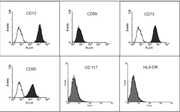
the culture medium every 2–3 days. Cells between passages 6 and 9 were used in all experiments. Immunophenotyping of MSCs was done using a BD FACSCalibur flow cytometer to determinate the presence/absence of the fol-lowing cell markers: CD13, CD69, CD73, CD90, CD117 and HLA-DR (Figure 1). All reagents used here were from Sigma Chemical Co. (St Louis, MO, USA), unless other-wise stated. Plasticware was from TPP (Trasadingen, Swit-zerland).
The ovarian cancer cell line OVCAR-3 was pur-chased from ATCC (American Type Culture Collection, Manassas, Virginia, USA) and cultured with DMEM sup-plemented with 10% of FBS and 1% penicillin/streptomy-cin (Cell Culture Medium 2 – CCM2) at 37 °C in a humidified culture chamber with 5% CO2. Cells were
ex-panded according to the experiments requirement.
Cisplatin (CIS, CAS No.15663-27-1) was obtained as the clinical preparation Platistine® (Pfizer Ltda., São Paulo, Brazil). Ethyl methanesulfonate (EMS, CAS No.62-50-0) was purchased from Sigma–Aldrich. Solu-tions of CIS and EMS were made with CCMs immediately before use.
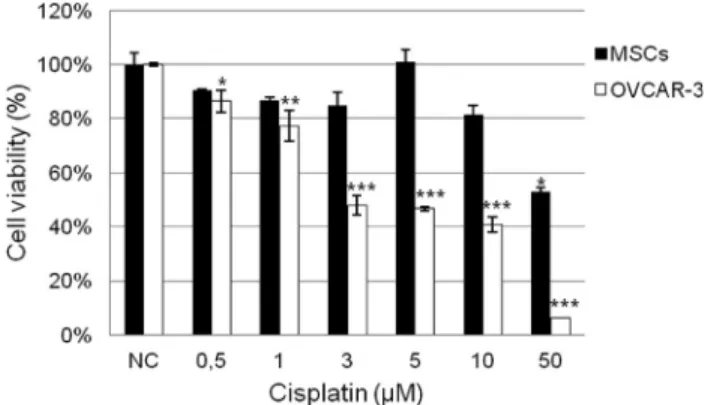
For the MTT assay evaluation of CIS cytotoxicity, cells were seeded in 96-well plates at a density of 3 x 10³ cells/well for MSCs and 5 x 10⁴cells/well for OVCAR-3 cells and treated the following day with CIS at 0.5, 1, 3, 5, 10 and 50mM dosages.CCMs, 1 and 2, were used as nega-tive control (NC). After 72 hours, CCMs were removed and 20mL of 3-(4,5-dimethylthiazol-2-yl)-2,5-diphenyl tetra-zolium bromide (MTT) solution (5 mg/mL) was added to each well and incubated at 37°C for 2 h. Formazan crystals resulting from the cleavage of MTT were dissolved in 100mL DMSO for 5 min with shaking. Each plate was read immediately in a microplate reader (Thermo Scientific, Waltham, MA) at a wavelength of 540 nm. Three inde-pendent experiments were performed in triplicate for each type of cell culture. Cell viability is expressed in Figure 2 as percentage of the viability of untreated cells. Since the per-centage of viable cells was calculated considering the aver-age absorbance from cells of NCs, standard deviations of these groups of treatment are variable. The determination of the 50% inhibition concentration (IC50) of CIS for each
cell type was carried out by the sigmoidal fitting method (Sebaugh, 2011).
To detect DNA strand breaks and alkali labile as well as incomplete excision repair sites, we used the alkaline single-cell microgel electrophoresis (Comet) assay as de-scribed previously (Ticeet al., 2000). MSCs and OVCAR-3 cells were treated for 1 and 24 h with three concentrations of CIS, 3, 5 and 10mM. These drug dosages were chosen considering viability of at least 70% of cells treated for 24h with CIS (data not shown). We used ethyl metha-nesulfonate (EMS) 5 mM as positive control (PC). After treatments, viable cells were trypsinized, resuspended in
0.5% low melting agarose (Invitrogen Co, Carlsbad, CA) and distributed onto slides (Knittel Glaser, Braunschweig, Germany) previously coated with 1.5% normal melting agarose (Invitrogen Co, Carlsbad, CA). After cell lysis for 24 h in alkaline lysis buffer (10% DMSO, 1% Triton-X, 2.5 M NaCl, 10 mM Tris, 100 mM EDTA, pH 10), slides were placed in a horizontal gel electrophoresis chamber and cov-ered with alkaline buffer (5 mM NaOH and 200 mM EDTA) at pH >13. After a 20 min period for DNA denatur-ation, electrophoresis was performed under standard condi-tions (1 V/cm, 300 mA, distance between electrodes 36 cm) for 20 min. Following neutralization at pH 7.5 (0.4 M Tris), cells were stored until analysis. All preparation steps were performed under red or yellow light to avoid DNA damage by UV light.
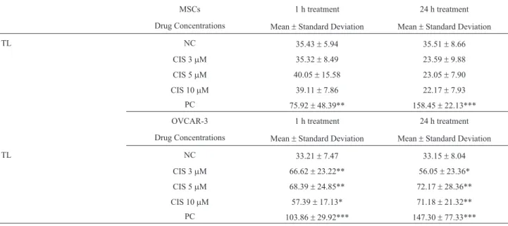
The slides were analyzed in an Olympus System Mi-croscope (Model BX41) equipped with a Olympus Re-flected Fluorescence System (Model U-RFL-T) and Olympus U-TV0.35XC-2 Camera (Tokyo, Japan). After coding and blinding of the slides, they were stained with ethidium bromide solution and the comets were determined by an image analysis system (Comet Assay IV, Perceptive Instruments, UK). Four slides with 25 cells (total of 100 cells) for every test sample were counted and analyzed for the Tail Length (TL) parameter to quantify the induced DNA damage. Data from TL are given in Table 1. Results are given as mean ± standard deviation (SD).
Statistical analysis of the obtained data was per-formed using the SPSS software, version 13.0. To analyze differences, One Way ANOVA with Dunnett post hoc test was applied, where the drug treatments were compared against the negative control. Differences were considered statistically significant when the P-value was less than 0.05.
After 72 h of treatment, MSCs, but not OVCAR-3 cells, showed resistance to the increasing concentrations of CIS when evaluated by MTT assay (Figure 2, black bars). We assumed a reduction above 50% of cell viability for treated cells in relation to NC as indication of cytotoxicity. The cell percentage for OVCAR-3 cells was highly re-duced, in comparison with the NC, showing their sensitiv-ity to the drug (Figure 2, white bars). In addition, due to the high cytotoxic effect of CIS on OVCAR-3 cells, we calcu-lated the IC50value, that was found to be 2.86mM.
We assessed genotoxic potential of CIS by Comet as-say during two times of exposure (1 and 24 h) by tail length (TL) analysis, as migration distance of DNA inmm. After the two times of exposure to the drug, no DNA damage was observed in MSCs for all tested dosages, with exception of PC (Table 1). However, CIS was able to induce DNA dam-age in OVCAR-3 cells during the two times of exposure (Table 1). After 1 h of treatment, all dosages of CIS caused a significant increase in DNA migration (TL) in OVCAR-3 cells in comparison to NC. TL was also significantly higher
in OVCAR-3 cells treated with CIS after 24 h of exposure, at 3, 5 and 10mM dosages, in relation to NC.
In this study we demonstrated the resistance of hu-man adipose-derived MSCs to the exposure of increasing concentrations of CIS during 72 h ofin vitro cultivation (Figure 2, black bars). Our data are in agreement with re-sults obtained by Lianget al.(2011), which showed the re-sistance and recovery of human adipose-derived MSCs to CIS exposure. They also demonstrated that MSCs retain their phenotypical characteristics, such as a fibroblast-like morphology and stem cell marker expression, as well as their multilineage differentiation capacity.
In contrast, CIS was able to dramatically reduce the viability of OVCAR-3 cells after 72 h of treatment (Figure 2, white bars). The IC50value of CIS for OVCAR-3 cells
was 2.86mM, which means that CIS was, at least, 17 fold more cytotoxic for OVCAR-3 cells than for MSCs, since the highest tested dosage of CIS (50mM) was not able to re-duce MSC viability above 50%. The sensitivity of OVCAR-3 cells to CIS is in accordance with previously data demonstrated by several studies (Smithet al., 2005; Karacaet al., 2013).
After confirming the resistance of MSCs to CIS we chose three dosages (3, 5 and 10 mM) to evaluate the genotoxic potential of this drug on MSCs and OVCAR-3 cells. Using the Comet assay, we were able to demonstrate, for the first time, the absence of DNA damage caused by CIS on MSCs (Table 1) after 1 and 24 h of treatment, in our experimental conditions. In contrast, CIS significantly in-creased DNA migration of OVCAR-3 cells comets (Table 1), showing its genotoxic effect. In addition, our results demonstrate that the concentrations of CIS used in this study were not associated with retarded DNA migration, as
expected by interstrand DNA cross-links, observed for treatments with concentrations above 50mM of CIS (Al-meidaet al., 2006; Panget al., 2007).
CIS is a strong genotoxic and mutagenic agent (Roos and Kaina, 2013). It is able to induce DNA damage in a broad range of eukaryotic cells, from Drosophila melanogasterto humans, eitherin vitro andin vivo. Be-cause of its capacity to Be-cause DNA adducts, CIS induced DNA strand breaks in D. melanogaster somatic cells in vivo, evaluated by the Comet assay (Garcia Saret al., 2012) and SMART test (Danesi et al., 2010). A large panel of mammalian cells have already been exposed to CIS, and its genotoxic potential has been confirmed on cells from ham-ster (Brozovicet al., 2009), mice (Narayana, 2012), rats (Mendonçaet al., 2010) and human normal and cancer cells (Blasiaket al., 2000; Shimabukuroet al., 2011). Although CIS is a widely used drug for the treatment of a broad range of cancers, tumor resistance to CIS is an issue to be sur-passed. The main mechanisms of cell resistance to CIS are described as: decreasing intracellular accumulation of CIS, increasing intracellular trapping of CIS, increased repair of DNA damage or increased tolerance of DNA damage and, finally, the mixture of a variety of others and before men-tioned mechanisms (Borstet al., 2008).
Here we demonstrated that human MSCs are strongly resistant to CIS exposure, considering this drug as a cyto-toxic and genocyto-toxic agent, but the mechanisms underlying this property are still poorly understood. It was shown that human bone marrow MSCs can be isolated from patients after high-dose or standard chemotherapy and the cells re-tained their MSCs characteristics (Mueller et al., 2006). These authors also demonstrated that MSCs have an ele-vated threshold for CIS-induced apoptosis, which was characterized by a lack of caspase-9 activity in apoptotic
Table 1- Results of the Comet assay on MSCs and OVCAR-3 cells after treatment with CIS.
MSCs 1 h treatment 24 h treatment
Drug Concentrations Mean±Standard Deviation Mean±Standard Deviation
TL NC 35.43±5.94 35.51±8.66
CIS 3mM 35.32±8.49 23.59±9.88
CIS 5mM 40.05±15.58 23.05±7.90
CIS 10mM 39.11±7.86 22.17±7.93
PC 75.92±48.39** 158.45±22.13***
OVCAR-3 1 h treatment 24 h treatment
Drug Concentrations Mean±Standard Deviation Mean±Standard Deviation
TL NC 33.21±7.47 33.15±8.04
CIS 3mM 66.62±23.22** 56.05±23.36*
CIS 5mM 68.39±24.85** 72.17±28.36**
CIS 10mM 57.39±17.13* 71.18±21.32**
cells and an increased p53 expression, independent of apoptosis induction (Muelleret al., 2006). p73 also seems to play a role in MSC resistance to CIS, since the over-expression induction of this apoptosis regulator sensibilizes human bone marrow MSCs to CIS treatment (Lianget al., 2010). Prendergastet al.(2011) showed that CIS can acti-vate DNA damage response pathways, including induction of p53 and p21, and activation of PI3 kinase-related protein kinase (PIKK)-dependent phosphorylation of histone H2AX on serine 139, and replication protein A2 on serine4/serine8, in human bone marrow MSCs. Taken to-gether, these findings indicate that the resistance of MSCs to CIS results from complex cellular pathways, involving alteration of apoptosis regulation and activation of mole-cules engaged in DNA repair process. Our data suggest that the tolerance of MSCs to DNA damage, potentially induced by CIS, could also be related to the resistance of these cells to the drug. Finally, we emphasize the need for further in-vestigations aiming to elucidate the mechanisms responsi-ble for MSC resistance to drugs, since the importance of these cells in the tumor microenvironment context is well known.
Acknowledgments
The authors thank CNPq, FAPERGS and ULBRA for financially support for this study.
References
Almeida GM, Duarte TL, Steward WP and Jones GD (2006) De-tection of oxaliplatin-induced DNA crosslinks in vitro and in cancer patients using the alkaline comet assay. DNA Re-pair (Amst) 5:219-225.
Blasiak J, Kowalik J, Malecka-Panas E, Drzewoski J and Woje-wodzka M (2000) DNA damage and repair in human lym-phocytes exposed to three anticancer platinum drugs. Teratog Carcinog Mutagen 20:119-131.
Borst P, Rottenberg S and Jonkers J (2008) How do real tumors become resistant to cisplatin? Cell Cycle 7:1353-359. Brozovic A, Damrot J, Tsaryk R, Helbig L, Nikolova T, Hartig C,
Osmak M, Roos WP, Kaina B and Fritz G (2009) Cisplatin sensitivity is related to late DNA damage processing and checkpoint control rather than to the early DNA damage re-sponse. Mutat Res 670:32-41.
da Silva Meirelles L, Chagastelles PC and Nardi NB (2006) Mesenchymal stem cells reside in virtually all post-natal or-gans and tissues. J Cell Sci 119:2204-2213.
da Silva Meirelles L, Caplan AI and Nardi NB (2008) In search of the in vivo identity of mesenchymal stem cells. Stem Cells 26:2287-2299.
Danesi CC, Bellagamba BC, Dihl RR, de Andrade HH, Cunha KS, Spano MA, Reguly ML and Lehmann M (2010) Mutagenic evaluation of combined paclitaxel and cisplatin treatment in somatic cells of Drosophila melanogaster. Mutat Res 696:139-143.
Dasari S and Tchounwou PB (2014) Cisplatin in cancer therapy: molecular mechanisms of action. Eur J Pharmacol 740:364-378.
Doorn J, Moll G, Le Blanc K, van Blitterswijk C and de Boer J (2012) Therapeutic applications of mesenchymal stromal cells: paracrine effects and potential improvements. Tissue Eng Part B Rev 18:101-115.
Galluzzi L, Senovilla L, Vitale I, Michels J, Martins I, Kepp O, Castedo M and Kroemer G (2012) Molecular mechanisms of cisplatin resistance. Oncogene 31:1869-1883.
Garcia Sar D, Aguado L, Montes Bayon M, Comendador MA, Blanco Gonzalez E, Sanz-Medel A and Sierra LM (2012) Relationships between cisplatin-induced adducts and DNA strand-breaks, mutation and recombination in vivoin so-matic cells of Drosophila melanogaster, under different conditions of nucleotide excision repair. Mutat Res 741:81-88.
Gomez-Ruiz S, Maksimovic-Ivanic D, Mijatovic S and Kalu-derovic GN (2012) On the discovery, biological effects, and use of Cisplatin and metallocenes in anticancer chemother-apy. Bioinorg Chem Appl 2012:140284.
Houthuijzen JM, Daenen LG, Roodhart JM and Voest EE (2012) The role of mesenchymal stem cells in anti-cancer drug re-sistance and tumour progression. Br J Cancer 106:1901-196. Jamieson ER and Lippard SJ (1999) Structure, recognition, and processing of Cisplatin-DNA adducts. Chem Rev 99:2467-2498.
Karaca B, Atmaca H, Bozkurt E, Kisim A, Uzunoglu S, Karabulut B, Sezgin C, Sanli UA and Uslu R (2013) Combination of AT-101/cisplatin overcomes chemoresistance by inducing apoptosis and modulating epigenetics in human ovarian can-cer cells. Mol Biol Rep 40:3925-3933.
Klopp AH, Gupta A, Spaeth E, Andreeff M and Marini F (2011) Concise review: dissecting a discrepancy in the literature: do mesenchymal stem cells support or suppress tumor growth? Stem Cells 29:11-19.
Kucerova L and Skolekova S (2013) Tumor microenvironment and the role of mesenchymal stromal cells. Neoplasma 60:1-10.
Li J, Law HK, Lau YL and Chan GC (2004) Differential damage and recovery of human mesenchymal stem cells after expo-sure to chemotherapeutic agents. Br J Haematol 127:326-334.
Liang W, Lu C, Li J, Yin JQ and Zhao RC (2010) p73alpha regu-lates the sensitivity of bone marrow mesenchymal stem cells to DNA damage agents. Toxicology 270:49-56.
Liang W, Xia H, Li J and Zhao RC (2011) Human adipose tissue derived mesenchymal stem cells are resistant to several chemotherapeutic agents. Cytotechnology 63:523-530. Mendonça LM, dos Santos GC, dos Santos RA, Takahashi CS,
Bianchi M de L and Antunes LM (2010) Evaluation of curcumin and cisplatin-induced DNA damage in PC12 cells by the alkaline comet assay. Hum Exp Toxicol 29:635-643. Mueller LP, Luetzkendorf J, Mueller T, Reichelt K, Simon H and
Schmoll HJ (2006) Presence of mesenchymal stem cells in human bone marrow after exposure to chemotherapy: evi-dence of resistance to apoptosis induction. Stem Cells 24:2753-2765.
Narayana K (2012) Effects of L-ascorbic acid on two cycles of cisplatin-induced DNA double-strand breaks and phos-phorylation of p53 in the liver. Exp Toxicol Pathol 64:495-502.
anticancer complexes. Biochem Biophys Res Commun 363:235-240.
Prendergast AM, Cruet-Hennequart S, Shaw G, Barry FP and Carty MP (2011) Activation of DNA damage response path-ways in human mesenchymal stem cells exposed to cisplatin or gamma-irradiation. Cell Cycle 10:3768-3777.
Roos WP and Kaina B (2013) DNA damage-induced cell death: from specific DNA lesions to the DNA damage response and apoptosis. Cancer Lett 332:237-248.
Samples J, Willis M and Klauber-Demore N (2013) Targeting angiogenesis and the tumor microenvironment. Surg Oncol Clin N Am 22:629-639.
Sancho-Martinez SM, Prieto-Garcia L, Prieto M, Lopez-Novoa JM and Lopez-Hernandez FJ (2012) Subcellular targets of cisplatin cytotoxicity: an integrated view. Pharmacol Ther 136:35-55.
Schaffler A and Buchler C (2007) Concise review: adipose tis-sue-derived stromal cells—basic and clinical implications for novel cell-based therapies. Stem Cells 25:818-827. Sebaugh JL (2011) Guidelines for accurate EC50/IC50
estima-tion. Pharm Stat 10:128-134.
Shimabukuro F, Neto CF, Sanches Jr JA and Gattas GJ (2011) DNA damage and repair in leukocytes of melanoma patients exposed in vitro to cisplatin. Melanoma Res 21:99-105.
Smith JA, Ngo H, Martin MC and Wolf JK (2005) An evaluation of cytotoxicity of the taxane and platinum agents combina-tion treatment in a panel of human ovarian carcinoma cell lines. Gynecol Oncol 98:141-145.
Tice RR, Agurell E, Anderson D, Burlinson B, Hartmann A, Kobayashi H, Miyamae Y, Rojas E, Ryu JC and Sasaki YF (2000) Single cell gel/comet assay: guidelines for in vitro and in vivo genetic toxicology testing. Environ Mol Mutagen 35:206-221.
Unger FT, Klasen HA, Tchartchian G, de Wilde RL and Witte I (2009) DNA damage induced by cis- and carboplatin as in-dicator for in vitro sensitivity of ovarian carcinoma cells. BMC Cancer 9:1471-2407.
Zhu X, Shi W, Tai W and Liu F (2012) The comparison of biologi-cal characteristics and multilineage differentiation of bone marrow and adipose derived mesenchymal stem cells. Cell Tissue Res 350:277-287.
Zuk PA, Zhu M, Ashjian P, De Ugarte DA, Huang JI, Mizuno H, Alfonso ZC, Fraser JK, Benhaim P and Hedrick MH (2002) Human adipose tissue is a source of multipotent stem cells. Mol Biol Cell 13:4279-4295.
Associate Editor: Carlos R. Machado