*Correspondence: Lee Chen Chen. Departamento de Biologia Geral, Instituto de Ciências Biológicas, Universidade Federal de Goiás -Campus II - 74001-970 – Goiânia - GO, Brasil. E-mail: [email protected]
A
rti
Pharmaceutical Sciences vol. 46, n. 4, out./dez., 2010
Cytotoxic and genotoxic investigation on barbatimão
[
Stryphnodendron adstringens
(Mart.) Coville, 1910] extract
Juliana Brandstetter Vilar
1,*, Maria Inez Prudente D’Oliveira
1, Suzana da Costa Santos
2,
Lee Chen Chen
11Mutagenesis and Microorganisms Radiobiology Laboratory, Biological Sciences Institute, Federal University of Goiás, 2Molecular Bioactivity Laboratory, Chemical Institute, Federal University of Goiás
Stryphnodendron adstringens (Mart.) Coville, 1910 is a small tree, distributed widely throughout the Cerrado region of Brazil and named “barbatimão” by the Tupi-Guarani tribes, which presents astringent properties. Its ethnopharmacological uses comprise, among others, anti-inlammatory and wound healing action, and it is used in the treatment of diarrhea and gynecological problems. The phytotherapeutic use of ‘barbatimão’ is largely related to its tannin content, which is abundant in its bark. The main goal of the present study was to evaluate the cytotoxic, mutagenic, and genotoxic potential of the lyophilized solution of the stem bark of S. adstringens, using the Ames test, the Inductest and the SOS-Chromotest. S. adstringens presented cytotoxic activity in all tested systems, did not present mutagenic activity detectable by the Ames test and SOS-Chromotest, and showed some genotoxic effect on the SOS-Inductest. However, the metabolization of the extract by S9 fraction attenuated its genotoxic and cytotoxic activities.
Uniterms:Stryphnodendron adstringens/cytotoxicity. Stryphnodendron adstringens/genotoxicity. Barbatimão/phytochemical study.
Stryphnodendron adstringens (Mart.) Coville, 1910 é uma pequena árvore amplamente distribuída nas regiões de cerrado do Brasil, chamada de “barbatimão” pelas tribos Tupi-Guarani, que apresenta propriedade adstringente. Seu uso etnofarmacológico compreende, entre outros, efeitos antiinlamatório e cicatrizante, sendo empregada no tratamento de diarréias e problemas ginecológicos. Grande parte das aplicações do itoterápico de barbatimão está relacionada aos taninos, abundantes em sua casca. O objetivo do presente trabalho foi avaliar os potenciais citotóxico, mutagênico e genotóxico da solução lioilizada da casca de S. adstringens, utilizando Teste de Ames, SOS-Induteste e SOS-Cromoteste. S. adstringens
apresentou atividade citotóxica em todos os sistemas testados, não apresentou atividade mutagênica detectável pelo teste de Ames e SOS-Cromoteste e mostrou certo efeito genotóxico no SOS-Induteste. Porém, a metabolização do extrato pela fração S9 atenuou suas atividades genotóxica e citotóxica.
Unitermos: Stryphnodendron adstringens/citotoxicidade. Stryphnodendron adstringens/genotoxicidade.
Stryphnodendron adstringens/mutagênese. Barbatimão/estudo itoquímico.
INTRODUCTION
Since the origin of mankind and to this day, medi-cinal herbs have been used to treat diseases worldwide. Although countless plant species have long been used in folk medicine (González-Ávila et al., 2003), only recently have the pharmacology and toxicity of these plants begun
to receive attention from scientists. Many studies have been carried out to verify the claimed pharmacological and/or therapeutic properties of these plants, isolate their active constituents, and investigate their possible toxicities (Rebecca et al., 2002).
heal wounds, and treat various diseases, such as diarrhea, leucorrhea, and gynecological problems (Audi et al., 1999; Bersani-Amado et al., 1996; Lima, Martins, Souza Jr., 1998; Neves et al., 1992a,b; Panizza et al., 1988; Santos; Torres; Leonart, 1988). Due to its importance as a phyto-therapic agent, this plant was included in the Brazilian Pharmacopoeia and so far its pharmacological activities have been attributed mainly to the tannins present in the bark (Brandão et al., 2006; Santos et al., 2002).
According to Santos and coworkers (2002), the condensed tannins from Stryphnodendron genus present molecules composed of prodelphinidin units, formed by gallocathechin and/or epigallocatechin, which con-tain ortho-trihydroxyl groups in the β-ring. Free radical scavenging activity is enhanced by the presence of three β-ring hydroxyl groups in the condensed tannin structu-re (De Bruyne et al., 1999a,b; Hagerman et al., 1998). Ortho-trihydroxyl groups in β-ring potentiate the antivi-ral activity of condensed tannins, while galloylation of condensed tannins in a speciic position (3-position), as found in this genus, presents anticancer activity (Dufresne; Farnworth, 2001). On the other hand, some authors have reported that the consumption of herbal tea rich in tannins has been proven to develop esophagus cancer and present chirrogenic, abortive, hepatocarcinogenic and hepatotoxic effects (Angell, Kassirer, 1998; Zhu et al., 1997).
Based on these facts, the aim of the present study was to evaluate the cytotoxic and genotoxic effects of the lyophilized solution of the stem bark of Stryphnodendron adstringens (Mart.) Coville, 1910 using the Ames Test, the SOS-Inductest, and the SOS-Chromotest.
MATERIAL AND METHODS
Plant material and preparation of Barbatimão extract (BE)
The ethanolic extract of “barbatimão”, obtained from bark decoction, was purchased from Klein Labora-tories Ltda. (lt 001, Porto Alegre, RS, Brazil). The aqueous extract was obtained from the ethanolic extract, according to the technique of vaporization under low pressure us-ing a rotary vacuum evaporator, at 40 °C. After complete
evaporation of ethanol, we added an equal volume of distilled water and lyophilized this solution, obtaining the lyophilized “barbatimão” extract (BE).
Strains
Escherichia coli SOS-Inductest tester strains, WP2s(λ) (uvrA)(AmpS) and RJF013 (uvrD3)(AmpR),
and Escherichia coli SOS-Chromotest tester strain PQ37 (rfa)(uvrA)(AmpR)(sulA: lacZ) were kindly provided by
the Laboratório de Radiobiologia Molecular from the Instituto de Biofísica Carlos Chagas Filho, Universidade Federal do Rio de Janeiro (Rio de Janeiro, RJ, Brazil).
Salmonella typhimurium strains TA97a (hisD6610) (rfa) (uvrB)(AmpR), TA98 (hisD3052)(rfa)(uvrB)(AmpR),
TA100 (hisG46)(rfa)(uvrB)(AmpR), and TA102 (hisG428)
(rfa)(uvrB)(AmpRTetR), as described by Maron and Ames
(1983), were kindly supplied by Dr. B. N. Ames, from the University of California (Berkeley, CA, USA).
Microsomal fraction
The postmicrosomal fraction S9 prepared from livers of Sprague-Dawley rats pre-treated with polychlo-rinated biphenyl mixture (Aroclor 1254) was purchased from Molecular Toxicology Inc. (MoltoxTM, Annapolis,
MD, USA). The S9 metabolic activation mixture was prepared according to Quillardet and Hofnung (1985) for the SOS-Chromotest and according to Maron and Ames (1983) for the SOS-Inductest and Salmonella/microsome assay.
Ames Test
Following the Salmonella typhimurium histidine point mutation assay, proposed by Maron and Ames (1983), a 0.1 mL bacterial suspension (1-2 x 10⁹ cells/
mL) of each Salmonella typhimurium strain (TA97a, TA98, TA100, and TA102) with 0.0, 0.25, 0.5, 1.0, 5.0, or 10 mg/plate BE was incubated, with or without S9 mix, at 37 °C for 25 min. Subsequently, 2.0 mL of top agar
(0.6% agar Difco, 0.5% NaCl, 50 µM L-histidine, 50 µM biotin, 45 °C) was added to test tubes and poured onto
Petri dishes with minimal agar (1.5% agar, 2% glucose, and Vogel-Bonner E medium). All assays included nega-tive (distilled water) and posinega-tive [15 µg 4-nitroquinoline 1-oxide (4NQO) per plate for TA97a and TA98, 1.5 µg of sodium azide for TA100 and 2.5 µg mitomycin C for TA102, data not shown] controls. After incubation at 37 °C
for 48 h, colonies (His+ revertants) were counted and the
results expressed as mutagenic index (MI = number of His+ induced in the sample/number of spontaneous His+
in the negative control). Bacterial survival was determined for TA98.
SOS-Inductest Prophage λ induction
growth phase culture (2x10⁸ cells/mL) of Escherichia coli WP2s(λ), grown in LB medium [1% bacto tryptone
(Difco), 0.5% bacto yeast extract (Difco) and 1% NaCl] was centrifuged (5000 rpm, 20 min), resuspended, and incubated at 37°C for 25 min with 0.0, 0.25, 0.5, 1.0, 5.0,
or 10 mg/tube BE (with or without S9 mix). The mixture was then diluted in M9 buffer (0.6% Na2HPO4, 0.3% KH2PO4, 1% NH4Cl, and 0.05% NaCl) and 0.1 mL of this
lysogenic culture [WP2s(λ)] was added to 0.3 mL
indica-tor strain (RJF013), after which 2.0 mL top agar at 45 °C
(0.6% agar Difco, 0.5% NaCl) was added to the mixture and poured onto Petri dishes with LB. All assays were performed in duplicate and included negative (distilled water) and positive [Cyclophosphamide (CP) and ultra-violet radiation, λ = 254nm, data not shown] controls. The
results were expressed as induction factor (IF = plaques of treated culture/plaques of untreated culture). Bacterial survival was determined for WP2s(λ).
SOS-Chromotest
The SOS-Chromotest was carried out as described by Quillardet and Hofnung (1985) with subsequent modifications (von der Hude et al., 1988). According to the standard procedure, BE was incubated with a 10-fold diluted exponential phase culture (108 cells/mL) of
the tester strain (PQ37) and the culture diluted in fresh medium (LB) with or without S9 mix. After incubat-ing this mixture for 2 h with 0.0, 0.25, 0.5, 1.0, 5.0, or 10 mg/tube BE, it was divided into two samples, one used for the β-galactosidase (βg) assay and the other for
the alkaline phosphatase (ap) assay. Appropriate buffers were added to disrupt the cell membranes and speciic substrates for each enzyme (4 mg/mL o -nitro-phenyl-galactopyranoside and 4 mg/mL p-nitro-phenyl-phosphate for βg and ap activities, respectively). After adding the
buffers to stop the enzymatic reaction (NaCO3 for βg and
HCl and Tris-HCl for ap), the absorbance of each assay was read against a colorimeter blank. βg and ap activities were calculated according to the simpliied method recom-mended by Quillardet and Hofnung (1993): enzyme units (U) = (A420x 1000)/t (A420 = optical density at 420 nm; t = substrate conversion time in minutes). Negative (dis-tilled water) and positive (mitomycin - MMC, data not shown) controls were also included in these experiments.
Exponential phase cultures of TA98 or WP2s(λ)
were centrifuged, resuspended and incubated at 37 °C for
25 min with 0.0, 0.25, 0.5, 1.0, 5.0, or 10 mg/tube BE. Each mixture was then diluted in M9 buffer and 0.1 mL of each treatment poured onto Petri dishes with Nutrient Agar or LB, for TA98 and E. coli, respectively. The results were
expressed as a Survival Fraction (SF), obtained as the rate between colonies of the treated culture and colonies of the untreated culture.
RESULTS
Ames test
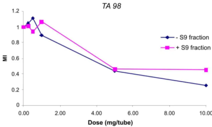
The mutagenic activity of BE was evaluated using the tester strains TA97a, TA98, TA100, and TA102 with or without S9 mix. All the results were generated by four independent experiments carried out in duplicate. A positive result is generally considered when (i) MI in one or more treatment groups is higher than in the control group by a gi-ven multiple, such as twice the negative control MI (positive results = MI in treatment group ≥ 2 spontaneous MI), and (ii) a dose-response relationship exists for at least several doses (Maron, Ames, 1983). Mutagenic Index (MI) as a function of concentrations of 0.25 to 10 mg/tube BE (with or without S9 fraction) is shown in Figures 1, 2, 3 and 4. All tested strains had similar proiles and the MI did not reach twice the respective numbers of spontaneous revertants.
The highest MI value was 1.74 (TA102, 0.5 mg/plate in the presence of metabolic activation), suggesting that neither BE nor its metabolites exhibited a mutagenic proile at any of the tested doses. Nevertheless, bacterial survival was considerably affected, as can be observed in Figure 5.
SOS-Inductest
To determine whether BE itself or its metabolic derivatives would act as inducing agents, the lysogens
FIGURE 1 - Mutagenic Index (MI) as a function of dose
were incubated in different doses of BE. The IF induced by BE, in the absence or presence of S9 fraction, is shown in Figure 6.
We observed an increase in the number of plaques as a function of BE dose and the maximum induction was observed at the dose of 5 mg/tube, which resulted in a 9-fold IF value compared to the negative control. The metabolized effect, however, did not increase the induc-tion of the prophage λ. The IF at the dose of 10 mg/tube
in the absence of S9 remained at the levels of the negative control. Bacterial survival was reduced by the treatment with BE, as observed in Figure 5.
SOS-Chromotest
When applying the SOS-Chromotest, genotoxic effects are assumed to exist whenever the induction
FIGURE 3 - Mutagenic Index (MI) as a function of dose
(mg/tube) of “barbatimão” extract (BE), with or without S9 fraction on TA100. The maximum coefficient of variation (CV) of experiments was 10%. Plots represent average of four independent experiments.
FIGURE 4 - Mutagenic Index (MI) as a function of dose (mg/
tube) of “barbatimão” extract (BE), with or without S9 fraction on TA102. The maximum coefficient of variation (CV) of experiments was 10%. Plots represent the average of four independent experiments.
FIGURE 2 - Mutagenic Index (MI) as a function of dose
(mg/tube) of “barbatimão” extract (BE), with or without S9 fraction on TA98. The maximum coeficient of variation (CV) of experiments was 10%. Plots represent average of four independent experiments.
FIGURE 5 - Survival Fraction (SF) as a function of dose (mg/
tube) of “barbatimão” extract (BE) on S. typhimurium TA98
and E. coli WP2s(λ). The maximum coeficient of variation
(CV) of experiments was 10%. Plots represent the mean of four independent experiments.
FIGURE 6 - Induction Factor (IF) as a function of dose (mg/
tube) of “barbatimão” extract (BE), with or without S9 fraction, on E. coli WP2s(λ). The maximum coefficient of variation
surpasses a deined level, and a positive result is gene-rally considered when (i) the enzymatic ratio increases at least 0.5 compared to the negative control, and (ii) a dose-response relationship exists for at least several doses. The decrease in the constitutive ap levels relects the cytotoxicity of a compound while the augmentation of the inducible SOS-dependent βg activity shows its
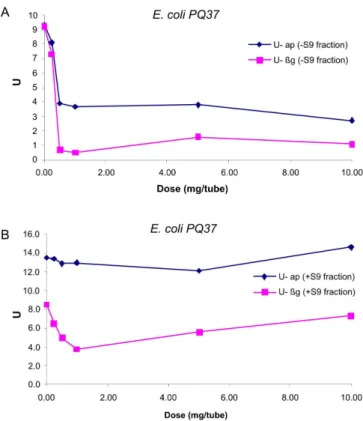
mutagenic potential (Von Der Hude et al., 1988). In the present study, BE did not induce SOS mutagenesis when the SOS-Chromotest was applied, neither in the absence nor in the presence of metabolic activation (Figures 7 A and B).
In the absence of the S9 mix, even at the initial do-ses, BE inhibited the ap enzyme while SOS-dependent βg
activity decreased at almost the same level. Nevertheless, when S9 mix was added to the complex, the cytotoxic effect of BE was attenuated and remained almost at the levels of ap U (enzymatic units) of the negative control. On the other hand, the enzyme U levels in the βg assay in
the presence of S9 fraction recovered slightly, and were clearly seen at the dose of 0.5 mg/tube).
FIGURE 7 - Enzymatic Units (U) of enzymes β-galactosidase
(βg) and alkaline phosphatase (ap) as a function of dose (mg/
tube) of “barbatimão” extract (BE), in the absence (A) or
presence (B) of S9 fraction on E. coli PQ37. The maximum
coeficient of variation (CV) of experiments was 10%. Plots represent average of four independent experiments.
DISCUSSION
Although there are differences in metabolism, DNA repair, and other physiological processes affecting chemi-cal mutagenesis according to species, the universality of DNA and the genetic code provides a rationale basis for using various non-human test systems aimed at predict-ing the intrinsic mutagenicity of the chemicals tested. It is known that a great variety of genetic events may lead to mechanisms that are likely to induce carcinogenicity and cytotoxicity. However, nowadays there is no speciic test that can be used alone to detect the full spectrum of differ-ent endpoints that reveal all manifestations of genotoxic actions (Dearield et al., 2002).
The Ames test was employed in this study, a test which is widely accepted to detect substances that can produce genetic damage leading to gene mutations. The
Salmonella strains we employed are able to detect both frameshift mutations (TA97a and TA98) and base-pair substitutions (TA100 and TA102) (Mortelmans; Zeiger, 2000). Nevertheless, BE did not induce direct or indirect mutagenicity (Figures 1, 2, 3, and 4). Cytotoxicity was also observed on TA98 (Figure 5).
Agents that inhibit DNA replication in E. coli trigger the expression of a series of cellular functions which nota-bly, lead to prophage λ induction. This property is explored
in the SOS-Inductest and has been used to screen potential chemical carcinogens and antitumor antibiotics (Anderson
et al., 1980). We observed an increase in IF as a function of BE dose, except for the dose of 10 mg/tube (Figure 6). A reasonable explanation for this inding is the high cytotoxicity observed at this concentration of BE (Figure 5). However, the metabolized effect did not increase the induction of prophage λ. Therefore these results suggest
that BE possesses direct mechanisms of genotoxicity and its metabolic activation by the post-microsomal S9 frac-tion is able to suppress the genotoxic effect, probably due to detoxiication of some extract components.
The SOS-Chromotest relects a possible aspect of the mutational process: error-prone DNA repair. It is also assumed that the majority of the cells respond when ex-posed to a mutagen (Rosenkranz, Mersch- Sundermann, Klopman, 1999). BE was not able to induce SOS mutagen-esis in the SOS-Chromotest, with or without the oxidative metabolism (Figures 7A and 7B). In the absence of S9 mix, BE early inhibited ap and SOS-dependent βg activity also
were in its absence. This could be explained by the fact that the number of mutational events, and consequently the enzyme U levels in the ßg assay, is affected by the number of viable cells. Our results suggest that BE possesses direct mechanisms of cytotoxicity and no genotoxic activity detectable by this test.
These data show that BE did not exhibit consider-able mutagenicity. However, it presented some genotox-icity revealed by an increase in prophage λ induction data
using the SOS-Inductest. A toxic effect was also observed for the strains employed in the three assays, especially on the SOS-Chromotest, since the constitutive enzyme ap was early inhibited. According to Houk and DeMarini (1988), differences in detection sensitivity could be at least partially explained by the fact that phage induction and the SOS response in general (i.e. prophage λ
induc-tion of the Inductest) are broader genetic endpoints than reverse mutation in bacteria (i.e. Ames test) because they occur due to a variety of mechanisms and involve a num-ber of classes of genetic damage, including alkylations, intercalations, strand scissions, oxidative deaminations, and crosslinks. In contrast, there is a close correlation between the results yielded by the SOS-Chromotest and the Ames test. The capacity of the Ames test to identify carcinogens is higher than that of the SOS-Chromotest. However, because the number of false positive com-pounds is lower in the SOS-Chromotest, it is suggested that both tests must be used so that one can comple-ment the other (Quillardet, Hofnung, 1993). Thus, the concurrent use of these tests improves the sensitivity of the screening procedure in the evaluation of plants com-monly used in folk medicine.
We suggest that the decrease in enzyme U levels observed in the SOS-Chromotest and the genotoxic activity detected in the SOS-Inductest could be at least partially affected by the tannin fractions commonly found in S. adstringens bark. These tannin fractions possess a well known ability to bind to proteins (leading to the formation of large protein aggregates) and precipitate (Naczk et al., 2001; Silber et al., 1998), properties that could affect both the enzymatic activity and the DNA replication by the formation of crosslinks with required proteins. Literature data also support that tannic acid and its hydrolyzed products lack mutagenic activity in
Salmonella tester strains (Chen, Chung, 2000). When the
S. adstringens extract was tested in eukaryotic systems, using the micronucleus test in mice bone marrow (Mota, 1997) and the SMART test in Drosophila melanogaster
(Sousa et al., 2003), conlicting results were obtained, inasmuch as the genotoxic effect of BE was reported in mice, but not in fruit lies. In contrast to our results,
the-se indings could relect some divergence in the in vivo
metabolism of the extract constituents.
In conclusion, according to our results, S. adstrin-gens presented cytotoxic activity in all tested systems, did not present mutagenic activity, but demonstrated some genotoxic effects. Moreover, the metabolization of the extract of this plant reduced its genotoxic and cytotoxic activities. Taken together, the data available on the geno-toxicity of S. adstringens are not fully conclusive and thus further in vitro and in vivo studies are needed to clarify its mechanisms of action and to better determine its risk for human consumption.
ACKNOWLEDGEMENTS
This study was supported by the Conselho Nacional de Desenvolvimento Científico e Tecnológico (CNPq, Brazil) and the Fundação de Apoio à Pesquisa de Goiás (FUNAPE).
REFERENCES
ANDERSON, W.A.; MOREAU, P.L.; DEVORET, R.; MARAL,
R. Induction of prophage λ by daunorubicin and derivates
correlation with antineoplastic activity. Mutat. Res., v.77,
n.3, p.197-208, 1980.
ANGELL, M.; KASSIRER, J. P. Alternative medicine: The risks
of untested and unregulated remedies. N. Engl. J. Med.,
v.339, n.12, p.839-844, 1998.
AUDI, E.A.; TOLEDO, D.P.; PERES, P.G.; KIMURA, E.; PEREIRA, W.K.V.; MELLO, J.C.P.; NAKAMURA, C.V.; ALVES-DO-PRADO, W.; CUMAN, R.K.N.; BERSANI-AMADO, C.A. Gastric antiulcerogenic effects of Stryphnodendron adstringens in rats. Phytother. Res., v.13, n.3, p.264-266, 1999.
BERSANI-AMADO, C.A.; NAKAMURA, C.V.; NAKAMURA, T.U.; MARTINEZ, M.; MELLO, J.C.P. Avaliação das atividades antiinflamatória e antibacteriana do extrato
bruto do Stryphnodendron adstringens (barbatimão). In:
SIMPÓSIO DE PLANTAS MEDICINAIS DO BRASIL,
14., 1996, Florianópolis. Resumos. Florianópolis: SPMB,
1996. p.14.
BRANDÃO, M.G.L.; COSENZA, G.P.; MOREIRA, R.A.; MONTE-MOR, R.L.M. Medicinal plants and other botanical
products from the Brazilian Oficial Pharmacopoeia. Braz.
CHEN, S.C.; CHUNG, K.T. Mutagenicity and antimutagenicity
studies of tannic acid and its related compounds. Food
Chem. Toxicol., v.38, n.1, p.1-5, 2000.
DEARFIELD, K.L.; CIMINO, M.C.; McCARROLL, N.E.; MAUER, I.; VALCOVIC, L.R. Genotoxicity risk
assessment: a proposed classiication strategy. Mutat. Res.,
v.521, n.1-2, p.121-135, 2002.
DE BRUYNE, T.; PIETERS, L.; DEELSTRA, H.; VLIETINCK, A.J. Condensed vegetable tannins: Biodiversity in structure
and biological activities. Biochem. Syst. Ecol., v.27, n.4,
p.445-459, 1999a.
DE BRUYNE, T.; PIETERS, L.; WITVROUW, M.; DE CLERCQ, E.; BERGHE, D.V.; VLIETINCK, A.J. Biological evaluation of proanthocyanidin dimers and
related polyphenols. J. Nat. Prod., v.62, n.7, p.954-958,
1999b.
DUFRESNE, C.J.; FARNWORTH, E.R. A review of latest research indings on the health promotion properties of tea.
J. Nutr. Biochem., v.12, n.7, p.404-421, 2001.
GONZÁLEZ-AVILA, M.; ARRIAGA-ALBA, M.; GARZA, M.; HERNÁNDEZ-PRETELÍN, C.M.; DOMÍNGUEZ-ORTÍZ, M.A.; FATTEL-FAZENDA, S.; VILLA-TREVIÑO, S. Antigenotoxic, antimutagenic and ROS scavenging
activities of a Rhoeo discolor ethanolic crude extract.
Toxicol. in Vitro, v.17, n.1, p.77-83, 2003.
HAGERMAN, A.E.; RIEDL, K.M.; JONES, G.A.; SOVIK, K.N.; RITCHARD, N.T.; HARTZFELD, P.W.; RIECHEL, T.L. High molecular weight plant polyphenolics (tannins)
as biological antioxidants. J. Agric. Food Chem., v.46, n.5,
p.1887-1892, 1998.
HOUK, V.S.; DEMARINI, D.M. Use of the microscreen phage-induction assay to assess the genotoxicity of 14 hazardous
industrial wastes. Environ. Mol. Mutagen., v.11, n.1,
p.13-29, 1988.
LIMA, J.C.S.; MARTINS, D.T.O.; SOUZA, JR.P.T.
Experimental evaluation of stem bark of Stryphnodendron
adstringens (Mart.) Coville for antiinlammatory activity.
Phytother. Res., v.12, n.3, p.218-220, 1998.
MARON, D.M.; AMES, B.N. Revised methods for the
Salmonella mutagenicity test. Mutat. Res., v.113, n.3-4,
p.173-215, 1983.
MOREAU, P.; BAILONE, A.; DEVORET, R. Prophage γ
induction in Escherichia coli K12 envA uvrB: a highly
sensitive test for potential carcinogenesis. Proc. Natl. Acad.
Sci. U S A, v.73, n.10, p.3700-3704, 1976.
MORTELMANS, K.; ZEIGER, E. The Ames Salmonella/
microsome mutagenicity assay. Mutat. Res., v.455, n.1-2,
p.29-60, 2000.
MOTA, I.C.S. Avaliação da atividade mutagênica de
Stryphnodendron adstringens Mart. através do teste de
micronúcleo em camundongos (Mus musculus) out bred linhagem Swiss Webster. Goiânia, 1997. 20 f. [Dissertation of Master’s in Biology Genetics. Universidade Federal de Goiás].
NACZK, M.; AMAROWICZ, R.; ZADERNOWSKI, R.;
SHAHIDI. F.Protein precipitating capacity of condensed
tannins of beach pea, canola hulls, evening primrose and
fava bean. Food Chem., v.73, n.4, p.467-471, 2001.
NEVES, M.C.L.; JORGE NETO, J.; IFA, D.R.; FRACASSO, J.F.; LEPERA, E.Z.P.; SILVA, R.F.P. Estudo dos efeitos farmacológicos de hamamelis e barbatimão. In: SIMPÓSIO DE PLANTAS MEDICINAIS DO BRASIL, 12., 1992,
Curitiba. Resumos. Curitiba: SPMB, 1992a. p.12.
NEVES, M.C.L.; JORGE NETO, J.; IFA, D.R.; FRACASSO, J.F.; LEPERA, E.Z.P.; SILVA, R. F.P. Estudo dos efeitos farmacológicos produzidos pelos extratos aquosos de hamamelis e barbatimão. In: REUNIÃO ANUAL DA FEDERAÇÃO DE SOCIEDADES DE BIOLOGIA
EXPERIMENTAL, 8., 1992, Caxambu. Resumos. São
Paulo: FeSBE, 1992b. p.7.
PANIZZA, S.; ROCHA, A.B.; GECCHI, R.; SOUZA E SILVA,
R.A.P. Stryphnodendron barbadetiman (Vellozo) Martius:
teor em tanino na casca e sua propriedade cicatrizante. Rev.
Ciênc. Farm., v.10, p.101-106, 1988.
QUILLARDET, P.; HOFNUNG, M. The SOS chromotest, a colorimetric bacterial assay for genotoxins: procedure.
Mutat. Res., v.147, n.3, p.65-78, 1985.
QUILLARDET, P.; HOFNUNG, M. The SOS chromotest: a
REBECCA, M.A.; ISHII-IWAMOTO, E.M.; GRESPAN, R.; CUMAN, R.K.N.; CAPARROZ-ASSEF, S.M.; MELLO, J.C.P.; BERSANI-AMADO, C.A. Toxicological studies on
Stryphnodendron adstringens. J. Ethnopharmacol., v.83, n.1-2, p.101-104, 2002.
ROSENKRANZ, H.S.; MERSCH- SUNDERMANN, V.; KLOPMAN, G. SOS-chromotest and mutagenicity in
Salmonella: evidence for mechanistic differences. Mutat.
Res., v.431, n.1, p.31-38, 1999.
SANTOS, C.A.M.; TORRES, K.R.; LEONART, R. Plantas
medicinais: herbarium, flora et scientia. 2.ed. Curitiba: Scientia et Labor, 1988. 160 p.
SANTOS, S.C.; COSTA, W.F.; RIBEIRO, J.P.; GUIMARÃES, D.O.; FERRI, P.H.; FERREIRA, H.D.; SERAPHIN, J.C.
Tannin composition of barbatimão species. Fitoterapia,
v.73, n.4, p.292-299, 2002.
SILBER, M.L.; DAVITT, B.B.; KHAIRUTDINOV, R.F.; HURST, J.K. A mathematical model describing tannin–
protein association. Anal. Biochem., v.263, n.1, p.46-50,
1998.
SOUSA, N.C.; CARVALHO, S.; SPANÓ, M.A.; GRAF, U. Absence of genotoxicity of a phytotherapeutic extract from
Stryphnodendron adstringens (Mart.) Coville in somatic
and germ cells of Drosophila melanogaster. Environ. Mol.
Mutagen., v.41, n.4, p.293-299, 2003.
VON DER HUDE , W.; BEHM, C.; GÜRTLER, R.; BASLER,
A. Evaluation of the SOS Chromotest. Mutat. Res., v.203,
n.2, p.81-94, 1988.
ZHU, M.; PHILLIPSON, J.D.; GREENGRASS, P.M.; BOWERY, N.E.; CAI, Y. Plant polyphenols: biologically active compounds or non selective binders to protein?
Phytochemistry, v.44, n.3, p.441-447, 1997.
Received for publication on 18th May 2009.